MCCC2 / Methylcrotonoyl-CoA carboxylase beta chain, mitochondrial · IHC design guide

Design Immunohistochemistry for MCCC2

This guide uses high-staining hepatocytes and adipocytes with no detected staining as IHC tissue controls (HPA tissue IHC). Interpret the reported cytoplasmic pattern alongside MCCC2’s mitochondrial matrix location, and account for medium staining/RNA consistency when assessing results (HPA tissue IHC; UniProt).

Evidence assembled Oct 2026 · For research use; verify linked source records and product datasheet before use
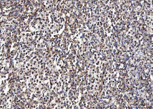
Immunohistochemistry protocol sheet for MCCC2 (IHC for MCCC2): expected localisation Cytoplasmic tissue staining (HPA tissue IHC); mitochondrial matrix location (UniProt), antibody A06515-2, validated IHC image, and IHC protocol steps
Printable MCCC2 IHC protocol sheet — expected localisation Cytoplasmic tissue staining (HPA tissue IHC); mitochondrial matrix location (UniProt), antibody A06515-2, controls and protocol steps. Open the full MCCC2 IHC guide →

MCCC2 Immunohistochemistry Experimental Design Guide

Expected localisation, validated protocols, controls and antibodies — the at-a-glance facts below, then the full design guide.

Must know before staining
Expected localisation Cytoplasmic tissue staining (HPA tissue IHC); mitochondrial matrix location (UniProt)
Staining pattern Cytoplasmic staining in most tissues; high in hepatocytes (HPA tissue IHC)
Antigen retrieval EDTA pH 8.0 HIER, heat-mediated (datasheet A06515-2)
Positive control ⓘ Appendix+4 more · see all
Negative control ⓘ Adipose tissue+4 more · see all
Important caveats
Reasons your staining may differ from the expected pattern.
Fixation Keep fixation consistent across sections (standard IHC practice; not target-specific)
Caveat Staining/RNA consistency is medium; verify tissue signal (HPA tissue IHC)
Regulation Liver-enhanced RNA expression (HPA tissue RNA)
Isoform / epitope Two isoforms; mature chain spans residues 23–563; epitope impact unknown (UniProt)
Section 1

Recommended MCCC2 IHC & IF Protocols

The catalog antibody uses EDTA pH 8.0 retrieval (datasheet: A06515-2). Published IHC protocols for prostate, kidney, and colorectal tissue follow (PMC7395692; PMC13347088; PMC10571261).

Recommended immunohistochemistry (IHC-P) protocol parameters
SampleParaffin-embedded human breast cancer tissue; fixative not specified (datasheet A06515-2)
FixationImage fixative and duration unreported (datasheet A06515-2); verify before use.
Sectioning4–5 µm sections on charged slides (standard)
DeparaffinisationXylene, graded ethanol series to water (standard)
Antigen retrievalHeat retrieval: EDTA pH 8.0 (datasheet A06515-2); 20 min, 95–100 °C (standard)
Peroxidase block3% H2O2, 10 min, room temperature (standard)
Blocking10% goat serum (datasheet A06515-2)
Primary antibodyRabbit anti-MCCC2, 2-5 μg/ml (datasheet A06515-2)
Primary incubationOvernight at 4 °C (datasheet A06515-2)
DetectionHRP-conjugated secondary, DAB chromogen (datasheet A06515-2)
CounterstainHematoxylin, blue, dehydrate and mount (standard)
Expected resultMCCC2-positive staining in glandular cells of appendix (HPA tissue IHC: High). HPA tissue profile: Cytoplasmic expression in most tissues. No signal in the no-primary control.
💡Decision noteStart with heat-mediated EDTA pH 8.0 for the catalog antibody (datasheet: A06515-2); the published protocols use citrate retrieval (PMC7395692; PMC13347088; PMC10571261).
Section 2

What Is the Expected MCCC2 Staining Pattern?

MCCC2 is a mitochondrial matrix protein with no transmembrane segment (UniProt Q9HCC0: location and topology). In paraffin sections, expect cytoplasmic staining in glandular cells, hepatocytes, cardiomyocytes, and kidney collecting ducts (HPA: tissue IHC). HPA describes cytoplasmic expression in most tissues; its tissue profile is Enhanced, with medium agreement between staining and RNA data and external verification pending (HPA: tissue IHC reliability).

What am I looking at on my slide?
Cytoplasmic staining in liver hepatocytes and colon or duodenum glandular cells.This matches HPA High protein staining in those cells (HPA: tissue IHC). Read a mitochondrial matrix protein's chromogenic signal within the cytoplasm; IHC alone does not prove organelle identity (UniProt Q9HCC0: location; standard IHC interpretation).
Predominantly nuclear, membranous, or extracellular staining in otherwise positive cells.These compartments disagree with the mitochondrial matrix assignment and lack support in the HPA cytoplasmic profile (UniProt Q9HCC0: location and topology; HPA: tissue IHC). Reassess specificity and detection controls before scoring them as MCCC2 (standard IHC practice).
Strong staining in adipocytes, skeletal myocytes, or smooth muscle cells.HPA reports MCCC2 as Not detected in these cell types (HPA: tissue IHC). Unexpected signal may reflect cross-reactivity or endogenous detection activity; check controls before calling it target expression (standard IHC practice).
Diffuse color across cells and tissue, including areas expected to be negative.An indiscriminate signal does not match HPA's cell-specific High and Not detected observations (HPA: tissue IHC). Background from the detection system or insufficient blocking is possible; compare controls and staining conditions (standard IHC practice).
No signal in liver hepatocytes or heart cardiomyocytes.Both are High in HPA tissue IHC, so an absent signal warrants a technical check (HPA: tissue IHC). Check the IHC-validated antibody's documented procedure and positive control before interpreting a sample as negative (standard IHC practice).
💡Expected MCCC2 appearanceCall a positive result when hepatocytes or other HPA High cell types show clear cytoplasmic signal; isolated nuclear staining or strong signal in HPA Not detected cell types is suspect (HPA: tissue IHC; UniProt Q9HCC0: location).
How each factor affects the staining
Compartment and topologyMCCC2 is assigned to the mitochondrial matrix and has no transmembrane segment (UniProt Q9HCC0: location and topology). Judge chromogenic IHC chiefly by cytoplasmic distribution and the stained cell type (HPA: tissue IHC; standard IHC interpretation).
Choice of tissue controlLiver hepatocytes, heart cardiomyocytes, and appendix glandular cells are High; adipocytes and skeletal myocytes are Not detected (HPA: tissue IHC). These observations offer contrasting controls, but HPA's Enhanced profile awaits external verification (HPA: reliability).
RNA and protein evidenceLiver is tissue enhanced at the RNA level, while hepatocytes are High by tissue IHC (HPA: RNA specificity and tissue IHC). Use the observed protein pattern for slide interpretation; RNA enrichment does not specify chromogenic intensity in every liver cell (standard IHC interpretation).
Antibody validationHPA038300, HPA038301, and HPA061546 each have Enhanced IHC status (HPA: antibody validation). This supports the reported pattern, while the tissue profile's medium RNA agreement and pending external verification remain relevant limits (HPA: tissue IHC reliability).
Sequence and processingUniProt lists two isoforms and a processed chain spanning residues 23–563 (UniProt Q9HCC0: isoforms and processing). No epitope position is supplied, so these records cannot establish which isoforms the IHC-validated antibody detects (UniProt Q9HCC0; supplied antibody data).
IF/ICC Q&AQ: What localisation should IF/ICC show? A: Mitochondrial signal, as reported in HPA's enhanced subcellular data (HPA: ICC-IF). That observation supports the localisation expectation; it does not establish an IHC-P retrieval or detection setting (HPA: ICC-IF; standard assay distinction).
Why is my staining missing, weak or wrong?
SituationLikely causeNext action
Liver control is blank.HPA reports High hepatocyte staining, making a technical failure plausible (HPA: tissue IHC).Check the IHC-validated antibody's IHC-P procedure, dilution, retrieval, detection reagents, and control section (standard IHC practice); no target-specific retrieval condition is supplied.
Color is widespread, including adipocytes.Adipocytes are Not detected in HPA; excess chromogen or detection background is possible (HPA: tissue IHC; standard IHC practice).Compare a no-primary control, review blocking and detection, and adjust development time using the documented IHC-P procedure (standard IHC practice).
Strong nuclear signal dominates.Nuclear staining conflicts with the matrix location and HPA cytoplasmic profile (UniProt Q9HCC0: location; HPA: tissue IHC).Check localisation in a known-positive tissue and compare the no-primary control before scoring; review antibody specificity if nuclear signal persists (standard IHC practice).
Smooth muscle stains as strongly as adjacent glands.HPA lists smooth muscle cells as Not detected but several glandular cell populations as High (HPA: tissue IHC).Confirm cell identity on the counterstained section and compare detection controls; treat persistent smooth muscle staining as unverified (standard IHC practice).
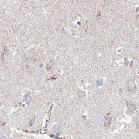
Only faint staining appears in hippocampal neurons.HPA reports Low staining in hippocampal neuronal cells, so faint signal may fit the reference (HPA: tissue IHC).Check a High control, such as liver hepatocytes, before changing staining conditions or calling the run insensitive (HPA: tissue IHC; standard IHC practice).
Expected cytoplasmic staining varies between sections.The supplied sources establish tissue patterns but give no MCCC2-specific fixation sensitivity or retrieval setting (HPA: tissue IHC; UniProt Q9HCC0).Compare section and run controls, then follow the IHC-validated antibody's documented IHC-P procedure consistently (standard IHC practice); avoid assigning the variation to MCCC2-specific fixation effects.

Sample controls for MCCC2 IHC & IF

🧪Run colon first: glandular cells should stain strongly (HPA: Colon glandular cells, High). Use adipose tissue as the negative tissue; adipocytes should be unstained (HPA: Adipose tissue adipocytes, Not detected). On the colon slide, identify unstained nonglandular cells by morphology as internal negative cells; HPA does not specify a negative cell type within colon.
Positive control tissue: Appendix (Glandular cells, HPA High)
Negative control tissue: Adipose tissue (HPA Not detected)
ICC-IF cell lines (HPA subcellular resource): HPA ICC-IF images show MCCC2 in A-431, U-251MG, U2OS, Hep-G2, MCF-7, with annotated localisation: Mitochondria (enhanced) (HPA subcellular).
Technical controls: Include a no-primary, secondary-only control, a matched rabbit IgG isotype control (selected-SKU caption: rabbit primary antibody), and MCCC2-knockout material as a biological negative. Block endogenous peroxidase in colon sections before HRP/DAB detection (selected-SKU caption: HRP/DAB).
⚠️Feasibility: A target-specific fixation window and fixation effect are unreported in the supplied evidence; the selected-SKU paraffin-section caption does not state a fixative (selected-SKU caption: fixative unreported). Heat retrieval in EDTA at pH 8.0 was used for the illustrated paraffin section, but whether retrieval is required is unreported (selected-SKU caption: heat-mediated EDTA retrieval). Frozen-section performance and any ease advantage for IF are unreported; IF/ICC offers a mitochondrial localization readout (HPA: mitochondria, enhanced; UniProt Q9HCC0: mitochondrial matrix).

HPA tissue IHC evidence for MCCC2

Comprehensive Human Protein Atlas IHC scoring per tissue (reliability: Enhanced — Medium consistency between antibody staining and RNA expression data. Pending external verification.). Rows are taken directly from the HPA tissue chart — click any row's HPA link to view the source.

Positive expression · recommended positive controls

TissueCell typeLevelEvidenceSource
Appendix Glandular cells High Protein (IHC) HPA →
Colon Glandular cells High Protein (IHC) HPA →
Duodenum Glandular cells High Protein (IHC) HPA →
Endometrium Glandular cells High Protein (IHC) HPA →
Gallbladder Glandular cells High Protein (IHC) HPA →

Undetected expression · recommended negative controls

TissueCell typeLevelEvidenceSource
Adipose tissue Adipocytes Not detected Protein (IHC) HPA →
Cerebellum Cells in granular layer Not detected Protein (IHC) HPA →
Cerebral cortex Endothelial cells Not detected Protein (IHC) HPA →
Ovary Ovarian stroma cells Not detected Protein (IHC) HPA →
Skeletal muscle Myocytes Not detected Protein (IHC) HPA →
Section 3

Advanced MCCC2 IHC Tips

Use compartment, cell type and control tissue together when optimising MCCC2 staining; the paraffin-section conditions below come from the selected antibody’s tissue image (datasheet A06515-2).

Which retrieval conditions should I start with for paraffin-section MCCC2 IHC?
Use heat-mediated antigen retrieval in EDTA at pH 8.0 for paraffin-section MCCC2 IHC (datasheet A06515-2). Run a known positive tissue alongside the test section, and keep heating, cooling and section thickness consistent while comparing retrieval runs (HPA: high in liver hepatocytes; standard IHC practice). The selected image used 2 μg/ml primary antibody overnight at 4°C, so changing retrieval and antibody concentration together would obscure the cause of weak staining (datasheet A06515-2; standard IHC practice). If staining remains weak, compare a citrate-buffer retrieval condition as a fallback on matched sections, while checking whether stronger retrieval damages morphology or raises background (standard IHC practice).
How should I troubleshoot weak staining when fixation may be the cause?
The selected tissue image describes a paraffin-embedded section but does not state its fixative, so MCCC2-specific fixation sensitivity is unknown (datasheet A06515-2). Record the fixative and processing history for each block, then compare matched sections using the documented EDTA pH 8.0 retrieval and the same antibody incubation (datasheet A06515-2; standard IHC practice). Include a positive control such as liver hepatocytes to distinguish a specimen-wide technical failure from low signal in the test tissue (HPA: high in liver hepatocytes; standard IHC practice). Review morphology and staining across blocks processed differently before attributing a difference to fixation; the available evidence does not establish a target-specific fixation effect (standard IHC practice; datasheet A06515-2).
Where should convincing MCCC2 staining appear in a tissue section?
Expect cytoplasmic staining with a mitochondrial distribution: MCCC2 is assigned to the mitochondrial matrix, and tissue IHC shows cytoplasmic expression in most tissues (UniProt Q9HCC0 subcellular location; HPA: tissue profile). At routine chromogenic resolution, assess cytoplasmic signal within identified cells rather than requiring individually resolved mitochondria (standard IHC practice). Liver hepatocytes and heart cardiomyocytes provide high-expression reference cell populations, whereas adipocytes and skeletal-muscle myocytes are reported as not detected in their respective HPA tissue images (HPA: liver, heart muscle, adipose tissue, skeletal muscle). Compare these compartments on sections retrieved in EDTA at pH 8.0; isolated nuclear or extracellular DAB should prompt a specificity and background check (datasheet A06515-2; UniProt Q9HCC0 subcellular location; standard IHC practice).
Can the antibody distinguish MCCC2 isoforms or detect processed protein equally?
MCCC2 has 2 annotated isoforms, but the supplied antibody image does not map its epitope or establish isoform selectivity (UniProt Q9HCC0 isoforms; datasheet A06515-2). The annotated mature chain spans residues 23–563, and MCCC2 has no transmembrane segment; neither annotation establishes which sequence the antibody recognizes in a retrieved section (UniProt Q9HCC0 processing and topology). Acetylation or succinylation is annotated at several residues, including alternative modifications at 70 and 495, but their effect on IHC binding is untested here (UniProt Q9HCC0 modified residues; datasheet A06515-2). For isoform or modification claims, obtain the antibody’s mapped immunogen and validate staining with an independent, appropriately controlled assay (standard IHC practice).
How can I assess MCCC2 by multiplex IF alongside tissue IHC?
For multiplex IF, pair MCCC2 with a marker that identifies the expected cell population, such as hepatocytes in liver, and assess signal within that population (HPA: high in liver hepatocytes; standard IF practice). Assign MCCC2 a far-red fluorophore, around 647 nm, when shorter-wavelength tissue autofluorescence interferes, and include single-stain controls to assess bleed-through (standard IF practice). Because MCCC2 is in the mitochondrial matrix, optimise permeabilisation for antibody access across the plasma membrane and mitochondrial membranes while preserving organelle structure (UniProt Q9HCC0 subcellular location; standard IF practice). HPA reports mitochondrial ICC/IF localisation, but the selected paraffin-section IHC conditions do not establish an IF fixation or permeabilisation protocol (HPA: subcellular localisation; datasheet A06515-2).
What should I check when MCCC2 DAB staining looks diffuse or excessive?
First compare a no-primary control with the test section to assess detection-reagent and endogenous-peroxidase background; block endogenous peroxidase before DAB development (standard chromogenic IHC practice). The selected image used 10% goat serum, 2 μg/ml primary antibody overnight at 4°C, and a peroxidase-conjugated secondary for 30 minutes at 37°C (datasheet A06515-2). If diffuse colour persists, compare lower primary concentration or shorter chromogen development on matched sections, changing one variable at a time (standard IHC practice). Judge improvement by preserved cytoplasmic staining in an expected positive population and reduced signal in no-primary controls, rather than by overall colour intensity alone (HPA: tissue profile; standard IHC practice).
How should I score MCCC2 staining across specimens with different cell mixtures? ⚠ ANSWER MARKED FOR VERIFICATION
Define the cell population and cytoplasmic compartment before scoring, since HPA reports high staining in some cell types and no detectable staining in others (HPA: tissue IHC). For an H-score, record the percentage of target cells at each intensity from 0–3 and sum percentage times intensity; alternatively report percentage positive cells (standard IHC quantification practice). If measuring stained-cell density per mm², normalise to evaluable tissue area and report the counted cell population so changes in tissue composition remain visible (standard IHC quantification practice). Use identical retrieval, DAB development, thresholds and exclusion rules across specimens, and exclude folds, edges and necrotic areas before analysis (standard IHC practice).
How can I distinguish a true MCCC2-positive cell from an artefact?
A credible result places cytoplasmic signal in the relevant cell population, consistent with MCCC2’s mitochondrial-matrix location and the reported tissue IHC pattern (UniProt Q9HCC0 subcellular location; HPA: tissue profile). For example, hepatocyte staining is supported, whereas signal assigned solely to adipocytes deserves closer review because HPA reports high hepatocyte staining and no detectable adipocyte staining (HPA: liver hepatocytes and adipose-tissue adipocytes). Treat nuclear-only deposits, section-edge staining, necrotic regions and colour persisting without primary antibody as possible artefacts; check peroxidase blocking and the no-primary control (standard chromogenic IHC practice). HPA rates its tissue evidence “Enhanced” with medium staining–RNA consistency pending external verification, so corroborate unexpected cell-specific conclusions independently (HPA: tissue IHC reliability).
Boster reagents

Best MCCC2 / Methylcrotonoyl-CoA carboxylase beta chain, mitochondrial IHC Antibodies

Both anti-MCCC2 antibodies have human paraffin-section IHC images; A06515-2 also lists mouse and rat reactivity, while A06515-1 lists rat reactivity (catalog image captions and reactivity).

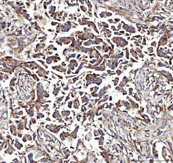
Real IHC data IHC analysis of MCCC2 using anti-MCCC2 antibody (A06515-2). MCCC2 was detected in a paraffin-embedded section of human breast cancer tissue. Heat mediated antigen retrieval was performed in EDTA buffer (pH 8.0, epitope retrieval solution). The tissue section was blocked with 10% goat serum. The tissue section was then incubated with 2 μg/ml rabbit anti-MCCC2 Antibody (A06515-2) overnight at 4°C. Peroxidase Conjugated Goat Anti-rabbit IgG was used as secondary antibody and incubated for 30 minutes at 37°C. The tissue section was developed using HRP Conjugated Rabbit IgG Super Vision Assay Kit (Catalog # SV0002) with DAB as the chromogen.
Anti-MCCC2 Antibody ®
Cat # A06515-2
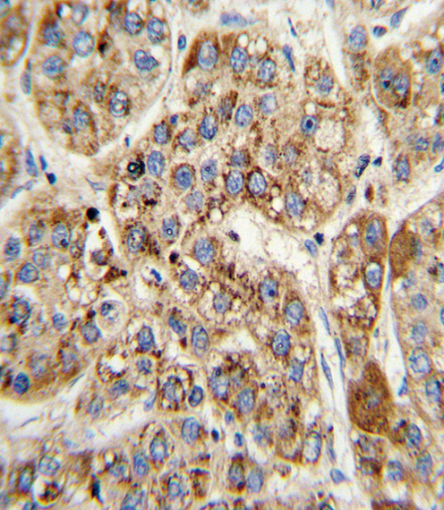
Real IHC data Formalin-fixed and paraffin-embedded human hepatocarcinoma with MCCC2 Antibody (Center), which was peroxidase-conjugated to the secondary antibody, followed by DAB staining. This data demonstrates the use of this antibody for immunohistochemistry; clinical relevance has not been evaluated.
Anti-MCCC2 Antibody (Center)
Cat # A06515-1

A06515-2 will render with an IHC image from a paraffin-embedded human breast cancer section; its listed IHC reactivity is human, mouse and rat (A06515-2 image caption and catalog reactivity). A06515-1 will render with an IHC-P image from formalin-fixed, paraffin-embedded human hepatocarcinoma; its listed reactivity is human and rat (A06515-1 image caption and catalog reactivity).

Which to pick: For tissue IHC, choose A06515-2 if its documented EDTA retrieval at pH 8.0 and 2 μg/ml starting concentration suit the experiment; its paraffin-section caption does not report a fixative (A06515-2 image caption). Choose A06515-1 for a documented paraffin-section, paraffin-embedded human tissue example, or A06515-2 when mouse reactivity is needed (A06515-1 image caption; catalog reactivity). Neither SKU lists IF/ICC validation or an IF dilution, so neither is a validated IF/ICC choice here (catalog applications and dilutions). The selected A06515-2 tissue-IHC caption documents paraffin sections, but does not specify the fixative (selected-SKU IHC image A06515-2).

Each figure is that product's own IHC / IF validation image from its datasheet.

References

  1. UniProt Consortium. UniProt entry Q9HCC0 (MCCB_HUMAN, Methylcrotonoyl-CoA carboxylase beta chain, mitochondrial).
  2. Human Protein Atlas. MCCC2 tissue IHC expression (reliability: Enhanced).
  3. Human Protein Atlas. MCCC2 subcellular location (ICC-IF): Localized to the mitochondria..
  4. Human Protein Atlas. MCCC2 antibody validation summary (3 antibodies).
  5. Inhibition of MCCC2 Impedes TNBC Progression by Downregulating Leucine Metabolism-Dependent mTOR Signaling. Breast cancer (Dove Medical Press) 2026 — PMC12988748.
  6. Methylcrotonoyl-CoA Carboxylase 2 Promotes Proliferation, Migration and Invasion and Inhibits Apoptosis of Prostate Cancer Cells Through Regulating GLUD1-P38 MAPK Signaling Pathway. OncoTargets and therapy 2020 — PMC7395692.
  7. Methylcrotonyl-CoA carboxylase 2 supports leucine catabolism to promote mitochondrial biogenesis and alleviate cisplatin-induced acute kidney injury. Kidney research and clinical practice 2026 — PMC13347088.
  8. MCCC2 is a novel mediator between mitochondria and telomere and functions as an oncogene in colorectal cancer. Cellular & molecular biology letters 2023 — PMC10571261.
  9. PubMed PMID:11170888 — UniProt-cited evidence.
  10. PubMed PMID:11181649 — UniProt-cited evidence.
  11. PubMed PMID:11406611 — UniProt-cited evidence.