This website uses cookies to ensure you get the best experience on our website.


- Table of Contents
Plan MCU staining in paraffin sections using the cytoplasmic tissue pattern reported by HPA (HPA tissue IHC). The guide pairs that observation with MCU’s inner mitochondrial membrane location (UniProt) and the catalog antibody’s IHC dilution of 2–5 μg/ml (datasheet A00685-1).

Expected localisation, validated protocols, controls and antibodies — the at-a-glance facts below, then the full design guide.
| Expected localisation | Cytoplasmic staining in most tissues (HPA tissue IHC) | |
| Staining pattern | Cytoplasmic stain; high in several glandular cell types (HPA tissue IHC) | |
| Antigen retrieval | EDTA pH 8.0 HIER, heat-mediated (datasheet A00685-1) | |
| Positive control | Adrenal gland+4 more · see all | |
| Negative control | None in HPA (detected in all 45 tissues); use no-primary + isotype controls |
| Fixation | Keep paraffin-section fixation consistent across samples (standard IHC practice; not target-specific) | |
| Caveat | Staining and RNA expression show low consistency (HPA tissue IHC) | |
| Regulation | No expression regulator annotated (UniProt) | |
| Isoform / epitope | 3 isoforms; epitope coverage is unspecified (UniProt) |
The catalog antibody’s IHC-P protocol is followed by three published MCU staining protocols for paraffin sections (PMC7680189; PMC8805845; PMC8195654).
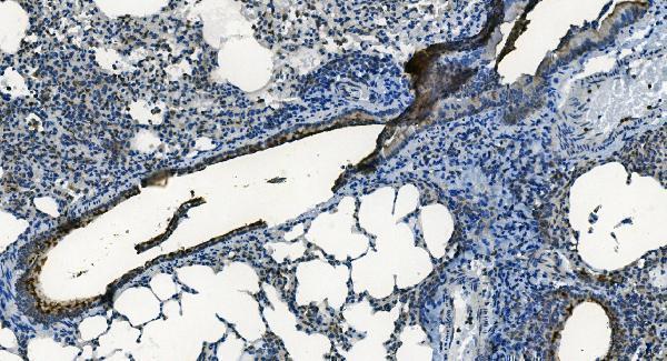
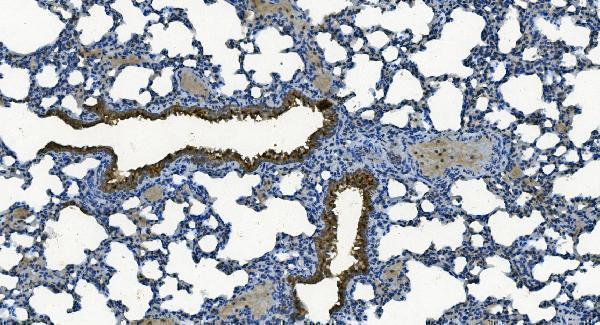
| Sample | Paraffin-embedded mouse lung tissue; fixative not specified (datasheet A00685-1) |
| Fixation | Image fixative and duration unreported (datasheet A00685-1); verify before use. |
| Sectioning | 4–5 µm sections on charged slides (standard) |
| Deparaffinisation | Xylene, graded ethanol series to water (standard) |
| Antigen retrieval | Heat retrieval: EDTA pH 8.0 (datasheet A00685-1); 20 min, 95–100 °C (standard) |
| Peroxidase block | 3% H2O2, 10 min, room temperature (standard) |
| Blocking | 10% goat serum (datasheet A00685-1) |
| Primary antibody | Rabbit anti-MCU, 2-5μg/ml (datasheet A00685-1) |
| Primary incubation | Overnight at 4 °C (datasheet A00685-1) |
| Detection | Streptavidin-biotin complex (SABC), DAB chromogen (datasheet A00685-1) |
| Counterstain | Hematoxylin, blue, dehydrate and mount (standard) |
| Expected result | MCU-positive staining in glandular cells of adrenal gland (HPA tissue IHC: High). HPA tissue profile: Cytoplasmic expression in most tissues. No signal in the no-primary control. |
MCU is an inner mitochondrial membrane protein with two transmembrane segments (UniProt Q8NE86 topology). In paraffin sections, expect cytoplasmic staining in many cell types, including strong staining in selected glandular cells and neurons (HPA tissue IHC). HPA rates its tissue staining Approved but reports low consistency with RNA expression; interpret intensity alongside controls rather than assuming every stained cell has high MCU expression (HPA tissue IHC).
| Cytoplasmic staining in adrenal, appendix, breast, or colon glandular cells. | This matches the High staining reported for those cell populations (HPA tissue IHC). A granular pattern is compatible with mitochondrial localisation (UniProt Q8NE86; HPA subcellular ICC-IF), but chromogenic IHC may not resolve individual mitochondria (general IHC practice). |
| Predominantly nuclear staining, or a sharp cell-surface outline without cytoplasmic signal. | These patterns conflict with MCU's inner mitochondrial membrane location (UniProt Q8NE86 topology). Check counterstain and chromogen distribution, then compare an appropriate control before calling the pattern specific (general IHC practice). |
| Strong staining confined to cells expected to stain weakly, while an expected High population is blank. | Consider cross-reactivity or endogenous detection activity (general IHC practice). HPA reports Low staining in liver cholangiocytes and soft-tissue fibroblasts, but neither is a proven negative control; cell identity and staining conditions still need checking (HPA tissue IHC). |
| Diffuse staining across tissue, extracellular spaces, and areas without clear cell boundaries. | This distribution is difficult to reconcile with cytoplasmic MCU staining (HPA tissue IHC; UniProt Q8NE86). Review background in reagent controls and check blocking, washes, and chromogen development (general IHC practice). |
| No detectable staining in a documented High population. | A blank result in colon glandular cells or cerebral-cortex neurons conflicts with the reported tissue pattern (HPA tissue IHC). First verify tissue identity and control performance; then assess retrieval, antibody conditions, and detection as general IHC variables. |
| Cell population and comparator selection | HPA reports High staining in bronchial basal cells, caudate neurons, cerebellar granular-layer cells, and several glandular populations (HPA tissue IHC). Its Low categories are relative comparators, not validated negatives; choose the same cell population when comparing runs. |
| Antibody evidence and interpretation limit | HPA016480 is IHC Approved, while HPA notes low consistency between antibody staining and RNA expression (HPA antibody validation; HPA tissue IHC). This supports using the reported pattern as a guide, with additional controls for an unexpected result. |
| Topology and epitope accessibility | MCU has two membrane-spanning segments and matrix-facing regions on either side of a short intermembrane segment (UniProt Q8NE86 topology). Without an antibody epitope or target-specific retrieval study, these facts do not predict which retrieval condition will work best. |
| Processing and isoforms | UniProt lists a 51–351 mature chain and three isoforms (UniProt Q8NE86). Epitope coverage across the isoforms is unspecified here; avoid attributing a staining difference to processing or an isoform without antibody-specific evidence. |
| IF/ICC Q&A: what localisation should be expected? | Mitochondrial signal is expected in IF/ICC; HPA supports that location and lists images from A-431, U-251MG, and U2OS (HPA subcellular ICC-IF). That observation helps assess localisation but does not establish an IHC-P staining condition. |
| Situation | Likely cause | Next action |
|---|---|---|
| Expected High cells are blank, and the run control is also blank. | The run may have a detection or reagent failure (general IHC practice). HPA's High calls describe observed staining, not guaranteed intensity in every section (HPA tissue IHC). | Check reagent order, expiry, and detection-control performance; repeat with a documented High population in the same run (general IHC practice; HPA tissue IHC). |
| Expected High cells are blank, but the run control stains. | Section identity, retrieval, antibody conditions, or local tissue preservation may differ (general IHC practice). No MCU-specific fixation sensitivity is reported in this payload. | Confirm the cell population, then compare controlled retrieval and antibody conditions using the same detection system (general IHC practice). |
| Colour is concentrated in nuclei rather than cytoplasm. | The compartment conflicts with the mitochondrial location assigned to MCU (UniProt Q8NE86; HPA subcellular ICC-IF); counterstain or nonspecific signal may complicate reading (general IHC practice). | Inspect the chromogen and counterstain separately, compare controls, and require cytoplasmic staining before assigning MCU positivity (general IHC practice; HPA tissue IHC). |
| Weakly rated cells stain strongly while expected High cells do not. | Cross-reactivity or endogenous detection activity is possible (general IHC practice). HPA's Low designations are observations, not proof that those cells lack MCU (HPA tissue IHC). | Verify cell identity, examine reagent controls, and repeat alongside an HPA High comparator before interpreting the discrepancy (general IHC practice; HPA tissue IHC). |
| Broad, hazy colour obscures individual cells. | Background from blocking, washing, or excessive development can impair interpretation (general IHC practice); it does not establish broader MCU distribution. | Review reagent controls and adjust blocking, washes, or development within the established IHC workflow (general IHC practice). Score only identifiable cellular staining. |
| Staining is cytoplasmic but lacks visible mitochondrial puncta. | Chromogenic sections may not resolve mitochondria individually (general IHC practice). HPA reports cytoplasmic tissue staining, while IF/ICC supports mitochondrial localisation (HPA tissue IHC; HPA subcellular ICC-IF). | Assess whether the expected cell populations stain and whether controls are clean. Do not reject an otherwise consistent IHC result solely for lacking visible puncta (HPA tissue IHC; general IHC practice). |


Comprehensive Human Protein Atlas IHC scoring per tissue (reliability: Approved — Low consistency between antibody staining and RNA expression data.). Rows are taken directly from the HPA tissue chart — click any row's HPA link to view the source.
| Tissue | Cell type | Level | Evidence | Source |
|---|---|---|---|---|
| Adrenal gland | Glandular cells | High | Protein (IHC) | HPA → |
| Appendix | Glandular cells | High | Protein (IHC) | HPA → |
| Breast | Glandular cells | High | Protein (IHC) | HPA → |
| Bronchus | Basal cells | High | Protein (IHC) | HPA → |
| Caudate | Neuronal cells | High | Protein (IHC) | HPA → |
| Tissue | Cell type | Level | Evidence | Source |
|---|---|---|---|---|
| None in HPA: MCU is detected in all 45 scored tissues. Use a no-primary (secondary-only) and an isotype control instead. | ||||
Troubleshoot MCU staining in paraffin sections by checking retrieval, staining controls, mitochondrial localisation and cell type before comparing chromogenic signal across samples.
A00685-1 has real IHC images from paraffin sections of mouse and rat lung and human liver and bladder cancer tissue (catalog IHC captions). No IF images are provided (catalog IF images).
A00685-1 shows IHC staining in paraffin sections of mouse and rat lung and human liver and bladder cancer tissue (catalog IHC captions). Its listed applications include IHC but exclude IF/ICC; its listed reactivity is human, monkey, mouse, and rat (catalog applications and reactivity).
Which to pick: Choose A00685-1 for paraffin-section tissue IHC, starting within its 2–5 μg/ml IHC range (catalog IHC captions; datasheet: 2–5 μg/ml); the fixative is unreported (catalog IHC captions). No IF/ICC antibody can be recommended from this payload because A00685-1 has no listed IF/ICC application or IF image (catalog applications; catalog IF images). For work across species, A00685-1 has IHC images for human, mouse, and rat tissue, while monkey is listed only under reactivity; it is rabbit hosted, and its clonality is unreported (catalog IHC captions; catalog reactivity; catalog host and clone).