This website uses cookies to ensure you get the best experience on our website.


- Table of Contents
Plan MDC1 staining in paraffin sections around its general nuclear tissue pattern (HPA tissue IHC). Start with the catalog antibody at 0.5–1 μg/ml (datasheet: A01252-2), then compare nuclear staining across consistently processed sections (standard IHC practice).

Expected localisation, validated protocols, controls and antibodies — the at-a-glance facts below, then the full design guide.
| Expected localisation | General nuclear tissue staining (HPA tissue IHC) | |
| Staining pattern | Nuclear staining across diverse tissue cell types (HPA tissue IHC) | |
| Antigen retrieval | Citrate pH 6 HIER, heat-mediated (datasheet A01252-2) | |
| Positive control | Adrenal gland+4 more · see all | |
| Negative control | None in HPA (detected in all 45 tissues); use no-primary + isotype controls |
| Fixation | Matched tissue-IHC evidence does not establish the fixation claim. Validate the specimen-specific method before use. (selected-SKU IHC image A01252-2) | |
| Caveat | DNA damage can produce discrete nuclear foci (UniProt) | |
| Regulation | Intensity varies by tissue cell type (HPA tissue IHC) | |
| Isoform / epitope | Four isoforms; confirm antibody epitope coverage (UniProt) |
The catalog antibody has a datasheet IHC protocol (datasheet: A01252-2). Published MDC1 IHC methods provide three additional examples for tumor and tissue sections (PMC4177045; PMC4446443; PMC4515811).
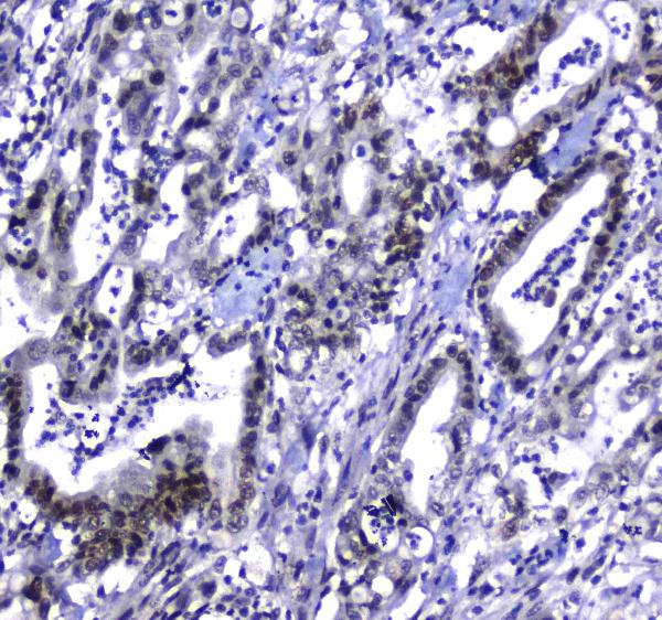
| Sample | Paraffin-embedded human colon cancer tissue; fixative not specified (datasheet A01252-2) |
| Fixation | Image fixative and duration unreported (datasheet A01252-2); verify before use. |
| Sectioning | 4–5 µm sections on charged slides (standard) |
| Deparaffinisation | Xylene, graded ethanol series to water (standard) |
| Antigen retrieval | Heat retrieval: Citrate pH 6, 20 min (datasheet A01252-2) |
| Peroxidase block | 3% H2O2, 10 min, room temperature (standard) |
| Blocking | 10% goat serum (datasheet A01252-2) |
| Primary antibody | Rabbit anti-MDC1, 0.5-1μg/ml (datasheet A01252-2) |
| Primary incubation | Overnight at 4 °C (datasheet A01252-2) |
| Detection | Streptavidin-biotin complex (SABC), DAB chromogen (datasheet A01252-2) |
| Counterstain | Hematoxylin, blue, dehydrate and mount (standard) |
| Expected result | MDC1-positive staining in glandular cells of adrenal gland (HPA tissue IHC: High). HPA tissue profile: General nuclear expression. No signal in the no-primary control. |
MDC1 should appear in nuclei across many cell types in paraffin sections: HPA describes general nuclear expression and reports high staining in several glandular, hematopoietic, respiratory epithelial and glial cell populations (HPA tissue IHC). It is a chromatin-associated nuclear protein with no transmembrane segment (UniProt Q14676). HPA rates the tissue IHC evidence Enhanced, while noting medium consistency between staining and RNA expression (HPA tissue IHC).
| Nuclear chromogenic staining in adrenal or breast glandular cells, or bone marrow hematopoietic cells. | This matches cell populations scored High by HPA (HPA tissue IHC). Compare signal within identified cells; a High category does not mean every nucleus in the section must stain equally. |
| Predominantly cytoplasmic or membranous staining with little nuclear signal. | That distribution conflicts with MDC1’s nuclear, chromatin-associated location (UniProt Q14676) and HPA’s general nuclear IHC profile (HPA tissue IHC). Treat it as suspect and review antibody specificity and detection controls (general IHC practice). |
| Strong color in a cell population expected to show little MDC1 staining, such as cardiomyocytes. | HPA scores cardiomyocytes Low (HPA tissue IHC). If the color lies outside nuclei or persists in a no-primary control, investigate cross-reactivity or endogenous detection activity before scoring it as MDC1 (general IHC practice). |
| Diffuse color across nuclei, cytoplasm and surrounding tissue, obscuring cell boundaries. | The lack of a distinct nuclear pattern makes specific MDC1 staining difficult to judge against HPA’s nuclear profile (HPA tissue IHC). Check background with a no-primary control, then review blocking, washing and chromogen development (general IHC practice). |
| No nuclear signal in a section containing adrenal glandular cells or bone marrow hematopoietic cells. | Those populations are scored High by HPA (HPA tissue IHC), so an entirely blank result warrants a technical check. HPA’s Enhanced rating supports the reported pattern but does not guarantee staining in every specimen (HPA tissue IHC). |
| Where should the IHC signal be scored? | Score nuclei in identifiable cells: HPA reports general nuclear expression (HPA tissue IHC), consistent with chromatin-associated MDC1 (UniProt Q14676). HPA’s Low categories for alveolar type I cells and cardiomyocytes are useful comparisons, not absolute negative controls (HPA tissue IHC). |
| How much confidence does the HPA validation provide? | The tissue profile is rated Enhanced, and HPA006915 and CAB080116 each have Enhanced IHC status (HPA tissue IHC; HPA antibodies). HPA also reports medium consistency between staining and RNA expression, so assess compartment and controls alongside intensity (HPA tissue IHC). |
| Are DNA damage foci required in routine tissue IHC? | MDC1 can relocalize to discrete nuclear foci after DNA damage through phosphorylated H2AX (UniProt Q14676). HPA describes general nuclear tissue staining (HPA tissue IHC); absence of resolvable foci alone should not overturn an otherwise appropriate nuclear IHC result. |
| What can sequence information tell us about antibody interpretation? | UniProt lists four isoforms, one protein chain spanning residues 1–2089, and no signal peptide, propeptide or transmembrane segment (UniProt Q14676). These facts support intracellular nuclear interpretation but do not identify the catalog antibody’s epitope or establish isoform coverage. |
| What should IF/ICC show? | HPA reports supported nucleoplasm and nuclear-body localization in ICC-IF, with images from A-431, U-251MG, U2OS and SiHa (HPA subcellular). That is a localization cross-check; this section makes no IF/ICC protocol recommendation. |
| Situation | Likely cause | Next action |
|---|---|---|
| Known HPA High cells are blank. | A failed IHC detection step, unsuitable working conditions or a missing positive population can yield an uninformative slide (general IHC practice); HPA lists the relevant cells as High (HPA tissue IHC). | Confirm the cell population is present, run a known-positive section and follow the catalog antibody’s IHC-P procedure for retrieval, dilution and detection; no target-specific fixation sensitivity is established here. |
| Color is mainly cytoplasmic or membranous. | The compartment conflicts with UniProt’s nuclear localization and HPA’s general nuclear IHC pattern (UniProt Q14676; HPA tissue IHC). | Check a no-primary control and the chromogenic detection workflow, then reassess nuclear signal with the counterstain visible (general IHC practice). |
| Low-scored cells appear strongly positive. | Cardiomyocytes and alveolar type I cells are scored Low, although Low does not mean uniformly absent (HPA tissue IHC). Misidentified cells or nonspecific color can distort interpretation (general IHC practice). | Confirm cell identity and nuclear placement; compare with a no-primary control and an HPA High population on a separately validated section (HPA tissue IHC; general IHC practice). |
| Background masks nuclear detail. | Widespread color cannot be reliably assigned to HPA’s reported nuclear MDC1 pattern (HPA tissue IHC). Excess detection signal or inadequate washing can raise background (general IHC practice). | Inspect the no-primary control; review blocking, washes and chromogen development, then score only nuclei that remain distinguishable (general IHC practice). |
| Nuclear staining is present but lacks discrete foci. | UniProt describes foci after DNA damage, while HPA reports general nuclear tissue expression (UniProt Q14676; HPA tissue IHC). | Assess compartment and cell identity against the tissue profile; do not require visible foci to call routine chromogenic IHC positive (HPA tissue IHC; general IHC practice). |
| Two antibodies give different nuclear intensities. | HPA reports Enhanced IHC status for HPA006915 and CAB080116, but the tissue profile has only medium staining-to-RNA consistency (HPA antibodies; HPA tissue IHC). The supplied sources do not resolve epitope or isoform coverage. | Compare matched positive and no-primary controls, then document each antibody’s nuclear pattern separately rather than treating equal intensity as a validation requirement (general IHC practice). |
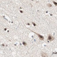


Comprehensive Human Protein Atlas IHC scoring per tissue (reliability: Enhanced — Medium consistency between antibody staining and RNA expression data.). Rows are taken directly from the HPA tissue chart — click any row's HPA link to view the source.
| Tissue | Cell type | Level | Evidence | Source |
|---|---|---|---|---|
| Adrenal gland | Glandular cells | High | Protein (IHC) | HPA → |
| Appendix | Glandular cells | High | Protein (IHC) | HPA → |
| Bone marrow | Hematopoietic cells | High | Protein (IHC) | HPA → |
| Breast | Glandular cells | High | Protein (IHC) | HPA → |
| Bronchus | Respiratory epithelial cells | High | Protein (IHC) | HPA → |
| Tissue | Cell type | Level | Evidence | Source |
|---|---|---|---|---|
| None in HPA: MDC1 is detected in all 45 scored tissues. Use a no-primary (secondary-only) and an isotype control instead. | ||||
Troubleshoot MDC1 staining by checking nuclear localisation, the documented paraffin section workflow, and controls that distinguish DNA damage foci from staining artefacts.
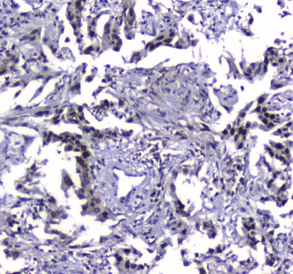
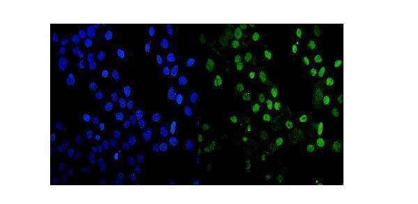
A01252-2 has IHC images from human paraffin sections of colon cancer, lung cancer and testis, and an IF/ICC image from A431 cells (catalog image captions).
A01252-2 is listed for human IHC, with images of paraffin sections from colon cancer, lung cancer and testis (catalog applications, reactivity and IHC captions). The same SKU is listed for IF/ICC, with an image from A431 cells (catalog applications and IF caption).
Which to pick: For tissue IHC, choose A01252-2: its images document human paraffin sections, including colon cancer; the fixative is unreported (catalog IHC captions). For IF/ICC, choose A01252-2 based on its A431 cell image; it is rabbit hosted, and clonality is unreported (catalog IF caption and product details). No cross species option is documented because A01252-2 lists only human reactivity (catalog reactivity).