MDH2 / Malate dehydrogenase, mitochondrial · IHC design guide

Design Immunohistochemistry for MDH2

Plan MDH2 paraffin-section IHC around the granular cytoplasmic staining reported across tissues (HPA tissue IHC). Compare cell type and staining intensity while treating mitochondrial matrix localization as a molecular expectation (UniProt).

Evidence assembled Oct 2026 · For research use; verify linked source records and product datasheet before use
Immunohistochemistry protocol sheet for MDH2 (IHC for MDH2): expected localisation Granular cytoplasm (HPA tissue IHC); mitochondrial matrix (UniProt), antibody A04803-2, validated IHC image, and IHC protocol steps
Printable MDH2 IHC protocol sheet — expected localisation Granular cytoplasm (HPA tissue IHC); mitochondrial matrix (UniProt), antibody A04803-2, controls and protocol steps. Open the full MDH2 IHC guide →

MDH2 Immunohistochemistry Experimental Design Guide

Expected localisation, validated protocols, controls and antibodies — the at-a-glance facts below, then the full design guide.

Must know before staining
Expected localisation Granular cytoplasm (HPA tissue IHC); mitochondrial matrix (UniProt)
Staining pattern Granular cytoplasmic staining across tissues (HPA tissue IHC)
Antigen retrieval EDTA pH 8.0 HIER, heat-mediated (datasheet A04803-2)
Positive control ⓘ Adrenal gland+4 more · see all
Negative control ⓘ None in HPA (detected in all 45 tissues); use no-primary + isotype controls
Important caveats
Reasons your staining may differ from the expected pattern.
Fixation Keep formalin fixation consistent across paraffin sections. (standard IHC practice; not target-specific)
Caveat Broad tissue staining makes negative controls harder to select (HPA tissue IHC)
Regulation RNA enhanced in skeletal muscle and tongue (HPA tissue RNA)
Isoform / epitope 2 isoforms; mature chain spans residues 25–338; epitope coverage unknown (UniProt)
Section 1

Recommended MDH2 IHC & IF Protocols

The catalog antibody’s IHC-P protocol is followed by three published MDH2 IHC methods with usable preparation or staining details (PMC10868282; PMC8586664; PMC5300292).

Recommended immunohistochemistry (IHC-P) protocol parameters
SampleParaffin-embedded human laryngeal carcinoma tissue; fixative not specified (datasheet A04803-2)
FixationImage fixative and duration unreported (datasheet A04803-2); verify before use.
Sectioning4–5 µm sections on charged slides (standard)
DeparaffinisationXylene, graded ethanol series to water (standard)
Antigen retrievalHeat retrieval: EDTA pH 8.0 (datasheet A04803-2); 20 min, 95–100 °C (standard)
Peroxidase block3% H2O2, 10 min, room temperature (standard)
Blocking10% goat serum (datasheet A04803-2)
Primary antibodyRabbit anti-MDH2, 1-2 μg/ml (datasheet A04803-2)
Primary incubationOvernight at 4 °C (datasheet A04803-2)
DetectionStreptavidin-biotin complex (SABC), DAB chromogen (datasheet A04803-2)
CounterstainHematoxylin, blue, dehydrate and mount (standard)
Expected resultMDH2-positive staining in glandular cells of adrenal gland (HPA tissue IHC: High). HPA tissue profile: Granular cytoplasmic expression in all tissues. No signal in the no-primary control.
💡Decision noteStart with heat-mediated EDTA pH 8.0 retrieval for the catalog antibody (datasheet A04803-2). Consider citrate pH 6.0 for the tissue-chip method (PMC5300292 methods).
Section 2

What Is the Expected MDH2 Staining Pattern?

MDH2 is a mitochondrial matrix protein with no transmembrane segment (UniProt P40926). In paraffin-section IHC, expect granular cytoplasmic staining across tissues, with high staining in several glandular and neural cell populations (HPA: tissue IHC). HPA rates the tissue pattern Enhanced, citing high consistency between antibody staining and RNA expression data (HPA: tissue IHC).

What am I looking at on my slide?
Granular cytoplasmic staining in adrenal glandular cells or bronchial respiratory epithelium.This matches high staining reported in those cells (HPA: tissue IHC). Granules are consistent with mitochondrial localisation (UniProt P40926; HPA: subcellular).
Predominantly nuclear or crisp plasma-membrane staining.This conflicts with the mitochondrial matrix assignment (UniProt P40926). Treat it as possible nonspecific staining or an imaging artefact; compartment alone cannot identify the cause.
Strong staining in smooth muscle cells while expected glandular cells stain weakly.HPA reports low staining in smooth muscle cells and high staining in several glandular populations (HPA: tissue IHC). Consider cross-reactivity or endogenous detection activity; neither is proven by this pattern.
Diffuse chromogen throughout cells, tissue spaces, or the whole section, obscuring granules.The distribution is difficult to reconcile with HPA's granular cytoplasmic profile (HPA: tissue IHC). Background may arise from detection or washing conditions (general IHC practice).
No detectable staining in a section expected to contain high-expressing cells.A blank adrenal glandular or bronchial epithelial population conflicts with HPA's high staining reports (HPA: tissue IHC). Check assay performance before interpreting it as absent MDH2.
💡Expected MDH2 appearanceCall a positive result when cytoplasmic granules are visible in the expected cells, including strong staining in HPA high populations (HPA: tissue IHC); dominant nuclear, membrane, or diffuse staining is discordant with the mitochondrial matrix location (UniProt P40926).
How each factor affects the staining
Subcellular assignment and topologyMDH2 is assigned to the mitochondrial matrix and has no transmembrane segment (UniProt P40926). Interpret IHC granules as a compartment-compatible pattern, not proof that each granule is a mitochondrion.
Tissue and cell contextHPA describes granular cytoplasmic expression in all tissues, with high staining in listed glandular, respiratory epithelial, glial, and neuronal populations and low staining in smooth muscle cells (HPA: tissue IHC). Compare cells within their tissue context.
Strength of pattern evidenceThe HPA tissue profile is rated Enhanced for agreement between antibody staining and RNA expression; four listed antibodies have Enhanced IHC status (HPA: tissue IHC; HPA: antibodies). These ratings support the reported pattern, but do not validate an unlisted catalog antibody.
Protein processing and isoformsUniProt annotates a mature chain spanning residues 25–338 and two isoforms (UniProt P40926). Without an epitope map for the antibody used, this record cannot establish which forms it detects or predict a form-specific tissue pattern.
Fixation and retrieval evidenceThe supplied UniProt and HPA records do not report MDH2-specific fixation sensitivity or antigen retrieval conditions. Treat retrieval optimisation as a general paraffin-IHC workflow decision, not a target-specific prediction.
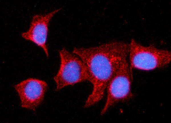
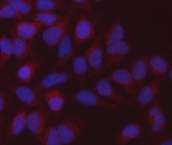
IF/ICC Q&A: what location should fluorescence show?Mitochondrial localisation is reported by HPA ICC-IF and agrees with the UniProt matrix assignment (HPA: subcellular; UniProt P40926). This supports an IF localisation check; it does not establish an IF protocol.
Why is my staining missing, weak or wrong?
SituationLikely causeNext action
High-staining reference cells show no chromogen.Possible assay failure; HPA reports high staining in adrenal glandular and bronchial respiratory epithelial cells (HPA: tissue IHC).Verify that the expected cells are present, then check primary-antibody and detection controls; review retrieval and dilution using the antibody's IHC-P instructions (general IHC practice).
Staining is mostly nuclear or outlines cell membranes.That distribution conflicts with MDH2's mitochondrial matrix location and HPA's granular cytoplasmic IHC profile (UniProt P40926; HPA: tissue IHC).Review morphology and counterstain, compare with a primary-omission control, and reassess antibody specificity if the pattern persists (general IHC practice).
Broad diffuse colour masks intracellular granules.Diffuse deposition can reflect excess detection background or inadequate washing (general IHC practice); it cannot be scored against HPA's granular profile (HPA: tissue IHC).Inspect the primary-omission control, washing, blocking, and chromogen development; adjust the workflow according to the detection system's instructions (general IHC practice).
Smooth muscle cells appear as strongly stained as nearby expected high-staining cells.HPA lists smooth muscle cells as low, while several other cell populations are high (HPA: tissue IHC). Cross-reactivity or endogenous detection activity is possible.Confirm cell identity on the counterstained section, inspect a primary-omission control, and compare with an independently validated antibody if available (general IHC practice).
Some tissue compartments stain weakly despite strong staining elsewhere.Intensity varies by cell population: HPA reports high staining in named populations and low staining in smooth muscle cells (HPA: tissue IHC).Score identified cell populations separately and compare each with its HPA entry; avoid treating a low-staining compartment alone as assay failure (HPA: tissue IHC).
IF/ICC shows fluorescence outside the expected mitochondrial distribution.A nonmitochondrial pattern conflicts with HPA's ICC-IF location and UniProt's matrix assignment (HPA: subcellular; UniProt P40926).Use the separate IF/ICC guide for its controls and imaging checks; this IHC section supplies a location expectation, not an IF protocol.

Sample controls for MDH2 IHC & IF

🧪Run adrenal gland first and expect glandular cells to stain (HPA: High in adrenal gland glandular cells). HPA detects MDH2 in all 45 scored tissues, so there is no supported negative tissue; use no-primary and isotype controls, and judge internal negative regions by background in acellular spaces rather than assuming any cell population is MDH2-negative (HPA: no negative tissue rows; UniProt P40926: mitochondrial matrix).
Positive control tissue: Adrenal gland (Glandular cells, HPA High)
Negative control tissue: None in HPA: MDH2 is detected in all 45 scored tissues. Use a no-primary (secondary-only) and an isotype control instead.
ICC-IF cell lines (HPA subcellular resource): HPA ICC-IF images show MDH2 in A-431, U2OS, U-251MG, with annotated localisation: Mitochondria (enhanced) (HPA subcellular).
Technical controls: Include a no-primary, secondary-only section, a host-species- and clonality-matched nonimmune IgG isotype control, and MDH2-knockout material processed in parallel as a biological negative. For chromogenic detection, quench endogenous peroxidase and assess endogenous biotin when using the caption’s streptavidin–biotin detection method (selected-SKU tissue-IHC caption: SABC with DAB).
⚠️Feasibility: No target-specific fixation window or fixation effect is reported in the supplied evidence, and the selected-SKU paraffin-section caption does not state the fixative (selected-SKU tissue-IHC caption: fixative unreported). Heat retrieval with EDTA at pH 8.0 was used in that caption; whether MDH2 staining depends on that retrieval condition is unreported (selected-SKU tissue-IHC caption: EDTA retrieval). Frozen-section performance and relative ease of IF are unreported, although mitochondrial localization can be checked by ICC-IF in the HPA-imaged cell lines (HPA subcellular: mitochondria, A-431, U2OS and U-251MG); check adrenal sections for endogenous biotin background with the caption’s SABC method (selected-SKU tissue-IHC caption: SABC).

HPA tissue IHC evidence for MDH2

Comprehensive Human Protein Atlas IHC scoring per tissue (reliability: Enhanced — High consistency between antibody staining and RNA expression data.). Rows are taken directly from the HPA tissue chart — click any row's HPA link to view the source.

Positive expression · recommended positive controls

TissueCell typeLevelEvidenceSource
Adrenal gland Glandular cells High Protein (IHC) HPA →
Appendix Glandular cells High Protein (IHC) HPA →
Breast Glandular cells High Protein (IHC) HPA →
Bronchus Respiratory epithelial cells High Protein (IHC) HPA →
Caudate Glial cells High Protein (IHC) HPA →

Undetected expression · recommended negative controls

TissueCell typeLevelEvidenceSource
None in HPA: MDH2 is detected in all 45 scored tissues. Use a no-primary (secondary-only) and an isotype control instead.
Section 3

Advanced MDH2 IHC Tips

Use compartment, cell type and control sections to assess MDH2 staining in paraffin-section IHC; the catalog image provides a starting retrieval and detection workflow.

What retrieval conditions should I try first for MDH2 in paraffin sections?
Start with heat-mediated retrieval in EDTA at pH 8.0 for paraffin sections (datasheet A04803-2). The selected image used this retrieval before 10% goat serum blocking and 2 μg/ml primary antibody overnight at 4°C (datasheet A04803-2). If staining is weak, adjust heating time on replicate sections while holding antibody concentration and detection conditions constant (standard IHC practice). Compare signal in intact cell cytoplasm with background on a no-primary control, because retrieval can also expose sites that bind detection reagents nonspecifically (standard IHC practice). Record the heating conditions for each run so a stronger signal can be judged against tissue preservation (standard IHC practice).
How should I troubleshoot fixation-related loss of MDH2 staining?
The selected paraffin-section caption does not state the fixative, so target-specific fixation sensitivity is unknown (datasheet A04803-2). Keep fixation history, section thickness and retrieval conditions documented when comparing specimens, because these variables can change antigen accessibility in paraffin-section IHC (standard IHC practice). First repeat the documented EDTA retrieval at pH 8.0 and 2 μg/ml primary antibody overnight at 4°C on a section with known staining (datasheet A04803-2). If one specimen remains weak, compare another section with similar processing history before attributing the difference to MDH2 abundance (standard IHC practice). Assess morphology alongside signal; damaged or poorly preserved regions are unreliable for scoring (standard IHC practice).
Where should convincing MDH2 staining appear in tissue?
Expect cytoplasmic granules consistent with mitochondria, since MDH2 is a mitochondrial matrix protein and tissue IHC shows granular cytoplasmic expression (UniProt P40926; HPA tissue IHC). Its lack of a transmembrane segment supports an internal mitochondrial location, although chromogenic IHC alone does not resolve the matrix from other mitochondrial structures (UniProt P40926 topology; standard IHC practice). Compare intact cells with adjacent tissue and a no-primary control before calling diffuse nuclear or surface staining positive (standard IHC practice). High staining has been reported in bronchial respiratory epithelial cells, while smooth muscle cells were reported as low, providing a tissue-context check when those cells are present (HPA tissue IHC). Score the relevant cell population separately (standard IHC practice).
Could isoforms or modifications explain discordant MDH2 staining?
MDH2 has 2 annotated isoforms, but the supplied antibody caption does not identify its epitope or establish isoform recognition (UniProt P40926; datasheet A04803-2). The mature chain spans residues 25–338, and modified residues include 33, 78, 91 and 165 (UniProt P40926). Without an epitope map, a change in staining cannot be assigned to cleavage, an isoform or a modification (standard IHC interpretation). Keep retrieval and antibody conditions constant across comparison sections, then assess whether differences persist in the expected granular cytoplasmic compartment (datasheet A04803-2; HPA tissue IHC; standard IHC practice). Request epitope information or use an independently characterized antibody if that distinction is essential (standard IHC practice).
How can IF help assess an ambiguous MDH2 IHC pattern?
IF can test whether MDH2 signal overlaps a mitochondrial marker, consistent with its matrix location and the reported mitochondrial IF localization (UniProt P40926; HPA subcellular). For multiplexing, pair that marker with a marker for the cell type being evaluated, and choose fluorophores whose signal can be distinguished from the tissue's measured autofluorescence (standard IF practice). Permeabilisation must allow antibody access across mitochondrial membranes to a matrix epitope; its extent should be checked against preserved mitochondrial morphology (UniProt P40926 topology; standard IF practice). Include single-stain and no-primary controls to assess channel bleed-through and background (standard IF practice). Treat this as a separate IF assessment, since the supplied antibody workflow documents chromogenic paraffin-section IHC only (datasheet A04803-2).
What should I check when MDH2 DAB staining looks widespread?
Broad staining alone is not evidence of background: tissue IHC reports granular cytoplasmic MDH2 expression across tissues (HPA tissue IHC). Examine whether signal remains granular within intact cells or also covers empty spaces, tissue edges and the no-primary control (HPA tissue IHC; standard IHC practice). The selected workflow used 10% goat serum, 2 μg/ml primary antibody overnight at 4°C, and a biotin-based secondary detection system with DAB (datasheet A04803-2). Check peroxidase blocking and potential endogenous biotin signal with appropriate detection controls before changing the primary antibody concentration (standard chromogenic IHC practice). Compare suspect areas at the same development time and counterstain intensity as the control section (standard IHC practice).
How should I quantify MDH2 staining across paraffin sections? ⚠ ANSWER MARKED FOR VERIFICATION
Define the cell population and intact tissue area before scoring, then apply identical imaging and DAB development settings across compared sections (standard IHC practice). An H-score combines the percentage of cells at each intensity with intensity grades to give a 0–300 scale; record percent positive cells separately if that is the study endpoint (standard IHC practice). Alternatively, report positive-cell density per mm² of viable tissue when cell numbers differ across fields (standard IHC practice). Normalize each result to the relevant cell population or viable area, and use a consistent threshold informed by no-primary controls (standard IHC practice). Exclude necrosis and section edges from analysis, and report how regions were selected (standard IHC practice).
How do I distinguish true MDH2 signal from tissue artefact?
A plausible positive pattern is granular cytoplasmic staining in intact cells, consistent with mitochondrial matrix localization and the tissue IHC profile (UniProt P40926; HPA tissue IHC). Check the identity of stained cells: high signal is reported in bronchial respiratory epithelial cells, whereas smooth muscle cells are listed as low (HPA tissue IHC). Diffuse nuclear staining, edge-heavy staining or color concentrated in necrotic areas needs review against morphology and a no-primary control (standard IHC practice). Because the selected method uses DAB and biotin-based detection, assess endogenous peroxidase and biotin contributions with detection controls before assigning unexpected color to MDH2 (datasheet A04803-2; standard chromogenic IHC practice). Interpret intensity only within comparable cell populations and processing conditions (standard IHC practice).
Boster reagents

Best MDH2 / Malate dehydrogenase, mitochondrial IHC Antibodies

The catalog shows MDH2 IHC staining in human paraffin sections and IF/ICC staining in human cells (catalog image captions). Listed antibody reactivity extends to Mouse and Rat (catalog reactivity).

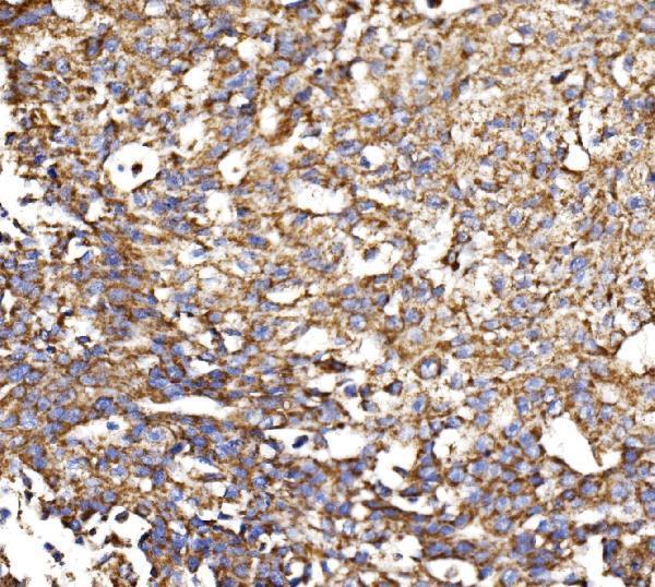
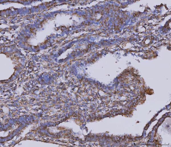
Real IHC data IHC analysis of MDH2 using anti-MDH2 antibody (A04803-2). MDH2 was detected in a paraffin-embedded section of human laryngeal carcinoma tissue. Heat mediated antigen retrieval was performed in EDTA buffer (pH 8.0, epitope retrieval solution). The tissue section was blocked with 10% goat serum. The tissue section was then incubated with 2 μg/ml rabbit anti-MDH2 Antibody (A04803-2) overnight at 4°C. Biotinylated goat anti-rabbit IgG was used as secondary antibody and incubated for 30 minutes at 37°C. The tissue section was developed using Strepavidin-Biotin-Complex (SABC) (Catalog # SA1022) with DAB as the chromogen.
Anti-MDH2 Antibody ®
Cat # A04803-2
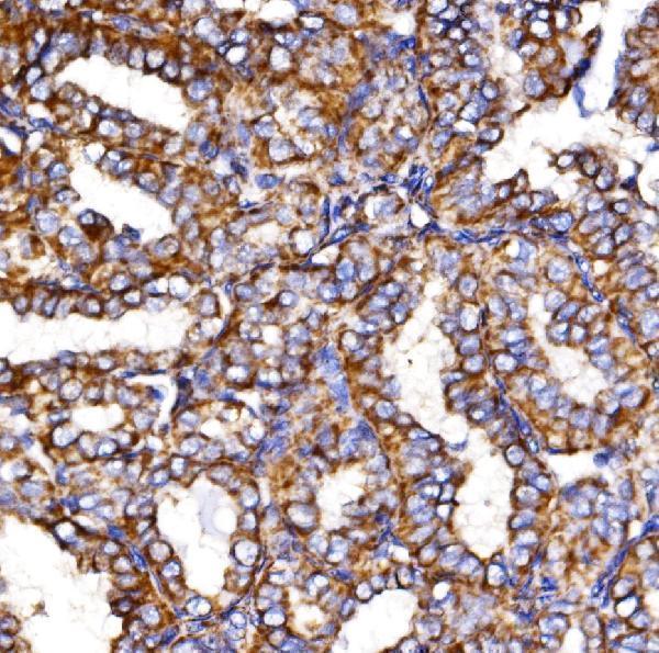
Real IHC data IHC analysis of MDH2 using anti-MDH2 antibody (M04803-1). MDH2 was detected in a paraffin-embedded section of human breast cancer tissue. Heat mediated antigen retrieval was performed in EDTA buffer (pH 8.0, epitope retrieval solution). The tissue section was blocked with 10% goat serum. The tissue section was then incubated with 2 μg/ml mouse anti-MDH2 Antibody (M04803-1) overnight at 4°C. Peroxidase Conjugated Goat Anti-mouse IgG was used as secondary antibody and incubated for 30 minutes at 37°C. The tissue section was developed using HRP Conjugated Mouse IgG Super Vision Assay Kit (Catalog # SV0001) with DAB as the chromogen.
Anti-MDH2 Antibody ® (monoclonal, 5D8C1)
Cat # M04803-1
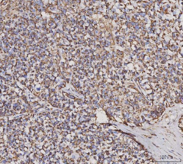
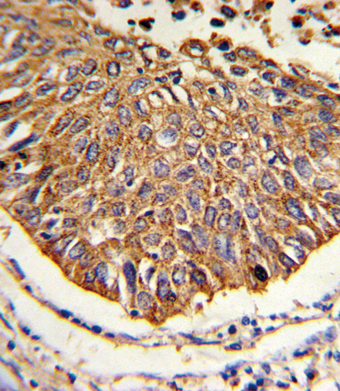
Real IHC data Formalin-fixed and paraffin-embedded human lung carcinoma reacted with MDH2 Antibody (Center), which was peroxidase-conjugated to the secondary antibody, followed by DAB staining. This data demonstrates the use of this antibody for immunohistochemistry; clinical relevance has not been evaluated.
Anti-MDH2 Antibody (Center)
Cat # A04803-1

A04803-2 has IHC images from human laryngeal carcinoma, Hashimoto thyroiditis, and liver cancer paraffin sections, plus IF/ICC in MCF-7 cells; M04803-1 has IHC images from human breast, cervical, and squamous cell carcinoma paraffin sections, plus IF/ICC in HeLa cells (catalog image captions). A04803-1 has an IHC image from formalin-fixed, paraffin-embedded human lung carcinoma (catalog IHC image caption).

Which to pick: For tissue IHC, A04803-2 offers a rabbit polyclonal antibody with human paraffin-section images, while M04803-1 offers a mouse monoclonal with human paraffin-section images; both captions specify EDTA retrieval at pH 8.0, but leave fixation unreported (catalog antibody details and IHC image captions). A04803-1 is another tissue IHC choice with an image from paraffin-section, paraffin-embedded human lung carcinoma (catalog IHC image caption). For IF/ICC, choose A04803-2 or M04803-1, each listed for IF/ICC and imaged in human cells; for cross-species IHC, A04803-2 lists Human, Mouse, and Rat reactivity, though its tissue IHC images show Human only (catalog applications, reactivity, and image captions). The selected A04803-2 tissue-IHC caption documents paraffin sections, but does not specify the fixative (selected-SKU IHC image A04803-2).

Each figure is that product's own IHC / IF validation image from its datasheet.

References

  1. UniProt Consortium. UniProt entry P40926 (MDHM_HUMAN, Malate dehydrogenase, mitochondrial).
  2. Human Protein Atlas. MDH2 tissue IHC expression (reliability: Enhanced).
  3. Human Protein Atlas. MDH2 subcellular location (ICC-IF): Localized to the mitochondria..
  4. Human Protein Atlas. MDH2 antibody validation summary (4 antibodies).
  5. Targeting oxidative phosphorylation to increase the efficacy of immune-combination therapy in renal cell carcinoma. Journal for immunotherapy of cancer 2024 — PMC10868282.
  6. Role of MDH2 pathogenic variant in pheochromocytoma and paraganglioma patients. Genetics in medicine : official journal of the American College of Medical Genetics 2018 — PMC7456538.
  7. LncRNA AC020978 facilitates non-small cell lung cancer progression by interacting with malate dehydrogenase 2 and activating the AKT pathway. Cancer science 2021 — PMC8586664.
  8. MDH2 Stimulated by Estrogen-GPR30 Pathway Down-Regulated PTEN Expression Promoting the Proliferation and Invasion of Cells in Endometrial Cancer. Translational oncology 2017 — PMC5300292.
  9. PubMed PMID:14702039 — UniProt-cited evidence.
  10. PubMed PMID:12853948 — UniProt-cited evidence.
  11. PubMed PMID:15489334 — UniProt-cited evidence.