This website uses cookies to ensure you get the best experience on our website.


- Table of Contents
Plan MED28 chromogenic IHC around the low nuclear and cytoplasmic staining reported across several tissues (HPA tissue IHC). The IHC-validated antibody A08611 has a starting concentration of 2.5 μg/mL for human tissue (datasheet).

Expected localisation, validated protocols, controls and antibodies — the at-a-glance facts below, then the full design guide.
| Expected localisation | Low nuclear and cytoplasmic tissue staining (HPA tissue IHC) | |
| Staining pattern | Low nuclear/cytoplasmic staining; medium in neuronal cells (HPA tissue IHC) | |
| Antigen retrieval | Tris-EDTA pH 9.0 HIER, 95–98 °C, 20 min (rule: nuclear antigen) | |
| Positive control | Breast+4 more · see all | |
| Negative control | Adrenal gland+4 more · see all |
| Fixation | Matched tissue-IHC evidence does not establish the fixation claim. Validate the specimen-specific method before use. (selected-SKU IHC image A08611) | |
| Caveat | Staining and RNA data show low consistency (HPA tissue IHC) | |
| Regulation | Higher expression in vascular tissues (UniProt) | |
| Isoform / epitope | One 1–178 chain; no isoforms annotated (UniProt) |
Compare the catalog antibody’s IHC-P protocol with two published MED28 IHC workflows (PMC2907343; PMC9444419).
| Sample | Tissue sections; selected-image fixative not specified (standard IHC workflow) |
| Fixation | Image fixative and duration unreported (datasheet A08611); verify before use. |
| Sectioning | 4–5 µm sections on charged slides (standard) |
| Deparaffinisation | Xylene, graded ethanol series to water (standard) |
| Antigen retrieval | Heat-induced epitope retrieval in Tris-EDTA buffer, pH 9.0, 20 min at 95–98 °C (standard rule: nuclear antigen) |
| Peroxidase block | 3% H2O2, 10 min, room temperature (standard) |
| Blocking | 10% normal serum of the secondary host, 30 min, room temperature (standard) |
| Primary antibody | Rabbit anti-MED28, 2.5 μg/mL (datasheet A08611) |
| Primary incubation | Overnight at 4 °C (standard) |
| Detection | HRP-polymer secondary, DAB chromogen 5–10 min (standard) |
| Counterstain | Hematoxylin, blue, dehydrate and mount (standard) |
| Expected result | MED28-positive staining in myoepithelial cells of breast (HPA tissue IHC: Medium). HPA tissue profile: Low nuclear and cytoplasmic expression in several tissues. No signal in the no-primary control. |
MED28 is reported in the nucleus, cytoplasm and at the membrane, despite having no transmembrane segment (UniProt Q9H204 localization and topology). In paraffin-section IHC, expect generally low nuclear and cytoplasmic staining, with medium staining reported in selected epithelial and neuronal cell populations (HPA tissue IHC). Treat the pattern as provisional: HPA rates its tissue IHC evidence Uncertain because antibody staining and RNA expression show low consistency (HPA tissue IHC reliability).
| Low nuclear and cytoplasmic staining across several tissues; medium staining in breast myoepithelial cells or bronchial respiratory epithelial cells. | This fits the reported tissue pattern (HPA tissue IHC: low nuclear and cytoplasmic expression; Medium in those cell populations). Score the named cells and compartments separately. A medium signal is an observed IHC result, not a definitive specificity control, because HPA rates the tissue IHC evidence Uncertain (HPA tissue IHC reliability). |
| Staining is confined to extracellular deposits or another compartment without an expected cellular pattern. | Investigate artefact before assigning that signal to MED28. Nuclear and cytoplasmic staining is reported in tissue IHC (HPA tissue IHC); UniProt also lists membrane localization and describes mainly membrane-associated cytoplasmic protein in one cited study (UniProt Q9H204 localization). Membrane-adjacent signal alone therefore does not establish an incorrect compartment. |
| Strong staining appears in cells listed as Not detected, such as adrenal glandular cells or bone-marrow hematopoietic cells. | Check cell identity, cross-reactivity and endogenous detection activity using appropriate controls (general IHC practice). HPA reports those specific cell populations as Not detected (HPA tissue IHC). That label is cell-specific and comes from IHC evidence rated Uncertain; it does not establish that every cell in either tissue must be negative (HPA tissue IHC reliability). |
| Diffuse chromogen covers tissue structures or obscures the nuclear and cytoplasmic pattern. | Treat the result as uninterpretable until background is resolved (general IHC practice). Review the no-primary control, blocking, detection chemistry, antibody concentration and wash conditions (general IHC practice). Diffuse background cannot be used to infer MED28 localization or tissue abundance from HPA's cell-specific observations (HPA tissue IHC). |
| No signal appears in a breast myoepithelial or cerebellar Purkinje cell population selected as a positive comparator. | Both populations have reported Medium staining (HPA tissue IHC). Confirm that the relevant cells are present and assess the staining run and detection controls (general IHC practice). A blank comparator warrants investigation, but one negative run does not overturn a target pattern whose tissue IHC reliability is Uncertain (HPA tissue IHC reliability). |
| Tissue IHC confidence | HPA describes low nuclear and cytoplasmic expression in several tissues, yet rates its tissue IHC evidence Uncertain because staining and RNA expression have low consistency (HPA tissue IHC). Use reported patterns as comparators, not absolute pass/fail thresholds. Both listed antibodies have Uncertain IHC status (HPA antibodies: HPA035900, HPA035901). |
| Cell population | A tissue name alone can conceal different cell results. HPA reports Medium staining in breast myoepithelial cells and bronchial respiratory epithelial cells, Low staining in lung alveolar cells, and Not detected in liver cholangiocytes (HPA tissue IHC). Compare like cells when judging intensity; do not transfer a cell-specific result to its whole tissue. |
| Localization and topology | Nuclear, cytoplasmic and membrane localizations are listed for MED28, while no transmembrane segment is annotated (UniProt Q9H204 localization and topology). HPA tissue IHC emphasizes low nuclear and cytoplasmic staining (HPA tissue IHC). Interpret a membrane-associated appearance cautiously against morphology and controls; topology alone does not predict an IHC compartment or retrieval condition. |
| Protein forms | The supplied record annotates one chain spanning residues 1–178, no isoforms, no signal peptide, no propeptide and no glycosylation sites (UniProt Q9H204 processing and isoforms). These annotations offer no basis to expect a distinct processed or shed staining pattern. They also do not identify the catalog antibody's epitope or establish fixation sensitivity. |
| IF/ICC question: should fluorescence match tissue IHC? | HPA reports supported nucleoplasmic localization in ICC-IF images from A-431, U-251MG and U2OS, while its tissue IHC profile is low nuclear and cytoplasmic with Uncertain reliability (HPA subcellular; HPA tissue IHC). Use the ICC-IF result as compartment context; assess IF/ICC workflow on its separate guide page. |
| Situation | Likely cause | Next action |
|---|---|---|
| The positive comparator is blank. | The selected cell population may be absent from the section, or the IHC run may have failed (general IHC practice). | Confirm the target cells on the counterstained section, then inspect run controls and detection steps (general IHC practice). Choose a population with reported Medium staining, such as breast myoepithelial cells, while retaining HPA's Uncertain reliability caveat (HPA tissue IHC). |
| A Not detected comparator stains strongly. | Possible cross-reactivity, endogenous detection activity or misidentified cells (general IHC practice); HPA lists adrenal glandular cells as Not detected (HPA tissue IHC). | Compare the no-primary and detection controls and verify the stained cell type (general IHC practice). Report the discrepancy by cell population rather than declaring the whole tissue positive; HPA's IHC assessment remains Uncertain (HPA tissue IHC reliability). |
| The whole section has diffuse background. | Excess antibody, insufficient blocking or wash carryover are possible general IHC causes (general IHC practice). | Review the no-primary control, blocking, wash steps and antibody titration (general IHC practice). Re-score MED28 only after cell boundaries and nuclear versus cytoplasmic staining can be resolved against the reported pattern (HPA tissue IHC). |
| Signal appears exclusively at cell edges. | MED28 has a reported membrane-associated cytoplasmic localization, but HPA tissue IHC describes low nuclear and cytoplasmic staining (UniProt Q9H204 localization; HPA tissue IHC). | Check morphology and controls before calling the edge signal specific (general IHC practice). Record its compartment separately and avoid assuming a transmembrane pattern: no transmembrane segment is annotated (UniProt Q9H204 topology). |
| Reported Medium populations stain only weakly. | The observed run may differ from HPA's examples, whose tissue IHC reliability is Uncertain (HPA tissue IHC reliability). | Compare equivalent cells and review run controls, antibody titration and detection conditions (general IHC practice). Document the observed intensity; do not force a Medium score solely because HPA reports Medium staining in that population (HPA tissue IHC). |
| A researcher expects the ICC-IF nucleoplasmic result to appear identically in paraffin IHC. | HPA supports nucleoplasmic localization in ICC-IF, while tissue IHC reports low nuclear and cytoplasmic staining and carries Uncertain reliability (HPA subcellular; HPA tissue IHC). | Interpret each assay against its own reported pattern and controls (general IHC practice). Use the separate IF/ICC guide for fluorescence workflow; do not treat ICC-IF support for HPA035900 as IHC validation (HPA antibodies: HPA035900 ICC Supported, IHC Uncertain). |


Comprehensive Human Protein Atlas IHC scoring per tissue (reliability: Uncertain — Low consistency between antibody staining and RNA expression data. Pending external verification.). Rows are taken directly from the HPA tissue chart — click any row's HPA link to view the source.
| Tissue | Cell type | Level | Evidence | Source |
|---|---|---|---|---|
| Breast | Myoepithelial cells | Medium | Protein (IHC) | HPA → |
| Bronchus | Respiratory epithelial cells | Medium | Protein (IHC) | HPA → |
| Caudate | Neuronal cells | Medium | Protein (IHC) | HPA → |
| Cerebellum | Purkinje cells | Medium | Protein (IHC) | HPA → |
| Cerebral cortex | Neuronal cells | Medium | Protein (IHC) | HPA → |
| Tissue | Cell type | Level | Evidence | Source |
|---|---|---|---|---|
| Adrenal gland | Glandular cells | Not detected | Protein (IHC) | HPA → |
| Bone marrow | Hematopoietic cells | Not detected | Protein (IHC) | HPA → |
| Endometrium | Cells in endometrial stroma | Not detected | Protein (IHC) | HPA → |
| Fallopian tube | Glandular cells | Not detected | Protein (IHC) | HPA → |
| Heart muscle | Cardiomyocytes | Not detected | Protein (IHC) | HPA → |
Troubleshoot MED28 staining in paraffin sections by checking retrieval, compartment-specific signal and cell-level controls before interpreting chromogenic intensity.
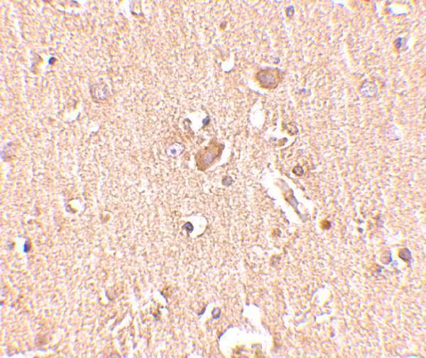
A08611 has a human brain tissue IHC image (A08611 image alt). A08611-2 lists human IF/ICC applications, but no IF image is supplied (catalog applications; image alts).
The only rendered card is A08611, which lists IHC-P and shows human brain tissue IHC at 2.5 μg/mL (catalog applications; A08611 image alt). A08611-2 lists human IF/ICC applications but has no IHC or IF figure in the payload, so it has no rendered card (catalog applications; image alts).
Which to pick: Choose A08611 for paraffin-section tissue IHC: IHC-P is listed, and its own image shows human brain tissue; the caption does not report the fixative (catalog applications; A08611 image alt). For human IF/ICC, A08611-2 lists both applications at 5 μg/mL, although no IF image is supplied (catalog applications; datasheet: 5 μg/mL; image alts). For mouse samples, A08611 lists Mouse reactivity, while A08611-2 lists Human only; neither antibody has a clone listed, and mouse IHC validation is unreported (catalog reactivity; catalog clone fields; A08611 image alt).