This website uses cookies to ensure you get the best experience on our website.


- Table of Contents
Plan chromogenic IHC for METTL14 in paraffin sections using the catalog antibody at 2–5 μg/ml (datasheet A07389-2). Expect widespread nuclear staining, with high signal in lung alveolar type I cells and testis pachytene spermatocytes (HPA tissue IHC).

Expected localisation, validated protocols, controls and antibodies — the at-a-glance facts below, then the full design guide.
| Expected localisation | Nuclear staining in tissue sections (HPA tissue IHC) | |
| Staining pattern | Widespread nuclear staining across cell types (HPA tissue IHC) | |
| Antigen retrieval | EDTA pH 8.0 HIER, heat-mediated (datasheet A07389-2) | |
| Positive control | Lung+4 more · see all | |
| Negative control | Liver |
| Fixation | Keep fixation consistent across sections (standard IHC practice; not target-specific) | |
| Caveat | Cholangiocytes may lack detectable signal (HPA tissue IHC) | |
| Regulation | High in pachytene spermatocytes (HPA tissue IHC) | |
| Isoform / epitope | No isoforms or processing annotated; no TM segment (UniProt) |
The catalog antibody’s IHC protocol uses EDTA pH 8.0 retrieval (datasheet A07389-2). The published protocols below provide three tissue-specific comparisons (PMC12885814; PMC13197395; PMC11711053).
| Sample | Paraffin-embedded human breast cancer tissue; fixative not specified (datasheet A07389-2) |
| Fixation | Image fixative and duration unreported (datasheet A07389-2); verify before use. |
| Sectioning | 4–5 µm sections on charged slides (standard) |
| Deparaffinisation | Xylene, graded ethanol series to water (standard) |
| Antigen retrieval | Heat retrieval: EDTA pH 8.0 (datasheet A07389-2); 20 min, 95–100 °C (standard) |
| Peroxidase block | 3% H2O2, 10 min, room temperature (standard) |
| Blocking | 10% goat serum (datasheet A07389-2) |
| Primary antibody | Rabbit anti-METTL14, 2-5μg/ml (datasheet A07389-2) |
| Primary incubation | Overnight at 4 °C (datasheet A07389-2) |
| Detection | Streptavidin-biotin complex (SABC), DAB chromogen (datasheet A07389-2) |
| Counterstain | Hematoxylin, blue, dehydrate and mount (standard) |
| Expected result | METTL14-positive staining in alveolar cells type I of lung (HPA tissue IHC: High). HPA tissue profile: Ubiquitous nuclear expression. No signal in the no-primary control. |
METTL14 is a nuclear protein with no transmembrane segment (UniProt Q9HCE5). In paraffin-section IHC, expect nuclear staining across many cell types, with particularly strong staining reported in lung alveolar type I cells and testis pachytene spermatocytes (HPA tissue IHC). HPA describes the tissue pattern as ubiquitous nuclear expression and rates its antibody evidence Supported, with medium consistency between staining and RNA data (HPA tissue IHC).
| Distinct nuclear staining in alveolar type I cells or pachytene spermatocytes, with other nuclei also stained. | This fits the reported high signal in those two cell populations and the broader nuclear pattern (HPA tissue IHC). Judge the named cells within their tissue context: HPA's High level describes their observed staining, not every cell in the section. Nuclear localization also agrees with UniProt Q9HCE5. |
| Predominantly cytoplasmic, membrane-like, or extracellular staining, with little nuclear signal. | That distribution conflicts with the nuclear assignment (UniProt Q9HCE5; HPA tissue IHC). Treat it as a warning for nonspecific staining or a detection artefact, rather than evidence for a new METTL14 compartment. Recheck the slide against a known nuclear-positive cell population and the negative controls (standard IHC practice). |
| Strong chromogenic signal in liver cholangiocytes, especially if nearby nuclei show no convincing stain. | HPA reports METTL14 as Not detected in cholangiocytes (HPA tissue IHC). Unexpected signal there raises concern for cross-reactivity or endogenous detection activity, but the HPA observation alone cannot identify which. Compare its compartment and distribution with a positive tissue and appropriate reagent controls (standard IHC practice). |
| A diffuse haze covers nuclei, cytoplasm, and spaces between cells; individual stained nuclei are hard to distinguish. | This is difficult to score as METTL14 because the expected localization is nuclear (UniProt Q9HCE5; HPA tissue IHC). Broad background may reflect detection reagents, insufficient blocking, or excessive staining conditions (standard IHC practice). Assess controls before interpreting small intensity differences between cell populations. |
| No nuclear signal in alveolar type I cells or pachytene spermatocytes on an otherwise assessable section. | Those populations are reported High in HPA tissue IHC, so a blank result calls for a technical check. Their reported staining does not guarantee that every specimen will match the reference. Verify tissue identity and preservation, the antibody and detection steps, and an independent positive control before calling the sample negative (standard IHC practice). |
| Cell population and tissue context | HPA reports High staining in lung alveolar type I cells and testis pachytene spermatocytes, Medium staining in several other listed populations, and no detected staining in liver cholangiocytes (HPA tissue IHC). Select and score the named cells rather than assigning one intensity to a whole organ. |
| Compartment and protein topology | UniProt places METTL14 in the nucleus and reports no transmembrane segment or signal peptide (UniProt Q9HCE5). A membrane outline or secreted-looking deposit therefore lacks support as the expected pattern. HPA's nucleoplasmic ICC-IF localization offers a compartment cross-check (HPA ICC-IF). |
| Strength of reference evidence | The tissue IHC profile is Supported, with medium consistency between antibody staining and RNA expression; HPA038002 is IHC Supported (HPA tissue IHC; HPA antibodies). Use the profile as a reference pattern, while investigating discordant results rather than treating its intensity labels as specimen-wide cutoffs. |
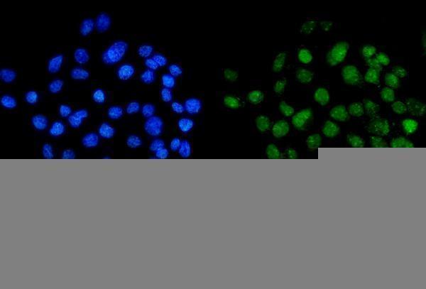
| IF/ICC cross-check: where should fluorescence appear? | HPA reports supported nucleoplasmic localization and ICC-IF images from A-431, U-251MG, and U2OS cells (HPA ICC-IF). Expect a nucleoplasmic pattern when comparing localization across methods. IF/ICC preparation and detection belong to the separate IF/ICC guide. |
| Situation | Likely cause | Next action |
|---|---|---|
| The known-positive nuclei are blank. | A missing detection step, unsuitable reagent condition, or poor tissue preservation can suppress visible staining (standard IHC practice). HPA reports High staining in alveolar type I cells and pachytene spermatocytes (HPA tissue IHC). | Confirm the named cells are present, inspect section quality, and check the catalog antibody's IHC-P instructions and the detection controls (standard IHC practice). Do not infer a METTL14-specific fixation effect: none is established by the supplied sources. |
| Signal is mostly outside nuclei. | The observed compartment disagrees with nuclear METTL14 (UniProt Q9HCE5; HPA tissue IHC). Background or nonspecific binding is a possible explanation, not a diagnosis from appearance alone (standard IHC practice). | Compare a known-positive tissue and negative reagent controls; assess nuclear and extranuclear staining separately (standard IHC practice). Treat extranuclear staining as unconfirmed unless independently supported. |
| Cholangiocytes stain strongly. | HPA lists liver cholangiocytes as Not detected (HPA tissue IHC). Cross-reactivity or endogenous chromogenic activity may account for unexpected staining (standard IHC practice). | Check the compartment, compare a known-positive population, and use suitable negative and endogenous-activity controls for the chosen detection system (standard IHC practice). Do not generalize the cholangiocyte result to every liver cell. |
| Background obscures nuclear boundaries. | Widespread colour development can conceal the nuclear pattern expected for METTL14 (UniProt Q9HCE5; HPA tissue IHC). Excess primary or detection reagent, inadequate blocking, or residual endogenous activity are general IHC possibilities (standard IHC practice). | Review reagent-only controls, blocking, antibody concentration, and development time under the chosen IHC-P workflow (standard IHC practice). Score intensity only where nuclei can be distinguished from surrounding background. |
| A specimen stains more weakly than the HPA example. | HPA intensity levels are observations in specified cell populations; its tissue evidence has medium consistency with RNA expression (HPA tissue IHC). Cell mix, section quality, and assay conditions may complicate comparison (standard IHC practice). | Compare the same cell type and compartment, include a known-positive section in the run, and document the observed nuclear intensity (standard IHC practice). Avoid assigning a universal positive cutoff from the HPA labels. |
| IF/ICC shows diffuse fluorescence instead of nucleoplasmic enrichment. | HPA's supported ICC-IF location is the nucleoplasm (HPA ICC-IF). Diffuse fluorescence alone does not establish a different METTL14 location; background remains possible (standard IF practice). | Compare with appropriate fluorescence controls and the HPA nucleoplasmic reference (HPA ICC-IF; standard IF practice). Use the separate IF/ICC guide for method choices; this IHC-P section supplies no IF protocol. |


Comprehensive Human Protein Atlas IHC scoring per tissue (reliability: Supported — Medium consistency between antibody staining and RNA expression data. External characterization data supports antibody staining.). Rows are taken directly from the HPA tissue chart — click any row's HPA link to view the source.
| Tissue | Cell type | Level | Evidence | Source |
|---|---|---|---|---|
| Lung | Alveolar cells type I | High | Protein (IHC) | HPA → |
| Testis | Pachytene spermatocytes | High | Protein (IHC) | HPA → |
| Adipose tissue | Adipocytes | Medium | Protein (IHC) | HPA → |
| Appendix | Glandular cells | Medium | Protein (IHC) | HPA → |
| Bone marrow | Hematopoietic cells | Medium | Protein (IHC) | HPA → |
| Tissue | Cell type | Level | Evidence | Source |
|---|---|---|---|---|
| Liver | Cholangiocytes | Not detected | Protein (IHC) | HPA → |
Troubleshoot METTL14 staining in paraffin sections by checking retrieval, nuclear localisation, controls and cell-level scoring before interpreting chromogenic signal.
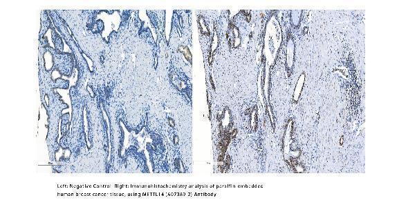
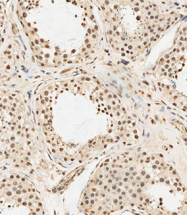
IHC examples cover human breast cancer and testis sections (catalog IHC captions); IF/ICC imagery covers A431 cells (A07389-2 IF caption). Listed reactivity includes human, mouse, rat and monkey (catalog reactivity).
A07389-2 has IHC imagery from paraffin-embedded human breast cancer tissue and IF/ICC imagery from A431 cells (A07389-2 image captions). M07389 has IHC imagery from paraformaldehyde-fixed, paraffin-embedded human testis tissue (M07389 IHC caption).
Which to pick: For tissue IHC, choose A07389-2 for paraffin-embedded human breast cancer sections (A07389-2 IHC caption: 2 μg/ml; fixative unreported) or M07389 for paraformaldehyde-fixed, paraffin-embedded human testis sections (M07389 IHC caption: 1:250). For IF/ICC, A07389-2 has an A431 cell image and a listed 5 μg/ml dilution (A07389-2 IF caption and catalog dilution). For broader listed species reactivity, M07389-1 is a monoclonal antibody with IHC and IF/ICC applications and human, mouse, rat and monkey reactivity (M07389-1 catalog applications, clone and reactivity); the payload provides no IHC image for it (M07389-1 catalog image alts).