This website uses cookies to ensure you get the best experience on our website.


- Table of Contents
Plan MICU1 chromogenic IHC on paraffin sections using the catalog antibody's IHC evidence (datasheet A01895). This guide covers the mainly cytoplasmic tissue pattern, high staining in glandular cells, and the reported low consistency between staining and RNA expression (HPA tissue IHC).

Expected localisation, validated protocols, controls and antibodies — the at-a-glance facts below, then the full design guide.
| Expected localisation | Mainly cytoplasmic in tissue (HPA tissue IHC); mitochondrial intermembrane space and inner membrane (UniProt) | |
| Staining pattern | Cytoplasmic staining in most tissues; high in glandular cells (HPA tissue IHC) | |
| Antigen retrieval | Citrate pH 6.0 HIER, heat-mediated (datasheet A01895) | |
| Positive control | Adrenal gland+4 more · see all | |
| Negative control | None in HPA (detected in all 45 tissues); use no-primary + isotype controls |
| Fixation | Keep fixation conditions consistent across sections (standard IHC practice; not target-specific) | |
| Caveat | Antibody staining and RNA expression show low consistency (HPA tissue IHC) | |
| Regulation | No expression inducer specified (UniProt) | |
| Isoform / epitope | 6 isoforms; mature chain starts at aa 34; epitope unspecified (UniProt) |
Compare the catalog antibody’s IHC-P protocol (datasheet: A01895) with published MICU1 IHC protocols for oral, liver, and breast specimens (PMC8195654; PMC7693594; PMC8805845; PMC11190767).
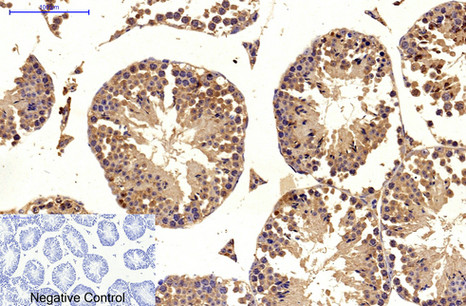
| Sample | Paraffin-embedded Mouse testis tissue; fixative not specified (datasheet A01895) |
| Fixation | Image fixative and duration unreported (datasheet A01895); verify before use. |
| Sectioning | 4–5 µm sections on charged slides (standard) |
| Deparaffinisation | Xylene, graded ethanol series to water (standard) |
| Antigen retrieval | Heat retrieval: Citrate pH 6.0, 20min (datasheet A01895) |
| Peroxidase block | 3% H2O2, 10 min, room temperature (standard) |
| Blocking | 10% normal serum of the secondary host, 30 min, room temperature (standard) |
| Primary antibody | Mouse monoclonal (clone Mix) anti-MICU1, 1:100 recommended; image 1:200 (datasheet A01895) |
| Primary incubation | Overnight at 4 °C (standard) |
| Detection | HRP-polymer secondary, DAB chromogen 5–10 min (standard) |
| Counterstain | Hematoxylin, blue, dehydrate and mount (standard) |
| Expected result | MICU1-positive staining in glandular cells of adrenal gland (HPA tissue IHC: High). HPA tissue profile: Mainly cytoplasmic expression in most tissues. No signal in the no-primary control. |
MICU1 localizes to the mitochondrial intermembrane space and inner membrane, with no transmembrane segment (UniProt Q9BPX6). In paraffin section IHC, expect mainly cytoplasmic staining (HPA tissue IHC), including strong staining in selected glandular, epithelial, hematopoietic, and Purkinje cells (HPA tissue IHC: High). HPA rates the tissue staining Approved but reports low consistency between antibody staining and RNA expression (HPA tissue IHC: reliability description).
| Cytoplasmic staining is strongest in colon or duodenal glandular cells and bronchial respiratory epithelial cells (HPA tissue IHC: High). | This matches the reported cell types and intensity. At routine chromogenic resolution, a mainly cytoplasmic appearance is compatible with mitochondrial MICU1 (HPA tissue IHC; UniProt Q9BPX6 location); it need not resolve into individual mitochondria (standard IHC practice). |
| Nuclear staining dominates while cytoplasmic staining is weak or absent (HPA tissue IHC: mainly cytoplasmic; UniProt Q9BPX6 location). | A predominantly nuclear pattern conflicts with the reported localization. Check the counterstain and compare primary antibody omission and known positive tissue controls before interpreting it as MICU1 (HPA tissue IHC; standard IHC practice). |
| Adipocytes stain as intensely as neighboring cell types despite their reported Low level (HPA tissue IHC: adipocytes Low). | The discrepancy raises concern for cross-reactivity or endogenous detection activity (standard IHC practice). Low is not absent: compare cellular distribution, background, and controls before calling adipocyte staining false (HPA tissue IHC: adipocytes Low). |
| Color covers stroma, empty spaces, and multiple cell types without a clear cellular pattern (standard IHC practice). | Treat widespread diffuse signal as background until controlled. HPA describes mainly cytoplasmic cellular staining, not a uniform field-wide deposit (HPA tissue IHC). Inspect primary antibody omission controls and the detection reagents (standard IHC practice). |
| No signal appears in colon glandular cells or cerebellar Purkinje cells, both reported High (HPA tissue IHC). | A blank known-positive specimen points to a possible assay failure, though HPA patterns do not guarantee every section or antibody will stain (HPA tissue IHC: Approved; low RNA consistency). Check tissue integrity and each detection step (standard IHC practice). |
| Cell type and comparison tissue (HPA tissue IHC) | Colon and duodenal glandular cells, bronchial respiratory epithelium, and Purkinje cells are reported High; adipocytes are Low (HPA tissue IHC). Use those levels as comparisons, not absolute positive or negative guarantees, given low staining–RNA consistency (HPA tissue IHC). |
| Antibody evidence (HPA antibodies; HPA tissue IHC) | HPA037480 is Approved for IHC; the supplied record does not label its IHC validation Enhanced (HPA antibodies). HPA's overall tissue reliability is Approved with low consistency between staining and RNA, so corroborate unexpected patterns with controls (HPA tissue IHC; standard IHC practice). |
| Subcellular resolution (UniProt Q9BPX6; HPA subcellular) | MICU1 occupies the mitochondrial intermembrane space and inner membrane (UniProt Q9BPX6). HPA ICC-IF supports mitochondrial localization (HPA subcellular); routine chromogenic IHC may present this as mainly cytoplasmic staining (HPA tissue IHC; standard IHC practice). |
| Does IF/ICC offer a localization check? (HPA subcellular) | Yes. HPA reports supported mitochondrial ICC-IF localization and images in A-431, A-549, and U2OS; HPA057525 has Supported ICC status (HPA subcellular; HPA antibodies). These data support compartment comparison but do not validate a new IHC protocol (standard IHC practice). |
| Isoforms and epitope coverage (UniProt Q9BPX6) | UniProt lists six MICU1 isoforms and a mature chain spanning residues 34–476 (UniProt Q9BPX6). The supplied evidence gives no antibody epitope, so isoform recognition and any effect of processing on this assay remain undetermined (UniProt Q9BPX6; HPA antibodies). |
| Situation | Likely cause | Next action |
|---|---|---|
| High HPA-listed cells show no detectable color (HPA tissue IHC: High). | The assay may have failed at antibody binding or chromogenic detection; HPA intensity alone cannot identify which step failed (standard IHC practice; HPA tissue IHC). | Run a reported High tissue alongside a primary antibody omission control, then review retrieval, antibody dilution, detection reagents, and counterstain (HPA tissue IHC; standard IHC practice). |
| The slide is strongly nuclear rather than mainly cytoplasmic (HPA tissue IHC; UniProt Q9BPX6 location). | Counterstain, nonspecific binding, or detection artifact may obscure the expected compartment (standard IHC practice). | Compare with the omission control and a reported High tissue; score MICU1 only where cellular color can be distinguished from nuclear counterstain (standard IHC practice; HPA tissue IHC). |
| Adipocytes or unstructured areas look as strong as reported High cells (HPA tissue IHC: adipocytes Low). | Cross-reactivity, endogenous detection activity, or excess background is possible; Low adipocyte staining does not imply zero signal (standard IHC practice; HPA tissue IHC). | Check omission and detection controls, then compare staining in adipocytes with High glandular or epithelial cells on suitably matched sections (standard IHC practice; HPA tissue IHC). |
| Brown color spreads across the section with poor cell boundaries (standard IHC practice). | Insufficient blocking or washing, excess antibody, or endogenous detection activity can create diffuse color (standard IHC practice). | Inspect controls, adjust blocking and washing, and titrate the IHC antibody within its validated conditions; the payload supplies no MICU1-specific dilution (standard IHC practice; HPA antibodies). |
| A punctate mitochondrial pattern is not individually resolved in chromogenic IHC (UniProt Q9BPX6 location). | Mitochondria may appear as a broader cytoplasmic signal at routine light-microscope resolution (HPA tissue IHC; standard IHC practice). | Judge cellular and compartment distribution first; consult HPA's supported mitochondrial ICC-IF localization for a separate resolution check (HPA tissue IHC; HPA subcellular). |
| A tissue differs from its HPA example despite clean controls (HPA tissue IHC). | HPA reports low consistency between antibody staining and RNA expression, and its listed levels describe observed examples rather than guaranteed outcomes (HPA tissue IHC). | Record the cell type, compartment, intensity, and controls; compare another reported High tissue before assigning a biological difference (HPA tissue IHC; standard IHC practice). |
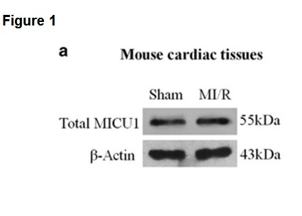

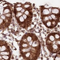


Comprehensive Human Protein Atlas IHC scoring per tissue (reliability: Approved — Low consistency between antibody staining and RNA expression data.). Rows are taken directly from the HPA tissue chart — click any row's HPA link to view the source.
| Tissue | Cell type | Level | Evidence | Source |
|---|---|---|---|---|
| Adrenal gland | Glandular cells | High | Protein (IHC) | HPA → |
| Appendix | Glandular cells | High | Protein (IHC) | HPA → |
| Bone marrow | Hematopoietic cells | High | Protein (IHC) | HPA → |
| Bronchus | Respiratory epithelial cells | High | Protein (IHC) | HPA → |
| Cerebellum | Purkinje cells | High | Protein (IHC) | HPA → |
| Tissue | Cell type | Level | Evidence | Source |
|---|---|---|---|---|
| None in HPA: MICU1 is detected in all 45 scored tissues. Use a no-primary (secondary-only) and an isotype control instead. | ||||
Troubleshoot MICU1 staining in paraffin sections by checking retrieval, compartment, controls and scoring before interpreting chromogenic signal.
A01895 has IHC data from paraffin-embedded mouse testis and IF data from HeLa cells and human appendix (A01895 image captions). Its listed reactivity is human, mouse and rat (A01895 catalog).
A01895 is the SKU with a rendered card; its IHC figure shows paraffin-embedded mouse testis (A01895 IHC caption). Its IF images show HeLa cells and human appendix (A01895 IF captions).
Which to pick: Choose A01895 for tissue IHC because its own figure shows paraffin-embedded mouse testis; the fixative is unreported (A01895 IHC caption). For IF/ICC, A01895 is the choice because IF is listed and cell imaging is shown (A01895 applications; A01895 IF caption). For cross-species work, A01895 and A01895-3 both list human, mouse and rat reactivity, but the supplied IHC figure establishes tissue staining only for A01895 in mouse testis (catalog reactivity; A01895 IHC caption).