This website uses cookies to ensure you get the best experience on our website.


- Table of Contents
High staining in lung macrophages and colon glandular cells (HPA tissue IHC) helps select positive controls. Start the catalog antibody at 1:100–1:300 for paraffin IHC (datasheet A30524).

Expected localisation, validated protocols, controls and antibodies — the at-a-glance facts below, then the full design guide.
| Expected localisation | Cytoplasmic staining in several tissues (HPA tissue IHC) | |
| Staining pattern | Cytoplasmic signal in colon glands and lung macrophages (HPA tissue IHC) | |
| Antigen retrieval | Tris-EDTA pH 8.0 HIER, heat-mediated (datasheet A30524) | |
| Positive control | Appendix+4 more · see all | |
| Negative control | Adipose tissue+4 more · see all |
| Fixation | Matched tissue-IHC evidence does not establish the fixation claim. Validate the specimen-specific method before use. (selected-SKU IHC image A30524) | |
| Caveat | Strong lung macrophage staining may affect scoring (HPA tissue IHC) | |
| Regulation | Isoform abundance varies by tissue (UniProt) | |
| Isoform / epitope | 5 isoforms; no propeptide; check epitope coverage (UniProt) |
The catalog antibody protocol is paired with one published MINK1 IHC protocol using lung sections (PMC9592806).
| Sample | Paraffin-embedded Human breast cancer tissue; fixative not specified (datasheet A30524) |
| Fixation | Image fixative and duration unreported (datasheet A30524); verify before use. |
| Sectioning | 4–5 µm sections on charged slides (standard) |
| Deparaffinisation | Xylene, graded ethanol series to water (standard) |
| Antigen retrieval | Heat retrieval: Tris-EDTA pH 8.0 (datasheet A30524); 20 min, 95–100 °C (standard) |
| Peroxidase block | 3% H2O2, 10 min, room temperature (standard) |
| Blocking | 10% normal serum of the secondary host, 30 min, room temperature (standard) |
| Primary antibody | Rabbit anti-MINK1, 1:100 - 1:300 (datasheet A30524) |
| Primary incubation | Overnight at 4 °C (standard) |
| Detection | HRP-polymer secondary, DAB chromogen 5–10 min (standard) |
| Counterstain | Hematoxylin, blue, dehydrate and mount (standard) |
| Expected result | MINK1-positive staining in glandular cells of appendix (HPA tissue IHC: High). HPA tissue profile: Cytoplasmic expression in several tissues. No signal in the no-primary control. |
MINK1 is primarily cytoplasmic in tissue IHC, with high staining reported in selected glandular, squamous epithelial, macrophage and trophoblastic cells (HPA: cytoplasmic expression in several tissues; HPA: High in listed cells). UniProt also places MINK1 at the Golgi and neuronal projections, and reports no transmembrane segment (UniProt Q8N4C8 subcellular location; topology). The HPA tissue IHC assessment is Approved, with medium consistency between staining and RNA expression (HPA: reliability).
| Cytoplasmic staining is strong in colon or appendix glandular cells, cervical squamous epithelial cells, lung macrophages, or placental trophoblasts (HPA: High in these cells). | This matches observed positive cell types and the reported cytoplasmic pattern (HPA: tissue IHC profile). Compare the stained cell population with tissue morphology; a positive call should describe the cells and compartment, not just the tissue name. High is the HPA observation for those specific cells, not a required intensity in every specimen (HPA: tissue IHC). |
| Signal is confined to nuclei or traces crisp cell borders while the cytoplasm is clear. | A predominantly nuclear or membrane-outline pattern conflicts with the reported tissue pattern and a protein without a transmembrane segment (HPA: cytoplasmic expression; UniProt Q8N4C8 topology). Check morphology, counterstain and detection controls before calling it MINK1. Golgi or neuronal-projection localisation is annotated, but neither annotation establishes a distinctive chromogenic pattern here (UniProt Q8N4C8 subcellular location). |
| Strong staining appears mainly in adipocytes, cardiomyocytes, or bone-marrow hematopoietic cells. | These particular cell populations were not detected by HPA tissue IHC (HPA: Not detected in adipocytes, cardiomyocytes and bone-marrow hematopoietic cells). Unexpected signal raises possible nonspecific antibody binding or detection activity; inspect a no-primary control and the positive tissue on the same run. An HPA negative observation is a comparison point, not proof that every sample must be negative (HPA: Approved; medium consistency). |
| Brown color spreads across stroma, empty spaces, or most cell types without clear cytoplasmic boundaries. | Treat this as background until morphology separates cellular staining from deposits. In chromogenic IHC, incomplete blocking, excess reagent or residual endogenous detection activity can produce broad color (standard IHC practice). The HPA description is cytoplasmic expression in several tissues, so diffuse color alone does not identify MINK1-positive cells (HPA: tissue IHC profile). |
| No staining is visible in a positive control such as colon glandular cells or lung macrophages. | HPA reports high staining in those defined cell populations (HPA: High in colon glandular cells; High in lung macrophages). If the expected cells are present, first suspect a run-level problem with antibody, retrieval or detection (standard IHC practice). Do not infer biological absence from a failed control; the supplied evidence does not establish MINK1-specific fixation sensitivity. |
| Which tissue and cell population are scored | HPA reports High staining in appendix, colon and duodenum glandular cells; cervix, esophagus and oral-mucosa squamous epithelial cells; lung macrophages; and placental trophoblasts (HPA: tissue IHC). It reports Not detected for selected other cell populations, including adipocytes and cardiomyocytes (HPA: tissue IHC). Score the named cells within each section; whole-tissue labels can obscure a mixed result. |
| Expected compartment and resolution | Cytoplasm is the documented tissue IHC pattern (HPA: tissue IHC profile). Golgi, axonal, dendritic and postsynaptic-density locations are UniProt annotations (UniProt Q8N4C8 subcellular location). Routine chromogenic sections may not resolve those structures, so do not require visible puncta to accept cytoplasmic staining (standard IHC practice). |
| Isoforms and antibody epitope | MINK1 has five listed isoforms; isoform 3 is ubiquitous, while isoform 1 is most abundant in skeletal muscle and isoform 2 exceeds isoform 1 in brain (UniProt Q8N4C8 tissue specificity; isoforms). The supplied antibody record gives no epitope or isoform coverage (HPA: HPA056296 validation record). Staining cannot therefore be assigned to a particular isoform from this evidence. |
| Strength of antibody and tissue evidence | The listed antibody HPA056296 has Approved IHC status; the tissue profile has medium staining–RNA consistency (HPA: antibody validation; tissue reliability). Neither entry reports Enhanced IHC validation (HPA: antibody validation). Use the observed pattern as a reference while retaining morphology and run controls when evaluating an unexpected result (standard IHC practice). |
| IF/ICC: what pattern can be expected? | A cytoplasmic location is supported by UniProt, which also annotates Golgi and neuronal projections (UniProt Q8N4C8 subcellular location). HPA supplies no main ICC-IF location and no cell-line ICC-IF images, so this payload cannot establish a verified fluorescent distribution (HPA: subcellular summary). Interpret any IF/ICC result on its own guide page. |
| Situation | Likely cause | Next action |
|---|---|---|
| The positive control is blank. | An antibody, retrieval or detection step may have failed (standard IHC practice). | Verify that the expected glandular cells or macrophages are present, then check the run reagents and detection controls (HPA: High in colon glandular cells and lung macrophages; standard IHC practice). |
| Only nuclei or cell borders stain. | The dominant compartment disagrees with the reported cytoplasmic tissue pattern (HPA: tissue IHC profile). | Review morphology and a no-primary control; repeat the stain if the pattern persists, and report the compartment actually seen (standard IHC practice). |
| Unexpected cells stain strongly in a comparison tissue. | Cross-reactivity or detection activity is possible; HPA's negative calls apply to named cells, not every cell in the organ (HPA: tissue IHC). | Identify the stained cell type and compare it with an HPA-listed positive population and a no-primary control before assigning MINK1 (HPA: tissue IHC; standard IHC practice). |
| The whole section has diffuse brown background. | Excess reagent, incomplete blocking or endogenous detection activity can obscure cellular signal (standard IHC practice). | Check no-primary and detection controls, then adjust blocking, washing or reagent concentration according to the validated IHC workflow (standard IHC practice). |
| Lung macrophages are dark in both test and no-primary sections. | A detection-system contribution is plausible when color remains without primary antibody (standard IHC practice). | Resolve the control signal before using macrophages as evidence of MINK1; HPA reports high macrophage staining only as a tissue IHC observation (HPA: High in lung macrophages; standard IHC practice). |
| Brain or skeletal-muscle staining seems inconsistent with expression notes. | UniProt describes tissue-dependent isoform abundance, while HPA reports Low staining in hippocampal neurons and Not detected in specified glial populations (UniProt Q8N4C8 tissue specificity; HPA: tissue IHC). | Score the actual cell population and compartment. Do not convert an isoform-expression note into a predicted IHC intensity without epitope or isoform-coverage evidence (HPA: tissue IHC; HPA: HPA056296 validation record). |
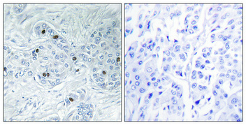
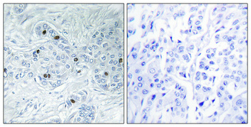


Comprehensive Human Protein Atlas IHC scoring per tissue (reliability: Approved — Medium consistency between antibody staining and RNA expression data.). Rows are taken directly from the HPA tissue chart — click any row's HPA link to view the source.
| Tissue | Cell type | Level | Evidence | Source |
|---|---|---|---|---|
| Appendix | Glandular cells | High | Protein (IHC) | HPA → |
| Cervix | Squamous epithelial cells | High | Protein (IHC) | HPA → |
| Colon | Glandular cells | High | Protein (IHC) | HPA → |
| Duodenum | Glandular cells | High | Protein (IHC) | HPA → |
| Esophagus | Squamous epithelial cells | High | Protein (IHC) | HPA → |
| Tissue | Cell type | Level | Evidence | Source |
|---|---|---|---|---|
| Adipose tissue | Adipocytes | Not detected | Protein (IHC) | HPA → |
| Adrenal gland | Glandular cells | Not detected | Protein (IHC) | HPA → |
| Bone marrow | Hematopoietic cells | Not detected | Protein (IHC) | HPA → |
| Caudate | Glial cells | Not detected | Protein (IHC) | HPA → |
| Cerebellum | Cells in granular layer | Not detected | Protein (IHC) | HPA → |
Troubleshoot MINK1 staining in paraffin sections by checking retrieval, cellular pattern, controls and scoring before interpreting chromogenic signal.
Both anti-MINK1 antibodies have IHC images from paraffin-embedded human breast cancer (each SKU’s image caption); both list IF and human/mouse reactivity (catalog entries).
A30524 lists IHC, IF and ICC, with an IHC image from paraffin-embedded human breast cancer (A30524 catalog entry and image caption). A06134-2 lists IHC and IF, with its own IHC image from paraffin-embedded human breast cancer (A06134-2 catalog entry and image caption).
Which to pick: Either SKU is supported for paraffin-section IHC by its own human breast cancer image; the fixative is unreported in both captions (A30524 and A06134-2 image captions). Choose A30524 when ICC is required because it lists ICC and is polyclonal; both SKUs list IF, while A06134-2 has no reported clonality (catalog entries). Both list human and mouse reactivity, but their IHC images show human tissue only (catalog entries and image captions).