This website uses cookies to ensure you get the best experience on our website.


- Table of Contents
Real validated MLKL Western blot protocols, expected-band and isoform facts, troubleshooting for weak or shifted signal, and recommended anti-MLKL WB antibodies. Everything you need to plan the experiment before you commit precious samples.

Expected bands, validated protocols, controls and antibodies — the at-a-glance facts below, then the full design guide.
| Expected band | ~54.5 kDa | |
| Observed band | ~54 kDa | |
| Gel | 5–20% (catalog M00535-1) | |
| Negative control | Suggested KO / knockdown lysate |
| PTM | Phosphorylated | |
| Caveat | Phosphorylation-state controls | |
| Gene-set association | MSigDB C7 membership | |
| Isoform | 2 isoform(s) |
The M00535-1 protocol combines labelled catalog values with standard starting conditions. Published comparisons retain their own sample, reagent and detection scope.
| Sample / lysate | human Hela, human PC-3, human A549 (catalog M00535-1) |
| Gel % | 5–20% (catalog M00535-1) |
| Load | 30 ug; reducing conditions (catalog M00535-1) |
| Transfer | a nitrocellulose membrane at 150 mA for 50-90 minutes (catalog M00535-1) |
| Membrane | nitrocellulose membrane (catalog M00535-1) |
| Blocking | 5% non-fat milk/TBS for 1.5 hour at RT (catalog M00535-1) |
| Primary antibody | M00535-1 · 1:500 (catalog M00535-1) |
| Primary incubation | overnight at 4°C (catalog M00535-1) |
| Secondary antibody | goat anti-rabbit IgG-HRP, 1:5000 (catalog M00535-1) |
| Secondary incubation | 1.5 hour at RT (catalog M00535-1) |
| Wash | TBS-0.1%Tween 3 times with 5 minutes each (catalog M00535-1) |
| Detection | ECL (catalog M00535-1) |
MLKL is predicted at 54.5 kDa and observed near 54 kDa; the cause of any difference is not established.
| Band near 54 kDa in whole-cell lysate | matches the empirical MLKL band |
| Single sharp band near 54 kDa | consistent with the predicted 54.5 kDa protein |
| Additional bands at different positions | could reflect isoforms 1 and 2; their migration is not established |
| More signal in a membrane fraction after necroptosis induction | consistent with MLKL translocation from cytoplasm to plasma membrane |
| UniProt predicted mass | places the 471-residue protein near 54.5 kDa |
| Empirical MLKL band | appears at approximately 54 kDa in reducing whole-cell blots |
| Isoform 1 | has no supplied isoform-specific mass or migration |
| Isoform 2 | could differ in size from isoform 1, but its mass and migration are not supplied |
| Situation | Likely cause | Next action |
|---|---|---|
| No band in lysate | MLKL expression or detection may be insufficient in the sampled cells | check sample loading and use a validated positive lysate |
| Band higher than expected | band identity or sample preparation may account for the difference; MLKL forms homotrimers, but their SDS-PAGE migration is not established | repeat under fully denaturing, reducing conditions and verify band identity |
| Band lower than expected | isoform identity or degradation may account for the difference; isoform masses are not supplied | compare with a validated positive sample and check sample integrity |
| Multiple bands | isoforms 1 and 2 are annotated, but distinct resolved bands are not established | verify band identity with an independent antibody or MLKL depletion |
| Weak or no signal | MLKL may be unevenly distributed between cytoplasm and membrane after necroptosis induction | check whole-cell recovery and compare cytoplasmic and membrane fractions |
Comprehensive Human Protein Atlas IHC scoring per tissue. Rows are taken directly from the HPA tissue chart — click any row's HPA link to view the source.
| Tissue | Cell type | Level | Evidence | Source |
|---|---|---|---|---|
| No high/medium HPA tissues identified in the supplied evidence. | ||||
| Tissue | Cell type | Level | Evidence | Source |
|---|---|---|---|---|
| No lower-expression tissue rows available in the supplied evidence. | ||||
Deeper troubleshooting and optimisation questions for MLKL, answered from its protein features.
Catalog antibodies with Western blot application and product-specific WB images. Evaluate suitability with the reported sample, controls and experimental conditions.
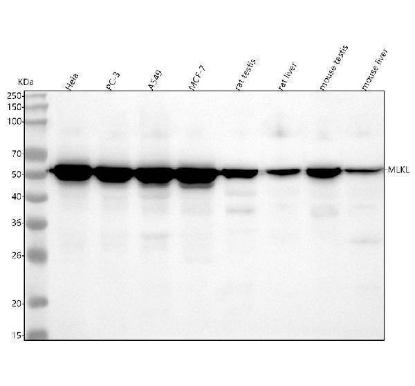
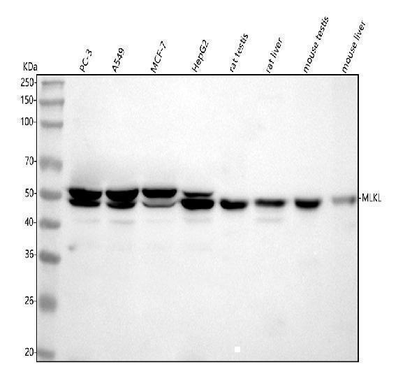
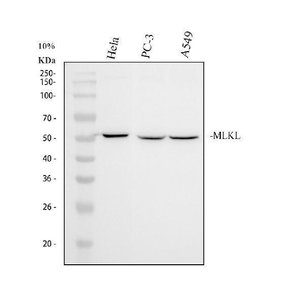
Three anti-MLKL antibodies have Western blot images. M00535-1 and A00535-3 show human cell and mouse and rat tissue lysates; M00535 shows human cell lysates. These images document the stated samples and conditions, not performance across every sample type.
Which to pick: For mouse or rat lysates, consider M00535-1 or A00535-3; both have WB images with testis and liver samples. For human cell lysates, all three have images. M00535-1 and M00535 show bands near the expected 54 kDa; A00535-3 shows one near 50 kDa.