This website uses cookies to ensure you get the best experience on our website.
- Table of Contents
29 Citations 4 Q&As
16 Citations 16 Q&As
1 Citations
Facts about Collagenase 3.
Can also degrade collagen type IV, type XIV and kind X. May also be the activating or degrading key regulatory proteins, such as TGFB1 and CTGF.
Human | |
---|---|
Gene Name: | MMP13 |
Uniprot: | P45452 |
Entrez: | 4322 |
Belongs to: |
---|
peptidase M10A family |
CLG 3; CLG3EC 3.4.24; collagenase 3; EC 3.4.24.-; EC 3.4.24.22; EC 3.4.24.24; EC 3.4.24.35; EC 3.4.24.65; EC 3.4.24.7; MANDP1; matrix metallopeptidase 13 (collagenase 3); matrix metalloproteinase 13 (collagenase 3); Matrix metalloproteinase-13; MMP13; MMP-13
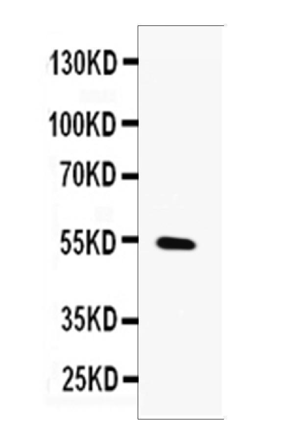
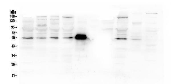
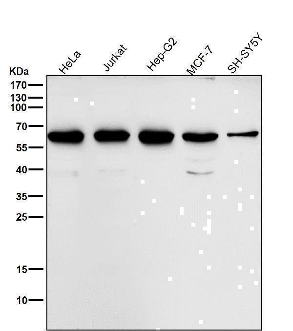
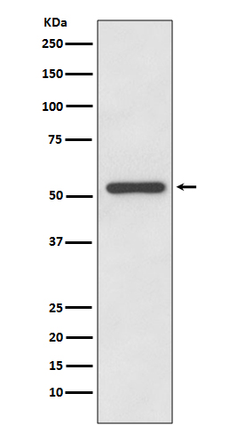
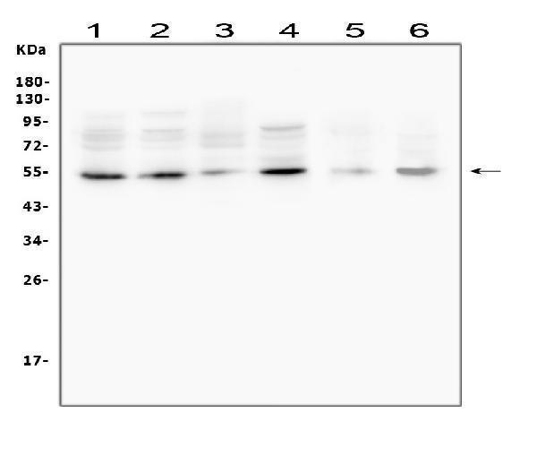
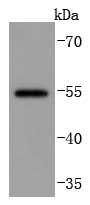
Mass (kDA):
53.82 kDA
Human | |
---|---|
Location: | 11q22.2 |
Sequence: | 11; NC_000011.10 (102942995..102955732, complement) |
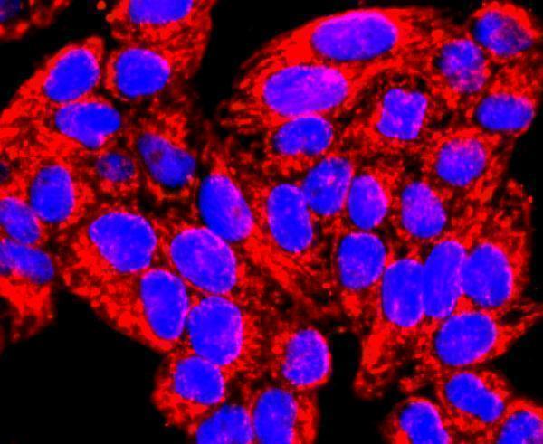
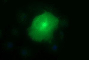
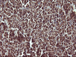
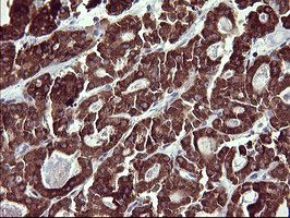
Detected in fetal cartilage and calvaria, in chondrocytes of hypertrophic cartilage in vertebrae and in the dorsal end of ribs undergoing ossification, as well as in osteoblasts and periosteal cells below the inner periosteal region of ossified ribs. Detected in chondrocytes from in joint cartilage that have been treated with TNF and IL1B, but not in untreated chondrocytes. Detected in T lymphocytes. Detected in breast carcinoma tissue.
Secreted, extracellular space, extracellular matrix. Secreted.
The MMP13 marker has many advantages. Boster Bio antibodies can be validated by using negative and positive samples on various platforms. They are highly specific and have high affinity. The first reviewers at Boster receive product credits. Boster is committed to providing superior antibodies to scientists across the world. Learn more about Boster's product's approval and the MMP-13 marker here.
A key component of Pyogenic Bacterium is the MMP13 protein. Boster antibodies are extremely specific to this protein and are highly active. The antibody was validated on immunohistochemistry, Western Blotting, and ELISA. The quality of Boster antibodies has been thoroughly examined by experts in science to assure high specificity and high affinity. The company offers generous rewards to the first reviewers of its products. These awards are acknowledged by scientists.
One study demonstrated that MMP-13 levels were highly predictive of invasive CSC and can be used to monitor the progression of disease. The results revealed that MMP-13 was more sensitive to cSCC than other biomarkers. The Weifang Municipal Science and Technology Project supported the research. The researchers believe that these new biomarkers can be an important tool in the fight against cancer.
The MMP13 marker can be used to study cellular activity of the MMP protein. Boster Bio offers a DNA kit that includes the MMP13 marker. The test uses the MMP13 marker to determine if you have the presence of psoriatic arthritis. The MMP13 protein plays a part in the breakdown of enzyme-producing proteins. However this enzyme doesn't directly cause inflammation in the cartilage of patients.
The MMP13 protein is manufactured by Boster Biological Technology, Ltd. which produces biotin-conjugated goat anti-rabbit secondary antibodies. The MMP-13 marker can be used to detect various forms of MMP in different tissues. At present, Boster Bio produces a biotin-conjugated MMP13 marker that can be used to detect a wide range of tumors and inflammatory processes.
Western Blot detection of MMP-13 can be an efficient method of determining the presence of this enzyme in a biological sample. There are many antibodies available on the market. One of the most sought-after is the Mouse Anti-Human MMP-13 Monoclonal Antibody (MAM-13). It detects the expression of MMP-13 in human ovarian cancer tissues.
MMP-13 protein is found in serum and BM and is extensively expressed in MM tissues. Additionally the elevated levels of MMP-13 in serum indicate the presence of active bone disease. Targeting the MMP-13 protein is much more efficient than its catalytic capability. Thus, the expression of MMP-13 can help researchers identify the presence of the agent responsible for causing disease. MMP-13 expression can be used to determine bone disease.
Detection of MMP-13 by western blots can be accomplished by using RNA extracted from BMSCs and the RPMI 8226 cells. RNA was then generated by SuperScript III reverse transcriptase (Invitrogen) and quantified by the Western blot. In addition the MMP-13 mRNA levels were determined using flow cytometry, or qRT-PCR.
Immunostaining cells are also able to detect the protein. The MMP-13 immunostaining reveals that the protein colocalizes with ED-1-positive perivascular cell lines and with type IV collagen, osteopontin, and type IV collagen. MMP-13 is also expressed by perivascular cells within the macrophage/microglia perivascular cell lineage. It is not known how MMP-13 affects BBB function However, it is well-known to be involved in atherosclerosis.
Monoclonal antibody to detect Human MMP-13 are employed in the ELISA. Its dilution is between 1 and 2 ug/ml and should be used in conjunction with other second-step agents compatible with it. The detection limit for Human MMP-13 may be as low as 20ng/lane when utilized with the chromogenic substratum. The sensitivity of this assay can increase up to 50 times with the chemiluminescent substrate.
Transfected pcDNA-derived mRNA to MMP-13 into HEK-293 cells. The cells were maintained in serum-free OptiMEM for 48 hrs. After transfection, cells were harvested and the media used for cell culture was filtered using ultrafiltration. The eluted proteins were subjected to Western analysis using a blot.
The MMP13 marker, a renowned instrument for detecting proteins, is used to identify the location and distribution of certain cells' components. Researchers can visualize the expression of proteins in tissue samples using antibodies that recognize specific proteins or antigens. However, successful immunohistochemistry depends on the correct preparation of the sample and staining techniques. Positive results are highly specific, provide high levels of signal, and can be readily interpreted.
MMP-13 can be found in a variety of pathological conditions such as head-and-neck SCC. It is associated with a poor prognosis. Since abnormal oral cavity cells are sloughed away into saliva which is a good diagnostic tool for assessing biomarkers. Additionally saliva collection is non-hazardous, noninvasive, and repeatable which makes it a great alternative to serum for disease monitoring.
The manufacturer is able to sell antibodies for MMP13 markers. Boster Bio MMP13 Antibody (catalog number A00420) can be stored at -20°C for one year or at 4degC for one month. Anti-MMP13 antibody contains four mg of Trehalose and 0.22mg Na2HPO4.
Apart from research on the function of MMP13 Other studies have indicated that circRNA-CER plays a role in breast cancer. MiR-136 inhibition reduced MMP13 expression in breast cancer cells. Furthermore, it facilitated the growth and migration of MCF-7 cells. This discovery could have many applications in the treatment of breast cancer. It opens the doorway to more effective cancer treatments.
Research in many diseases can be conducted using the MMP13 antibody. The antibodies are specifically designed for MMP-13 and can be used to identify illnesses that require the enzyme. It is useful for both veterinary and human purposes. The antibodies used in this study were made by combining Gal-3-FITC with DAPI and then processed using DIC imaging. Serial microphotographs are taken at intervals of one millimeter. Software was used to create Z stack projection.
The MMP13 antibody offers high-quality images for flow the cytometric analysis. The antibody is coupled to fluorescent probes for flow cytometric analysis. The probes that emit fluorescent light attach to the cell and interact over time with the laser beam. This produces an emission of light. The intensity of the fluorescence is directly related with the area of the pulse. The more channels you have, the bigger the area of the pulse.
The accuracy and specificity of ELISA testing for the MMP13 marker in serum are both high, suggesting that this marker can be used for the early detection of cancerous cSCC. The extent of invasiveness and lymph node involvement of the tumor is related to the level of MMP-13 in the serum. The sensitivity and specificity of ELISA for the MMP13 marker were 100 percent. This test can also be used to detect pre-invasive lesions or cancerous cells.
The MMP13 marker belongs to the metalloproteinase category. It degrades collagen type I, but it does not degrade casein or gelatin. In addition to destroying collagen, MMP13 is involved in bone disease called spondyloepimetaphyseal dysplasia Missouri type. As patients age, the epimetaphyseal alterations tend to improve. In contrast, patients with metaphyseal anadysplasia type 1 show extreme changes to the skeleton.
Patients with esophageal SCC had a low serum MMP-13 level. There was no correlation significant with their age or sex. The levels of MMP-13 in the serum of patients with neck and head SCC were not related to gender or age. The serum levels of MMP-1 2, - 2, and -9 in the same patients were not related. These results suggest that serum MMP-13 markers could be clinically significant in diagnosing and prognosing ESCC patients.
The MMP13 ELISA kit identifies MMP-13 in human samples. It has an antibody that is specifically for MMP-13. The antibody is biotinylated and captures MMP-13's protein. Streptavidin HRP is used to detect the MMP13 protein. The samples are then treated with a signal development agent. The results are highly reliable and reproducible.
The MMP13 ELISA kit has an excellent sensitivity and selectivity. This test analyzes the MMP13 marker levels in serum, synovial fluid, as well as body fluids. The ELISA MMP13 kit can be used to determine the presence of fibroblasts, cancer cells, and other diseases. A positive outcome in this test means that MMP-13 is present in the tissues of the patient.
To conduct an ELISA for the MMP13 marker, a small amount of the targeted tissue or fluid is in the sample to be tested. The reaction results in a color that is directly proportional to the amount of target protein in the original sample. The signal intensity is measured with a correction wavelength of 630nm. The result of the ELISA is a clear picture of the presence of MMP13.
PMID: 8207000 by Freije J.M.P., et al. Molecular cloning and expression of collagenase-3, a novel human matrix metalloproteinase produced by breast carcinomas.
PMID: 9562863 by Willmroth F., et al. A matrix metalloproteinase gene expressed in human T lymphocytes is identical with collagenase 3 from breast carcinomas.
*Showing only the more recent 20. More publications can be found for each product on its corresponding product page