This website uses cookies to ensure you get the best experience on our website.


- Table of Contents
Real validated MMP3 Western blot protocols, expected-band and isoform facts, troubleshooting for weak or shifted signal, and recommended anti-MMP3 WB antibodies. Everything you need to plan the experiment before you commit precious samples.
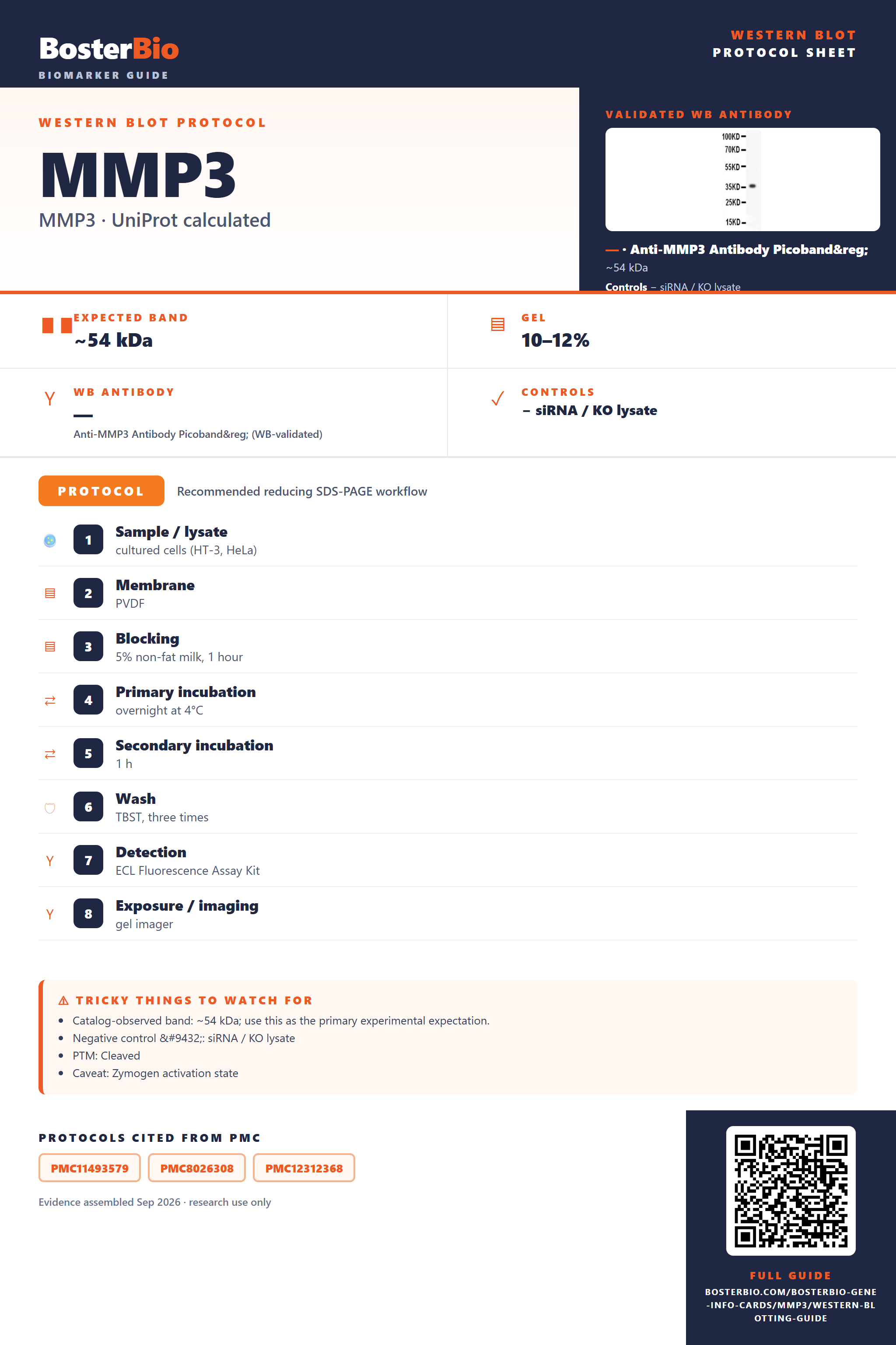
Expected bands, validated protocols, controls and antibodies — the at-a-glance facts below, then the full design guide.
| Expected band | ~54 kDa | |
| Observed band | 54 kDa | |
| Gel | 10–12% | |
| Negative control | siRNA / KO lysate |
| PTM | Cleaved | |
| Caveat | Zymogen activation state | |
| Regulation | Coagulation | |
| Isoform | 1 isoform(s) |
Literature-validated Western blot parameters for MMP3 — gel percentage, transfer, blocking, antibody incubation and detection, extracted from published methods.
| Sample / lysate | cultured cells (HT-3, HeLa) |
| Membrane | PVDF |
| Blocking | 5% non-fat milk, 1 hour |
| Primary incubation | overnight at 4°C |
| Secondary incubation | 1 h |
| Wash | TBST, three times |
| Detection | ECL Fluorescence Assay Kit |
| Exposure / imaging | gel imager |
MMP3 has a 54 kDa predicted and observed pro-enzyme band, though proteolytic removal of its ~9 kDa propeptide during activation can shift the mature band down to ~45 kDa.
| Single band at 54 kDa in tissue or whole-cell lysate | Matches the predicted mass of full-length pro-MMP3, consistent with an unmodified zymogen that carries no glycosylation |
| Smaller band near 45 kDa alongside or instead of the 54 kDa band | Reflects the mature, activated form of MMP3 after proteolytic removal of the ~9 kDa propeptide (residues 18-99) |
| Weak or absent band in whole-cell lysate alone | MMP3 is secreted into the extracellular space and ECM, so intracellular lysates capture little of the mature secreted pool |
| No shift in band position between reducing and non-reducing gels | The single disulfide bond is intrachain, stabilizing the hemopexin-like domain rather than linking two separate chains, so no dimer band forms |
| Sharp single band without smearing | MMP3 has no annotated glycosylation sites, so it migrates as a homogeneous, non-glycosylated species |
| Recombinant or truncated protein standard running near 36 kDa | Bacterially expressed or truncated constructs lack the full domain structure and any post-translational processing seen in native protein, so they run lower than the 54 kDa native form |
| Predicted mass (54 kDa, 477 aa) | Sets the baseline single-band size seen in native tissue and whole-cell lysates |
| Signal peptide cleavage (residues 1-17) | Removed co-translationally from the secreted precursor, so it does not affect the mass of the mature secreted species seen on blot |
| Propeptide cleavage (residues 18-99) | Removal of this ~9 kDa prodomain during zymogen activation converts the ~54 kDa proenzyme to a smaller ~45 kDa mature/active band |
| Intrachain disulfide bond (Cys290-Cys477) | Stabilizes the hemopexin-like domain fold within a single chain rather than linking separate chains, so it does not produce a higher-molecular-weight dimer band |
| Secreted/extracellular localization | Shifts most full-length protein into conditioned media or ECM fractions, reducing band intensity in standard whole-cell lysate preparations |
| Non-glycosylated recombinant/bacterial standard | Runs lower than native tissue-derived protein since it lacks any post-translational modification or full-length processing beyond the core expressed sequence |
| Situation | Likely cause | Next action |
|---|---|---|
| No band in lysate | MMP3 is secreted into the extracellular space/ECM, leaving little full-length protein inside whole-cell lysates | Test conditioned media, ECM extracts, or concentrated secretome samples alongside whole-cell lysate |
| Band lower than expected | The propeptide (residues 18-99) has been proteolytically removed, converting the ~54 kDa zymogen to the ~45 kDa mature active form | Confirm which form the antibody epitope targets and include pro- vs active-MMP3 controls to distinguish normal processing from degradation |
| Multiple bands | The ~54 kDa pro-form and the ~45 kDa mature/active form can both be present in samples with active MMP3 turnover | Compare relative band intensities across time points or use activator/inhibitor treatments to confirm a pro-to-active conversion pattern |
| Weak or no signal | Low steady-state levels of secreted MMP3 in standard lysates, or loss of antibody epitope if the propeptide or hemopexin region is removed | Increase protein loading, concentrate the secreted fraction, and verify the antibody epitope location relative to known processing sites |
| Fragments below expected size | Proteolytic cleavage at the propeptide junction or within the hemopexin-like domain can generate smaller fragments distinct from the full-length or mature bands | Add protease inhibitors during lysis and compare fresh versus stored samples to rule out post-lysis degradation |


Comprehensive Human Protein Atlas IHC scoring per tissue. Rows are taken directly from the HPA tissue chart — click any row's HPA link to view the source.
| Tissue | Cell type | Level | Evidence | Source |
|---|
| Tissue | Cell type | Level | Evidence | Source |
|---|
Deeper troubleshooting and optimisation questions for MMP3, answered from its protein features.
BosterBio's MMP3 antibodies are among the best-performing WB antibodies on the market — well cited, thoroughly validated, and orthogonally cross-validated against negative tissues and complementary methods.
The anti-MMP3 antibodies recommended below are top-performing, extensively cited reagents validated through rigorous testing, including orthogonal confirmation against negative tissue controls and complementary methods, ensuring specific, reproducible Western blot detection of MMP3 you can trust for your experiments.
Which to pick: Only one Boster anti-MMP3 antibody is catalogued, PB9267 (Picoband), so it's the clear choice. It has a genuine WB validation image showing a specific 36 kDa band on recombinant human MMP3 protein at 0.5 ug/ml.