This website uses cookies to ensure you get the best experience on our website.


- Table of Contents
Plan MNAT1 IHC-P staining around its nuclear pattern, using seminiferous duct cells as a positive reference (HPA tissue IHC). Start the catalog antibody at 0.5–1 μg/mL (datasheet PB9614) and assess staining by cell type.

Expected localisation, validated protocols, controls and antibodies — the at-a-glance facts below, then the full design guide.
| Expected localisation | Nuclear staining in tissue cells (HPA tissue IHC) | |
| Staining pattern | Variable nuclear staining, prominent in seminiferous ducts (HPA tissue IHC) | |
| Antigen retrieval | EDTA pH 8.0 HIER, heat-mediated (datasheet PB9614) | |
| Positive control | Testis+4 more · see all | |
| Negative control | Adipose tissue+4 more · see all |
| Fixation | Keep fixation conditions consistent across sections. (standard IHC practice; not target-specific) | |
| Caveat | Staining and RNA have medium consistency (HPA tissue IHC) | |
| Regulation | Expression regulation is unreported (UniProt) | |
| Isoform / epitope | 2 isoforms; epitope differences unspecified (UniProt) |
The catalog antibody protocol uses heat-mediated EDTA pH 8.0 retrieval (datasheet: PB9614). Published MNAT1 IHC examples cover colorectal cancer microarrays and paraffin-embedded laryngeal carcinoma sections (PMC6258412; PMC12274886).
| Sample | Paraffin-embedded mouse testis tissue; fixative not specified (datasheet PB9614) |
| Fixation | Image fixative and duration unreported (datasheet PB9614); verify before use. |
| Sectioning | 4–5 µm sections on charged slides (standard) |
| Deparaffinisation | Xylene, graded ethanol series to water (standard) |
| Antigen retrieval | Heat retrieval: EDTA pH 8.0 (datasheet PB9614); 20 min, 95–100 °C (standard) |
| Peroxidase block | 3% H2O2, 10 min, room temperature (standard) |
| Blocking | 10% goat serum (datasheet PB9614) |
| Primary antibody | Rabbit anti-MNAT1, 0.5-1μg/ml (datasheet PB9614) |
| Primary incubation | Overnight at 4 °C (datasheet PB9614) |
| Detection | Streptavidin-biotin complex (SABC), DAB chromogen (datasheet PB9614) |
| Counterstain | Hematoxylin, blue, dehydrate and mount (standard) |
| Expected result | MNAT1-positive staining in cells in seminiferous ducts of testis (HPA tissue IHC: High). HPA tissue profile: Nuclear expression at variable levels in several different cell types, most abundant in cells in seminiferous ducts, urinary bladder and gallbladder. No signal in the no-primary control. |
MNAT1 should appear predominantly in nuclei, with the strongest reported IHC staining in cells of testis seminiferous ducts and variable staining in other cell types (UniProt P51948: nucleus; HPA tissue IHC: High in cells in seminiferous ducts). It has no transmembrane segment (UniProt P51948 topology). HPA rates its tissue IHC profile Approved, with medium consistency between antibody staining and RNA expression (HPA tissue IHC reliability).
| Clear nuclear staining in cells of seminiferous ducts; weaker nuclear staining in some other tissues. | This matches HPA’s High testis signal and variable nuclear tissue profile (HPA tissue IHC). Compare the named cell type and its nuclei, since an entire tissue section is not uniformly positive (HPA tissue IHC). |
| Predominantly cytoplasmic or membrane-like staining, with little nuclear signal. | Treat this as a localization mismatch requiring investigation: MNAT1 is nuclear and lacks a transmembrane segment (UniProt P51948: subcellular location and topology). HPA also reports nucleoplasmic ICC-IF staining (HPA subcellular). |
| Strong staining in skeletal myocytes, adipocytes, or splenic red-pulp cells. | These cell types were Not detected by HPA tissue IHC (HPA tissue IHC). Consider cross-reactivity or endogenous chromogenic detection activity (general IHC practice); first verify which cells actually carry the stain. |
| A broad haze covers nuclei, cytoplasm, and surrounding tissue. | A haze without cell-specific nuclear contrast is difficult to score as MNAT1 (UniProt P51948: nucleus; HPA tissue IHC: nuclear profile). Review background controls, blocking, and detection conditions (general IHC practice). |
| No staining in cells of testis seminiferous ducts. | The expected High positive reference is absent (HPA tissue IHC). Before calling a sample negative, check section quality and control performance, then review the antibody’s IHC conditions and detection reagents (general IHC practice). |
| Subcellular location | Nuclear staining is the principal scoring feature (UniProt P51948: nucleus; HPA tissue IHC: nuclear profile). Membrane-like staining conflicts with the absence of a transmembrane segment (UniProt P51948 topology). |
| Cell-type variation | HPA reports Medium staining in lung macrophages and several glandular cell types, but Not detected in skeletal myocytes and splenic red-pulp cells (HPA tissue IHC). Judge staining by the identified cells. |
| Strength of tissue evidence | HPA calls the tissue IHC profile Approved while describing only medium consistency with RNA expression (HPA tissue IHC reliability). Its observed cell-level contrasts guide interpretation; they do not guarantee every specimen’s result. |
| Antibody-specific validation | HPA lists HPA000701 and CAB004495 as IHC Approved (HPA antibodies). That status belongs to those antibodies; it does not establish equivalent staining or an IHC dilution for another antibody. |
| Isoforms and processing | UniProt lists 2 isoforms and a single chain spanning residues 1–309, without a signal peptide or propeptide (UniProt P51948). The supplied record gives no antibody epitope, so isoform-specific detection cannot be predicted. |
| IF/ICC reference | HPA reports enhanced nucleoplasmic ICC-IF localization with HPA001154 (HPA subcellular; HPA antibodies). This supports the compartment expectation, but ICC-IF validation does not establish an IHC staining intensity or protocol. |
| Situation | Likely cause | Next action |
|---|---|---|
| Known-positive testis section has no nuclear signal. | A failed assay or poorly performing control is possible; HPA reports High staining in cells of seminiferous ducts (HPA tissue IHC). | Confirm those cells are present, inspect the positive control, and check the antibody’s IHC instructions, retrieval step, and detection reagents (general IHC practice). |
| Most of the section shows uniform brown haze. | Background from detection chemistry or insufficient blocking can obscure cell-specific contrast (general IHC practice). | Compare a no-primary control, review blocking and wash steps, and adjust detection conditions before scoring nuclear MNAT1 (general IHC practice; UniProt P51948: nucleus). |
| Cytoplasmic or membrane-like staining dominates. | The compartment conflicts with UniProt’s nuclear assignment and absence of a transmembrane segment (UniProt P51948: subcellular location and topology). | Compare a known-positive section and inspect nuclear counterstain alignment; review primary-antibody specificity and detection background (general IHC practice). |
| An HPA Not detected cell type stains strongly. | For example, HPA reports skeletal myocytes as Not detected; cross-reactivity or endogenous detection activity may explain unexpected chromogen (HPA tissue IHC; general IHC practice). | Identify the stained cells, examine a no-primary control, and confirm the pattern with an independently validated antibody if available (general IHC practice). |
| Signal varies between cells or tissue regions. | MNAT1 staining varies by cell type in HPA tissue IHC; HPA also reports only medium consistency with RNA expression (HPA tissue IHC reliability). | Score the intended cell population separately and compare it with the expected cell-level pattern before treating variation as assay failure (HPA tissue IHC). |
| Can IF/ICC settle an uncertain IHC compartment call? | HPA’s ICC-IF result is nucleoplasmic and enhanced, while its tissue IHC profile is nuclear and Approved (HPA subcellular; HPA antibodies; HPA tissue IHC). | Use the ICC-IF localization as a compartment cross-check; follow the separate IF/ICC guide for that application. Do not infer an IHC protocol from ICC-IF validation. |


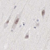


Comprehensive Human Protein Atlas IHC scoring per tissue (reliability: Approved — Medium consistency between antibody staining and RNA expression data.). Rows are taken directly from the HPA tissue chart — click any row's HPA link to view the source.
| Tissue | Cell type | Level | Evidence | Source |
|---|---|---|---|---|
| Testis | Cells in seminiferous ducts | High | Protein (IHC) | HPA → |
| Cerebellum | Purkinje cells | Medium | Protein (IHC) | HPA → |
| Gallbladder | Glandular cells | Medium | Protein (IHC) | HPA → |
| Lung | Macrophages | Medium | Protein (IHC) | HPA → |
| Parathyroid gland | Glandular cells | Medium | Protein (IHC) | HPA → |
| Tissue | Cell type | Level | Evidence | Source |
|---|---|---|---|---|
| Adipose tissue | Adipocytes | Not detected | Protein (IHC) | HPA → |
| Heart muscle | Cardiomyocytes | Not detected | Protein (IHC) | HPA → |
| Seminal vesicle | Glandular cells | Not detected | Protein (IHC) | HPA → |
| Skeletal muscle | Myocytes | Not detected | Protein (IHC) | HPA → |
| Soft tissue | Peripheral nerve | Not detected | Protein (IHC) | HPA → |
Troubleshoot MNAT1 staining in paraffin sections by checking retrieval, nuclear localisation, tissue controls and scoring (UniProt P51948; HPA tissue IHC).
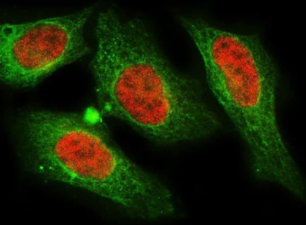
The catalog lists anti-MNAT1 antibodies for IHC and IF/ICC in human, mouse and rat (catalog: PB9614, A06523); the supplied figures show paraffin-section IHC and cell IF (image captions).
PB9614 is listed for IHC and IF/ICC in human, mouse and rat; its associated IHC captions show paraffin sections of human, mouse and rat testis but identify the antibody as PB9615 (catalog: applications/reactivity; IHC captions). A06523 is listed for IF/ICC in human, mouse and rat, with a HELA cell IF figure and no listed IHC application (catalog: applications/reactivity; IF caption).
Which to pick: For paraffin-section IHC, choose PB9614 based on its listed application and 0.5–1 μg/ml IHC range, while checking the PB9614/PB9615 caption discrepancy before relying on the images (catalog: PB9614 applications/dilution; IHC captions). The captions report paraffin sections and EDTA retrieval at pH 8.0, but do not report the fixative (IHC captions). For IF/ICC, A06523 has a HELA cell figure at 5 μg/ml; both SKUs list human, mouse and rat reactivity, while the supplied IF figures document cells only (catalog: applications/reactivity; IF captions).