This website uses cookies to ensure you get the best experience on our website.


- Table of Contents
Plan MRPL52 paraffin section IHC around the granular cytoplasmic pattern reported in tissue (HPA tissue IHC). This guide covers fixation consistency, antibody dilution, controls and interpretation using the IHC-validated antibody at 1:100–1:300 (datasheet).

Expected localisation, validated protocols, controls and antibodies — the at-a-glance facts below, then the full design guide.
| Expected localisation | Granular cytoplasm in tissue (HPA tissue IHC); mitochondrial protein (UniProt) | |
| Staining pattern | Granular cytoplasmic staining across cell types (HPA tissue IHC) | |
| Antigen retrieval | Citrate pH 6.0 HIER, 95–98 °C, 20 min (rule: cytoplasmic / membrane antigen) | |
| Positive control | Adrenal gland+4 more · see all | |
| Negative control | None in HPA (detected in all 45 tissues); use no-primary + isotype controls |
| Fixation | Keep fixation conditions consistent across sections. (standard IHC practice; not target-specific) | |
| Caveat | Staining and RNA show medium consistency (HPA tissue IHC) | |
| Regulation | No regulation annotated (UniProt) | |
| Isoform / epitope | 4 isoforms; epitope coverage is unspecified (UniProt) |
The catalog antibody’s IHC-P protocol is accompanied by one published MRPL52 IHC workflow for paraffin sections (PMC8210597).
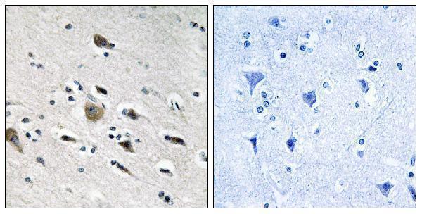
| Sample | Paraffin-embedded human brain tissue; fixative not specified (datasheet A15101) |
| Fixation | Image fixative and duration unreported (datasheet A15101); verify before use. |
| Sectioning | 4–5 µm sections on charged slides (standard) |
| Deparaffinisation | Xylene, graded ethanol series to water (standard) |
| Antigen retrieval | Heat-induced epitope retrieval in citrate buffer, pH 6.0, 20 min at 95–98 °C (standard rule: cytoplasmic / membrane antigen) |
| Peroxidase block | 3% H2O2, 10 min, room temperature (standard) |
| Blocking | 10% normal serum of the secondary host, 30 min, room temperature (standard) |
| Primary antibody | Rabbit anti-MRPL52, 1:100 - 1:300 (datasheet A15101) |
| Primary incubation | Overnight at 4 °C (standard) |
| Detection | HRP-polymer secondary, DAB chromogen 5–10 min (standard) |
| Counterstain | Hematoxylin, blue, dehydrate and mount (standard) |
| Expected result | MRPL52-positive staining in glandular cells of adrenal gland (HPA tissue IHC: High). HPA tissue profile: General cytoplasmic expression with a granular pattern. No signal in the no-primary control. |
MRPL52 is mitochondrial and has no annotated transmembrane segment (UniProt Q86TS9). In paraffin-section IHC, expect granular cytoplasmic staining (HPA: tissue IHC profile), including strong staining in adrenal glandular cells, bronchial respiratory epithelial cells, and cerebral cortical neurons (HPA: High in each). HPA rates its IHC evidence Approved, with medium consistency between staining and RNA data (HPA: tissue IHC reliability).
| Granular cytoplasmic staining in adrenal glandular cells or bronchial respiratory epithelial cells (HPA: High in both). | This fits the reported tissue pattern (HPA: granular cytoplasm) and mitochondrial location (UniProt Q86TS9). Compare cells within the same section before judging intensity; a uniformly dark field can obscure the granules (general IHC practice). |
| Predominantly nuclear staining, with little granular cytoplasmic signal (HPA: tissue IHC profile). | Question an IHC result dominated by nuclei because the reported tissue pattern is cytoplasmic (HPA: tissue IHC). Nucleoplasmic localisation is uncertain in ICC-IF (HPA: subcellular), so nuclear signal alone does not establish a true IHC pattern; inspect controls and counterstain (general IHC practice). |
| Strong staining in adipocytes or smooth muscle cells while nearby HPA high cell populations are weak (HPA: Low in adipocytes and smooth muscle cells). | That reversal warrants a specificity check: cross-reactivity or endogenous detection activity may contribute (general IHC practice). Low HPA staining does not mean these cells must be blank (HPA: Low); compare the same run with a no-primary control (general IHC practice). |
| Diffuse colour across cells and extracellular areas, without resolvable cytoplasmic granules (HPA: granular cytoplasmic profile). | Treat the pattern as background until controls support it. Review blocking, washes, and chromogen development (general IHC-P practice); broad colour alone cannot identify mitochondrial MRPL52 (UniProt Q86TS9; HPA: tissue IHC profile). |
| No detectable signal in adrenal glandular cells or bone marrow hematopoietic cells (HPA: High in both). | A missing expected signal calls for a run check before concluding absence. Confirm that the positive tissue and detection control stained, then review retrieval and antibody use under the assay conditions (general IHC-P practice). HPA High is an observed category, not a guarantee for every section (HPA: tissue IHC). |
| Tissue and cell context | HPA reports low RNA tissue specificity and a general granular cytoplasmic IHC pattern (HPA: tissue IHC). High staining spans glandular, epithelial, hematopoietic, glial, and neuronal cells in the listed tissues (HPA: High); Low calls include adipocytes, chondrocytes, smooth muscle, and vaginal squamous cells (HPA: Low). |
| Protein location and topology | MRPL52 belongs to the mitochondrial large ribosomal subunit and has no annotated transmembrane segment (UniProt Q86TS9). Interpret resolved cytoplasmic granules alongside the mitochondrial assignment; tissue IHC alone cannot prove that each granule is a mitochondrion (HPA: tissue IHC; general IHC interpretation). |
| Processing and isoforms | UniProt lists a chain at residues 24–123 and four isoforms, with no annotated glycosylation sites or modified residues (UniProt Q86TS9). The supplied evidence gives no antibody epitope, so these annotations cannot predict which isoforms the IHC antibody detects or how retrieval will affect staining. |
| Strength of IHC evidence | The tissue staining is Approved, with medium consistency between antibody staining and RNA expression data (HPA: tissue IHC reliability). One listed antibody, HPA012319, is IHC Approved; this supports using the reported pattern as a reference while leaving individual-slide specificity to controls (HPA: antibody validation; general IHC practice). |
| IF/ICC Q: Should nucleoplasmic signal count as expected? | Mitochondria are the supported main ICC-IF location; additional nucleoplasmic localisation is uncertain (HPA: subcellular). Assess a predominantly nuclear IF/ICC result cautiously and use its separate guide for assay design. This ICC-IF observation does not establish a nuclear IHC-P pattern (HPA: subcellular; HPA: tissue IHC). |
| Situation | Likely cause | Next action |
|---|---|---|
| HPA High reference tissue is blank or very faint (HPA: tissue IHC). | The run may have insufficient detection or a failed assay step (general IHC-P practice); HPA gives no MRPL52-specific fixation sensitivity. | Check a same-run positive control and the antibody's IHC-P instructions; verify retrieval, antibody concentration, and detection reagents within the assay workflow (general IHC-P practice). Do not infer biological absence from this slide alone. |
| Colour fills nuclei more than cytoplasm (HPA: granular cytoplasmic IHC profile). | Counterstain or nonspecific detection may complicate the readout (general IHC-P practice). ICC-IF nucleoplasm is an uncertain additional location, not confirmation of a nuclear IHC result (HPA: subcellular). | Compare with a no-primary control and inspect whether granular cytoplasmic signal is present in an HPA High cell population (general IHC practice; HPA: tissue IHC). |
| Adipocytes or smooth muscle dominate the stain (HPA: Low in both). | The pattern conflicts with those HPA Low calls, particularly if an HPA High population in the same run is weak (HPA: tissue IHC). Cross-reactivity or endogenous detection activity are possibilities (general IHC-P practice). | Run no-primary and appropriate detection controls, compare cell types within the section, and avoid scoring colour as MRPL52 solely by intensity (general IHC practice). |
| Every compartment has similar diffuse colour (HPA: granular cytoplasmic IHC profile). | Excess background can conceal the reported granular pattern (HPA: tissue IHC; general IHC-P practice). This appearance does not resolve cellular localisation. | Review blocking, wash stringency, antibody concentration, and chromogen development against the IHC-P workflow; compare with a no-primary control (general IHC-P practice). |
| Only scattered granules are visible, with uncertain cell boundaries (HPA: granular cytoplasmic IHC profile). | A granular pattern can be difficult to assign to a particular cell in a crowded or weakly counterstained field (general histology practice). HPA's profile alone cannot identify each stained cell (HPA: tissue IHC). | Use morphology and counterstain to identify the cell population, then compare with the HPA cell-level pattern and a same-run control before scoring (HPA: tissue IHC; general IHC practice). |


Comprehensive Human Protein Atlas IHC scoring per tissue (reliability: Approved — Medium consistency between antibody staining and RNA expression data.). Rows are taken directly from the HPA tissue chart — click any row's HPA link to view the source.
| Tissue | Cell type | Level | Evidence | Source |
|---|---|---|---|---|
| Adrenal gland | Glandular cells | High | Protein (IHC) | HPA → |
| Appendix | Glandular cells | High | Protein (IHC) | HPA → |
| Bone marrow | Hematopoietic cells | High | Protein (IHC) | HPA → |
| Bronchus | Respiratory epithelial cells | High | Protein (IHC) | HPA → |
| Caudate | Glial cells | High | Protein (IHC) | HPA → |
| Tissue | Cell type | Level | Evidence | Source |
|---|---|---|---|---|
| None in HPA: MRPL52 is detected in all 45 scored tissues. Use a no-primary (secondary-only) and an isotype control instead. | ||||
Troubleshoot MRPL52 staining by evaluating granular cytoplasmic signal, section quality, and controls together (HPA tissue IHC profile; UniProt Q86TS9).
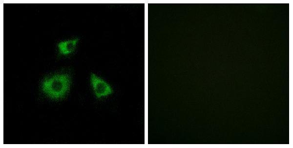
The catalog antibody A15101 has IHC data from paraffin-embedded human brain tissue (A15101 IHC image caption) and IF data from HUVEC cells (A15101 IF image caption).
A15101 is shown in IHC on paraffin-embedded human brain tissue with peptide blocking (A15101 IHC image caption). It is also shown in IF on HUVEC cells with peptide blocking (A15101 IF image caption) and lists Human, Mouse and Rat reactivity (A15101 catalog).
Which to pick: Choose A15101 for tissue IHC: its own image documents paraffin-embedded human brain tissue (A15101 IHC image caption); the fixative is unreported (A15101 IHC image caption). For IF/ICC, the same rabbit polyclonal antibody lists both applications (A15101 catalog), with an IF image from HUVEC cells (A15101 IF image caption). For cross-species work, A15101 lists Human, Mouse and Rat reactivity (A15101 catalog); the supplied IHC image documents human tissue only (A15101 IHC image caption).