This website uses cookies to ensure you get the best experience on our website.


- Table of Contents
Plan MSI1 chromogenic IHC in paraffin sections around cytoplasmic and nuclear tissue staining (HPA tissue IHC). This guide uses reported CNS and retinal staining for control selection (HPA tissue IHC) and the catalog antibody’s 0.5–1 μg/mL IHC-P range (datasheet A05052-1).

Expected localisation, validated protocols, controls and antibodies — the at-a-glance facts below, then the full design guide.
| Expected localisation | Cytoplasmic and nuclear tissue staining (HPA tissue IHC) | |
| Staining pattern | CNS glia and retinal ganglion cells; cytoplasmic/nuclear signal (HPA tissue IHC) | |
| Antigen retrieval | Citrate pH 6 HIER, heat-mediated (datasheet A05052-1) | |
| Positive control | Caudate+4 more · see all | |
| Negative control | Adipose tissue+4 more · see all |
| Fixation | Keep fixation consistent across paraffin sections (standard IHC practice; not target-specific) | |
| Caveat | Antibody staining and RNA have medium consistency (HPA tissue IHC) | |
| Regulation | Fetal and adult tissue expression differs (UniProt) | |
| Isoform / epitope | No annotated isoforms; one 1–362 chain (UniProt) |
The catalog antibody protocol uses citrate pH 6 retrieval (datasheet: A05052-1). The published IHC protocols below cover osteoarticular tissue, mouse brain, and cervical carcinoma tissue (PMC4433648; PMC11301063; PMC4279416).
| Sample | Paraffin-embedded human lung cancer tissues; fixative not specified (datasheet A05052-1) |
| Fixation | Image fixative and duration unreported (datasheet A05052-1); verify before use. |
| Sectioning | 4–5 µm sections on charged slides (standard) |
| Deparaffinisation | Xylene, graded ethanol series to water (standard) |
| Antigen retrieval | Heat retrieval: Citrate pH 6, 20 min (datasheet A05052-1) |
| Peroxidase block | 3% H2O2, 10 min, room temperature (standard) |
| Blocking | 10% goat serum (datasheet A05052-1) |
| Primary antibody | Rabbit anti-MSI1, 0.5-1μg/ml (datasheet A05052-1) |
| Primary incubation | Overnight at 4 °C (datasheet A05052-1) |
| Detection | Streptavidin-biotin complex (SABC), DAB chromogen (datasheet A05052-1) |
| Counterstain | Hematoxylin, blue, dehydrate and mount (standard) |
| Expected result | MSI1-positive staining in glial cells of caudate (HPA tissue IHC: Medium). HPA tissue profile: Cytoplasmic and nuclear expression in several tissues, including CNS and retina. No signal in the no-primary control. |
MSI1 should appear in cytoplasm and nuclei, including CNS glial cells, retinal ganglion cells, intestinal glandular cells, and pancreatic exocrine glandular cells (HPA: tissue IHC). It has no transmembrane segment, so an exclusively membrane-bound pattern is unexpected (UniProt O43347 topology). HPA rates tissue IHC reliability as Enhanced, while noting medium consistency between antibody staining and RNA expression (HPA: tissue IHC).
| Cytoplasmic and nuclear staining in caudate, cerebral cortex, or hippocampal glial cells. | This fits the reported MSI1 compartments and cell types; HPA records medium staining in these cells (HPA: tissue IHC). Score the relevant cells and compartments, since staining elsewhere in a section does not establish that its glial cells are positive (general IHC practice). |
| An exclusively sharp plasma-membrane outline, with no cytoplasmic or nuclear signal. | Treat this as an unexpected pattern and investigate an artefact or nonspecific reaction: MSI1 has no transmembrane segment (UniProt O43347 topology), and HPA describes cytoplasmic and nuclear tissue staining (HPA: tissue IHC). Compartment alone cannot identify the cause (general IHC practice). |
| Strong staining confined to a cell type reported as undetected, such as adipocytes in adipose tissue. | HPA reports MSI1 as not detected in these adipocytes (HPA: tissue IHC). Check for antibody cross-reactivity or endogenous chromogenic activity before calling the result positive (general IHC practice). An unexpected positive is a reason to investigate, not proof of either cause. |
| Broad, nearly uniform chromogen across cells and extracellular areas. | The distribution does not match HPA's cell-resolved cytoplasmic and nuclear MSI1 pattern (HPA: tissue IHC). Assess reagent background and endogenous detection activity with appropriate controls; diffuse deposit can obscure genuine stained cells (general IHC practice). |
| No staining in retinal ganglion cells or pancreatic exocrine glandular cells. | Both are reported at medium staining intensity (HPA: tissue IHC). Check slide quality and detection with a positive control before interpreting the negative; HPA's Enhanced rating does not guarantee that every preparation will yield a visible signal (HPA: tissue IHC; general IHC practice). |
| Which tissue and cell type is being scored? | HPA reports medium staining in retinal ganglion cells, CNS glial cells, small-intestinal and duodenal glandular cells, and pancreatic exocrine glandular cells; colon, rectum, and appendix glandular cells are Low (HPA: tissue IHC). A tissue name alone does not specify which cells should stain. |
| How much confidence does the tissue atlas provide? | HPA labels MSI1 tissue IHC Enhanced but describes only medium consistency between antibody staining and RNA expression (HPA: tissue IHC). Two listed rabbit polyclonal antibodies, HPA074923 and HPA075573, each have Enhanced IHC validation (HPA: antibodies). Interpret an individual slide alongside its controls. |
| Does protein architecture predict a surface stain or a shed fragment? | MSI1 has no transmembrane segment, signal peptide, propeptide, or annotated processed fragment; its listed chain spans residues 1–362 (UniProt O43347 topology and processing). These annotations support intracellular interpretation but cannot establish how a particular antibody performs in paraffin sections. |
| What pattern should IF/ICC show? | HPA reports approved nucleoplasm and cytosol localization in ICC-IF and lists AF22, SH-SY5Y, U2OS, and HAP1 image sets (HPA: subcellular; HPA: antibodies). Use this as a compartment comparison only; tissue IHC intensity and cell-type calls come from the HPA tissue record. |
| Situation | Likely cause | Next action |
|---|---|---|
| Expected CNS glial or retinal ganglion staining is absent. | The expected cell population may be absent from the viewed field, or the staining run may have failed; HPA reports medium signal in these cells (HPA: tissue IHC; general IHC practice). | Confirm cell identity and section quality, then review the run's positive control and the catalog antibody's IHC-P instructions before calling MSI1 negative (general IHC practice). |
| Colon, rectum, or appendix looks weaker than retina. | HPA calls glandular-cell staining Low in these intestinal sites but medium in retinal ganglion cells (HPA: tissue IHC). | Score each stated cell population against its own expected level; retain a suitable positive control to assess whether weak staining remains detectable (HPA: tissue IHC; general IHC practice). |
| Atypical cells stain while the expected cells do not. | Cell misidentification, cross-reactivity, or endogenous chromogenic activity are possible explanations; the HPA pattern is cell specific (HPA: tissue IHC; general IHC practice). | Recheck morphology and control slides, including a reagent control for detection background, before assigning the atypical signal to MSI1 (general IHC practice). |
| Chromogen is widespread or obscures cell boundaries. | Excess detection background or endogenous enzyme activity can create diffuse signal in chromogenic IHC (general IHC practice). | Inspect the reagent control and review blocking, washes, and detection settings in the validated IHC-P workflow; assess MSI1 only where cell boundaries and compartments remain readable (general IHC practice). |
| Staining appears only at the plasma membrane. | That pattern conflicts with the intracellular HPA tissue profile and MSI1's lack of a transmembrane segment (HPA: tissue IHC; UniProt O43347 topology). | Check a known-positive tissue for cytoplasmic or nuclear staining and review control slides for edge or detection artefacts before accepting the membrane signal (HPA: tissue IHC; general IHC practice). |
| A specimen reported as undetected shows a faint signal. | HPA lists adipose adipocytes and bronchial respiratory epithelial cells as Not detected; faint colour alone cannot identify its source (HPA: tissue IHC). | Compare the same cell type with the reagent control and an HPA-reported positive tissue, and record the discrepancy rather than treating faint colour as confirmed MSI1 expression (HPA: tissue IHC; general IHC practice). |
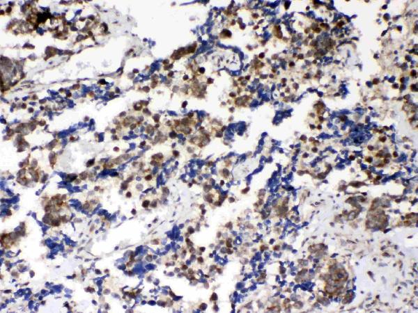
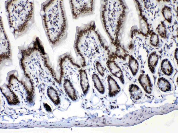
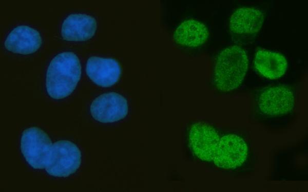
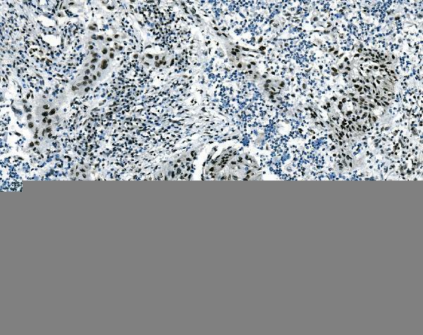
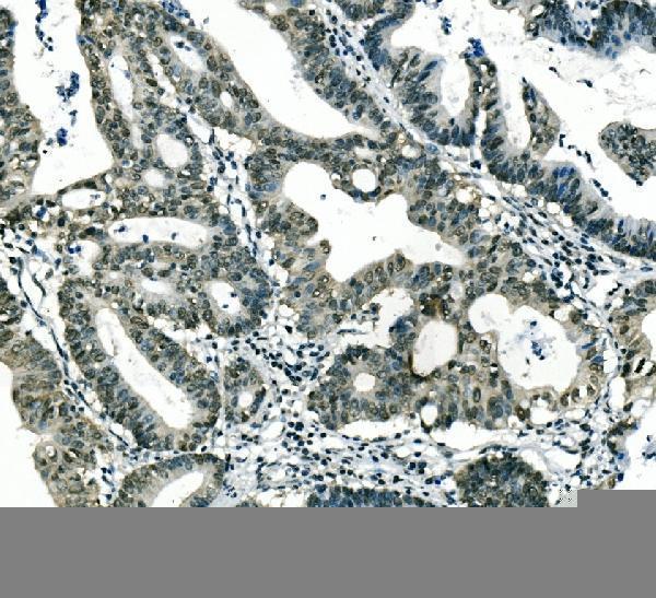
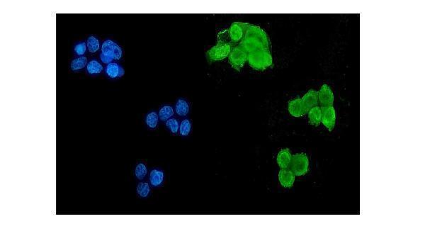


Comprehensive Human Protein Atlas IHC scoring per tissue (reliability: Enhanced — Medium consistency between antibody staining and RNA expression data.). Rows are taken directly from the HPA tissue chart — click any row's HPA link to view the source.
| Tissue | Cell type | Level | Evidence | Source |
|---|---|---|---|---|
| Caudate | Glial cells | Medium | Protein (IHC) | HPA → |
| Cerebellum | Cells in molecular layer | Medium | Protein (IHC) | HPA → |
| Cerebral cortex | Glial cells | Medium | Protein (IHC) | HPA → |
| Duodenum | Glandular cells | Medium | Protein (IHC) | HPA → |
| Hippocampus | Glial cells | Medium | Protein (IHC) | HPA → |
| Tissue | Cell type | Level | Evidence | Source |
|---|---|---|---|---|
| Adipose tissue | Adipocytes | Not detected | Protein (IHC) | HPA → |
| Adrenal gland | Glandular cells | Not detected | Protein (IHC) | HPA → |
| Bone marrow | Hematopoietic cells | Not detected | Protein (IHC) | HPA → |
| Breast | Adipocytes | Not detected | Protein (IHC) | HPA → |
| Bronchus | Respiratory epithelial cells | Not detected | Protein (IHC) | HPA → |
Troubleshoot MSI1 staining in paraffin section chromogenic IHC, using the catalog antibody’s tissue image and independent localisation evidence as reference points.
The catalog shows MSI1 IHC in paraffin sections of human lung cancer and IF/ICC in MCF7 cells; both antibodies list human, mouse, and rat reactivity (catalog image captions; catalog applications and reactivity).
A05052-1 will render with a human lung cancer IHC image; its other IHC captions show mouse intestine, rat intestine, and rat brain, and its IF captions show MCF7 cells and human and mouse tissue (A05052-1 image captions). M05052-1 will render with a human lung cancer IHC image; another IHC caption shows human rectum cancer, and its IF caption shows MCF7 cells (M05052-1 image captions).
Which to pick: For paraffin-section IHC, choose A05052-1 when evidence across species matters: its captions show human, mouse, and rat tissue with citrate retrieval (pH 6, 20 minutes); the mouse monoclonal M05052-1 (clone 2B9) has human tissue captions with EDTA retrieval (pH 8) (catalog IHC captions; catalog clone). For IF/ICC, A05052-1 has MCF7 and tissue IF examples, while M05052-1 has an MCF7 IF example; both list IF and ICC (catalog IF captions; catalog applications). The IHC captions describe paraffin sections but do not report the fixative (catalog IHC captions).