This website uses cookies to ensure you get the best experience on our website.


- Table of Contents
Plan chromogenic IHC for MTA1 using its nuclear and cytoplasmic tissue staining profile (HPA tissue IHC). This guide covers control tissue selection, fixation consistency, and interpretation of tumor staining (HPA tissue IHC; UniProt).

Expected localisation, validated protocols, controls and antibodies — the at-a-glance facts below, then the full design guide.
| Expected localisation | Nuclear and cytoplasmic staining in most tissues (HPA tissue IHC) | |
| Staining pattern | Nuclear and cytoplasmic; strongest in CNS and testis (HPA tissue IHC) | |
| Antigen retrieval | Citrate pH 6 HIER, heat-mediated (datasheet PA1483) | |
| Positive control | Cerebellum+4 more · see all | |
| Negative control | Adipose tissue+4 more · see all |
| Fixation | Matched tissue-IHC evidence does not establish the fixation claim. Validate the specimen-specific method before use. (selected-SKU IHC image PA1483) | |
| Caveat | Staining and RNA expression have only medium consistency (HPA tissue IHC) | |
| Regulation | Higher in tumors than adjacent tissue (UniProt) | |
| Isoform / epitope | 3 isoforms; epitope coverage is unreported (UniProt; datasheet PA1483) |
The catalog antibody’s IHC-P protocol (datasheet PA1483) is accompanied by four published MTA1 IHC protocols (PMC4148129; PMC2972736; PMC12261658; PMC3478212).
| Sample | Tissue sections; selected-image fixative not specified (standard IHC workflow) |
| Fixation | Image fixative and duration unreported (datasheet PA1483); verify before use. |
| Sectioning | 4–5 µm sections on charged slides (standard) |
| Deparaffinisation | Xylene, graded ethanol series to water (standard) |
| Antigen retrieval | Heat retrieval: Citrate pH 6, 20 min (datasheet PA1483) |
| Peroxidase block | 3% H2O2, 10 min, room temperature (standard) |
| Blocking | 10% goat serum (datasheet PA1483) |
| Primary antibody | Rabbit anti-MTA1, 0.5-1μg/ml (datasheet PA1483) |
| Primary incubation | Overnight at 4 °C (datasheet PA1483) |
| Detection | Streptavidin-biotin complex (SABC), DAB chromogen (datasheet PA1483) |
| Counterstain | Hematoxylin, blue, dehydrate and mount (standard) |
| Expected result | MTA1-positive staining in purkinje cells of cerebellum (HPA tissue IHC: High). HPA tissue profile: Nuclear and cytoplasmic expression in most tissues, most abundant in the CNS and testis. No signal in the no-primary control. |
MTA1 is mainly nuclear, with cytoplasmic staining also expected; nuclear envelope and cytoskeletal localization are reported (UniProt Q13330; HPA: nuclear and cytoplasmic tissue IHC). In paraffin sections, look for staining in the identified cells, especially Purkinje cells, cortical neurons, Hofbauer cells and Leydig cells (HPA: High). Tissue IHC reliability is Supported, with medium consistency against RNA data (HPA: tissue IHC). MTA1 has no transmembrane segment (UniProt Q13330 topology).
| Distinct nuclear staining, with or without cytoplasmic staining, in Purkinje cells, cortical neurons, Hofbauer cells or Leydig cells (HPA: High; HPA: tissue IHC pattern). | This matches the reported compartments and high staining in those cell types (HPA: tissue IHC). Judge each cell population separately: MTA1 is seen across many tissues, but staining level varies by cell type (HPA: tissue IHC). |
| Signal confined to extracellular material or cell borders, without convincing nuclear or cytoplasmic signal (HPA: tissue IHC pattern; UniProt Q13330 localization). | Treat this as a suspect pattern and compare it with a known-positive section and negative control (general IHC practice). Cytoplasmic or nuclear-envelope signal alone is not automatically misplaced, because both localizations are reported (UniProt Q13330; HPA: tissue IHC). |
| Strong staining in adipocytes or cardiomyocytes while the expected positive cell population also stains (HPA: Not detected in adipocytes and cardiomyocytes; HPA: High in selected cells). | Investigate nonspecific antibody binding or endogenous detection activity with appropriate controls (general IHC practice). A single unexpected positive cell does not establish cross-reactivity; the HPA observations are cell-specific reference patterns, not an absolute exclusion of MTA1 (HPA: tissue IHC). |
| Diffuse chromogen over cells and tissue spaces obscures nuclear boundaries (general IHC practice). | The section cannot support a confident compartment call (general IHC practice). Check the negative control, blocking, antibody concentration, washes and detection conditions before assigning MTA1 positivity (general IHC practice); HPA reports a nuclear and cytoplasmic pattern, not uniform haze (HPA: tissue IHC). |
| No staining in a section containing identifiable Purkinje cells, cortical neurons, Hofbauer cells or Leydig cells (HPA: High in these cell types). | First assess whether the IHC run worked using an appropriate positive control, then review retrieval, antibody and detection conditions (general IHC practice). An absent result in one section cannot establish that MTA1 is absent from the tissue (HPA: tissue IHC reference pattern). |
| Cell population and tissue context | HPA reports High staining in Purkinje cells, cortical neurons, Hofbauer cells and Leydig cells, but no detected staining in adipocytes or cardiomyocytes (HPA: tissue IHC). Identify the cell type before interpreting a tissue-level result (general IHC practice). |
| Compartment and topology | Nuclear, cytoplasmic, nuclear-envelope and cytoskeletal locations are reported (UniProt Q13330); tissue IHC describes nuclear and cytoplasmic staining (HPA: tissue IHC). MTA1 has no transmembrane segment, so a membrane-only pattern needs scrutiny (UniProt Q13330 topology). |
| Isoforms and antibody interpretation | UniProt lists 3 MTA1 isoforms (UniProt Q13330). The supplied record gives no antibody epitope or isoform coverage, so an unexpected pattern cannot be assigned to an isoform from these sources alone (UniProt Q13330; HPA: antibody summaries). |
| Evidence strength | The tissue pattern is Supported, with medium consistency against RNA data (HPA: tissue IHC). Two listed antibodies have Supported IHC status; that does not establish every observed stain as specific (HPA: antibody summaries; general IHC practice). |
| IF/ICC Q&A: Where should MTA1 appear? | Mainly in the nucleoplasm, with additional cytosolic signal (HPA: subcellular ICC-IF). This is an IF/ICC localization reference; interpret chromogenic paraffin sections against the tissue IHC pattern and its stated reliability (HPA: tissue IHC). |
| Situation | Likely cause | Next action |
|---|---|---|
| Known-positive cells show no signal (HPA: High in Purkinje cells, cortical neurons, Hofbauer cells and Leydig cells). | The run may have insufficient detectable signal; the supplied sources establish no MTA1-specific fixation or retrieval sensitivity (HPA: tissue IHC; UniProt Q13330). | Check a positive control and the detection reagents, then review retrieval and antibody conditions as general IHC variables (general IHC practice). Record the cell type examined. |
| Broad brown haze masks cells or persists in the negative control (general IHC practice). | Nonspecific binding or endogenous detection activity may contribute to chromogenic background (general IHC practice). | Review blocking, antibody concentration, washes and the relevant negative control; address endogenous activity for the detection system used (general IHC practice). |
| Only extracellular or crisp cell-border staining is seen (HPA: tissue IHC pattern; UniProt Q13330 localization). | The distribution does not fit the reported nuclear and cytoplasmic pattern or the absence of a transmembrane segment (HPA: tissue IHC; UniProt Q13330 topology). | Compare with known-positive cells and a negative control; reassess specificity before scoring those areas as MTA1 positive (general IHC practice). |
| Adipocytes or cardiomyocytes stain strongly (HPA: Not detected in these cell types). | Unexpected staining may reflect nonspecific binding or detection background (general IHC practice); HPA's cell-specific observation alone cannot identify the cause (HPA: tissue IHC). | Verify cell identity and examine the negative control and known-positive cells in the same run before interpreting the signal (general IHC practice). |
| A faint cytoplasmic component accompanies clear nuclear staining (HPA: nuclear and cytoplasmic tissue IHC). | Cytoplasmic localization is reported and need not be artefactual (UniProt Q13330; HPA: tissue IHC). | Score the nuclear and cytoplasmic components separately where boundaries are clear, and compare background against the negative control (general IHC practice). |
| Heart muscle gives a result that appears inconsistent across sources (UniProt Q13330: high expression in cardiac muscle; HPA: cardiomyocytes Not detected). | The supplied UniProt tissue statement and HPA cell-level IHC observation differ; neither establishes a fixation effect (UniProt Q13330; HPA: tissue IHC). | Report the observed cell type, compartment and controls, and state both reference findings when interpreting the result (UniProt Q13330; HPA: tissue IHC). |


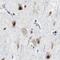


Comprehensive Human Protein Atlas IHC scoring per tissue (reliability: Supported — Medium consistency between antibody staining and RNA expression data.). Rows are taken directly from the HPA tissue chart — click any row's HPA link to view the source.
| Tissue | Cell type | Level | Evidence | Source |
|---|---|---|---|---|
| Cerebellum | Purkinje cells | High | Protein (IHC) | HPA → |
| Cerebral cortex | Neuronal cells | High | Protein (IHC) | HPA → |
| Placenta | Hofbauer cells | High | Protein (IHC) | HPA → |
| Testis | Leydig cells | High | Protein (IHC) | HPA → |
| Bronchus | Basal cells | Medium | Protein (IHC) | HPA → |
| Tissue | Cell type | Level | Evidence | Source |
|---|---|---|---|---|
| Adipose tissue | Adipocytes | Not detected | Protein (IHC) | HPA → |
| Cervix | Glandular cells | Not detected | Protein (IHC) | HPA → |
| Esophagus | Squamous epithelial cells | Not detected | Protein (IHC) | HPA → |
| Heart muscle | Cardiomyocytes | Not detected | Protein (IHC) | HPA → |
| Oral mucosa | Squamous epithelial cells | Not detected | Protein (IHC) | HPA → |
Troubleshoot MTA1 staining in paraffin-section chromogenic IHC by checking retrieval, tissue processing, cellular compartments, and controls before interpreting intensity.
The catalog antibody PA1483 has IHC images from human rectal cancer and rat ovary tissue (catalog image captions). No IF/ICC image is supplied (catalog image data).
PA1483 is the only card and is listed for IHC and IHC-F in human, mouse, and rat (catalog applications/reactivity). Its images show human rectal cancer IHC(P) and rat ovary IHC(P) and IHC(F); no mouse tissue image is supplied (catalog image captions).
Which to pick: Choose PA1483 for paraffin-section IHC: its captions show human rectal cancer and rat ovary tissue, and the fixative is unreported (catalog image captions). No SKU is listed for IF/ICC, and no IF image or dilution is supplied, so there is no supported IF/ICC pick here (catalog applications; catalog IF data). For cross-species tissue IHC, PA1483 lists human, mouse, and rat reactivity, with images for human and rat; its host is rabbit and its clonality is unreported (catalog reactivity; catalog image captions; catalog host/clone).