This website uses cookies to ensure you get the best experience on our website.
- Table of Contents
1 Citations 3 Q&As
3 Citations 5 Q&As
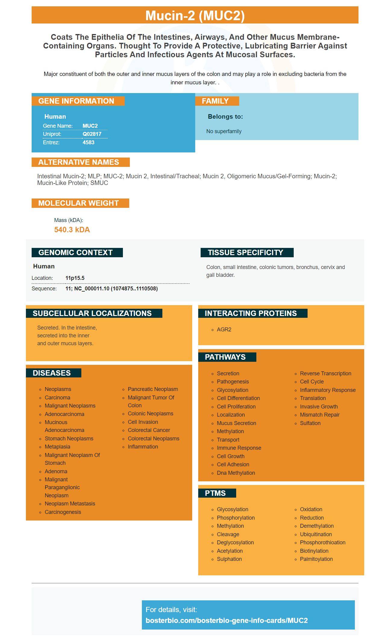
Facts about Mucin-2.
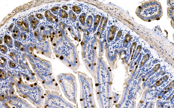
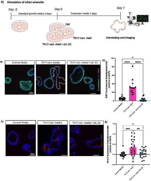
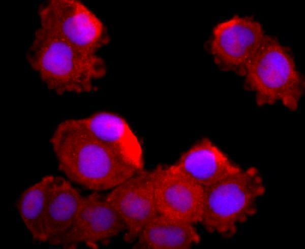
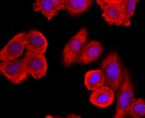
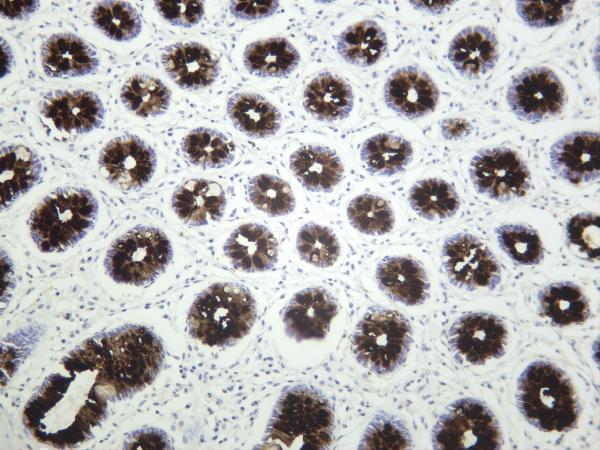
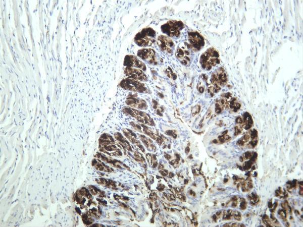
Major constituent of both the outer and inner mucus layers of the colon and may play a role in excluding bacteria from the inner mucus layer. .
Human | |
---|---|
Gene Name: | MUC2 |
Uniprot: | Q02817 |
Entrez: | 4583 |
Belongs to: |
---|
No superfamily |
Intestinal mucin-2; MLP; MUC-2; mucin 2, intestinal/tracheal; mucin 2, oligomeric mucus/gel-forming; mucin-2; mucin-like protein; SMUC
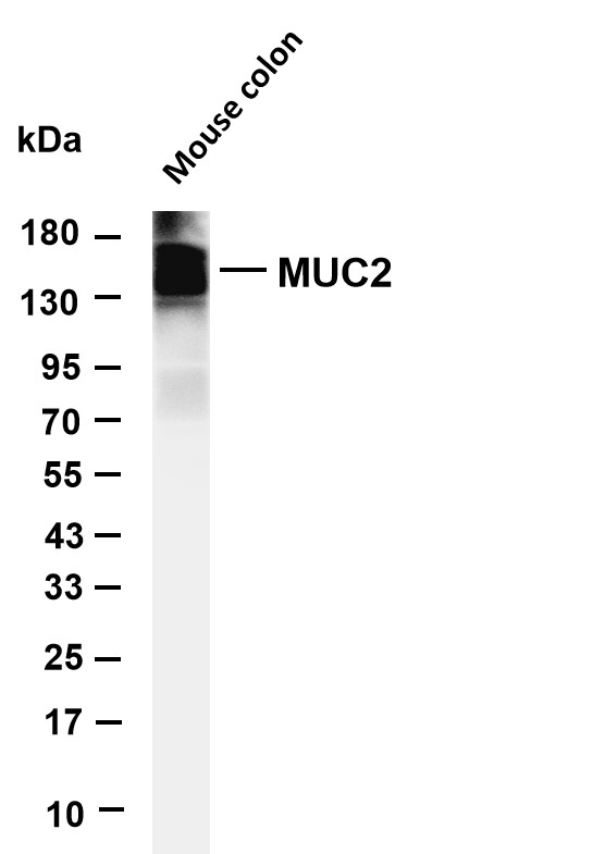
Mass (kDA):
540.3 kDA
Human | |
---|---|
Location: | 11p15.5 |
Sequence: | 11; NC_000011.10 (1074875..1110508) |
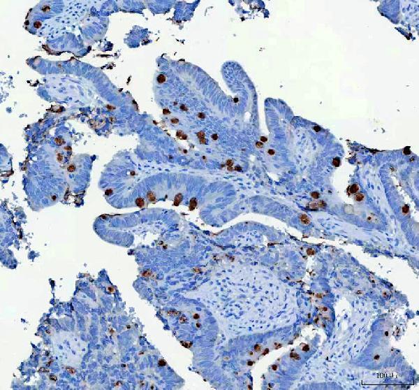
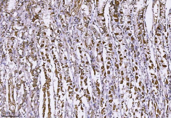
Colon, small intestine, colonic tumors, bronchus, cervix and gall bladder.
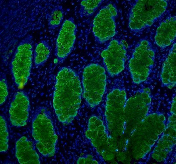
Secreted. In the intestine, secreted into the inner and outer mucus layers.
This article explains how you can make use of the Anti-MUC2 Monoclonal Antibody (AMO) and Picoband to detect MUC2 in cells. The steps involved in the process are described along with the verification of the results. The next article in this series will focus on the Stepwise IHC protocol for detecting MUC2 in cells. We will also discuss the MUC2 Marker in the context of immunohistochemistry.
Monoclonal antibodies that target MUC2 include the monoclonal antibodies that target MUC2 include the. MUC4 is a high-molecular-weight O-glycoprotein, encoded by a gene on chromosome 3 in the q29 region. It is made up of two distinct subunits: 850 kD mucin type and 80 kD membrane bound. Its 16-amino acid tandem repeats make up the sequence, which has the same structure as a growth factor.
This monoclonal antibody was created in the Hybridoma lab at North Carolina State University. The antibody binds to a fragment of Muc2 protein, and the concentration of the monoclonal antibody C4 binds to the peptide in a concentration-dependent manner. Monoclonal antibody C4 attaches to a muc2-apomucin-fragment that is in cold gelatin from a water fish.
The monoclonal antibody C4 recognizes both O and non-glycosylated Muc2. Both forms of Muc2 can be dissolved within the body. However the release by cytokine Muc2 could be due to the higher amount of sulphation in the latter. Mucins can be identified by a wide variety of techniques, including immunohistochemistry.
Boster Bio's monoclonal monoclonal anti-muc4 monoclonal antibodies a TFag at Se5 to recognize mucin glycopeptide antigen. This mAb is binds to gold nanoparticles. It is effective in anti-tumour vaccines. It is also utilized to detect pre-cancerous lesions. All the studies carried out with Anti-MUC2 Monoclonal Antibody from Boster Bio have shown that this anti-mucinate is extremely effective in treating different kinds of cancers.
This monoclonal antibody was made using affinity-purified polyclonal antibodies against MUC4 peptides. To maintain antigen-recognant molecular recognition it was tested in rabbits and mice with Complete Freund's Adjuvant aswell as in the absence of Freund's Adjuvant. The antibodies were then purified using Protein A chromatography.
The YY1 gene is involved in the regulation of the MUC2 gene. Its deregulation can lead to PEDV replication. This study utilized YY1-mRNA knockdown to examine the impact of the YY1 gene on MUC2 expression. Figure 6 shows how YY1 affects MUC2 expression. Prior to PCR amplifying using an antiYY1 antigen, a Rabbit IgG antibody and an Anti-RNA Polymerase II antibodies were added to the. The PCR products were then complete using the DL500 DNA marker.
A CpG island was detected in the MUC2 gene promoter region. This is an important aspect since DNA methylation can inhibit gene expression. 16 CG sites are found in the promoter regions of MUC2. DNA methylation blocks transcription factors from binding, and thus inhibits gene transcription. In turn MUC-15 (and mC-16) are positive indicators for MUC2 gene expression. It is interesting to note that DNA methylation can inhibit transcription of MUC2, thus reducing the ability to detect the disease.
MUC2 is a gene associated with a range of illnesses. Inflammatory bowel disease (IBD) and colorectal cancers are frequently associated with altered MUC2 levels. This marker has been associated with the development of colorectal cancer as well as the development of IBD. Similarly reduced MUC2 expression is associated with various diseases, including ulcerative colitis and chronic obstructive lung disease. It also increases mucus secretion during intestinal infection and inflammation of the bowel.
MUC2 has been implicated in the transmission of PEDV and in the regulation of the immune system. Further studies on the function of the MUC2 protein are expected to verify this theory. Its expression in IPECJ2 cells is an indication of how the intestinal mucosa is protected from external stimuli. 24 hours after infection, the MUC2 gene is re-activated. It also plays a significant role in protecting the intestinal mucosa from pathogenic microorganisms.
To perform IHC studies, tissue samples must be processed correctly. The final images must preserve the epitopes, as well as the morphology of the specimen. Tissue is typically processed with paraffin-fixed formalin block. This blocks all cellular processes and makes it easier to see them. The tissue is then cut into thin slices.
Before you begin the preparation for IHC analysis of MUC2 marker, ensure that the samples are properly secured. The slide can be cleaned by dipping it into water that has been distilled or 95% ethanol before drying it in an infrared oven. Make sure that the slide is clean before staining. Microscopic slides are 5 millimeters in diameter. The nervous tissue slides range from 20 to 100 millimeters. The bigger the slides, the better the track of the never fiber direction. Coverslips are treated in a similar way to that of microscopic slides but the coverslips are thinner.
Researchers can visualize the location of cells in complex tissues with an IHC test. Different antibodies can be used to target different regions of tissues. This allows researchers to analyze and compare expression patterns. In addition to the primary antibody, a secondary antibody is used to carry the pre-bound reporter molecule. These two antibodies must be used with the appropriate concentrations for each.
Expression of MUC2 was assessed in tumors, MDF, and ACF using a stepwise IHC protocol. Tumors, ACF, and MDF were identified and marked using a standard technique, and longitudinal sections of the tissues were mounted on electrostatic-treated slides. Each antibody was applied to one or more lesions on each animal. Each animal received three to five additional lesions.
In addition to its role in the immune system and the immune system, the MUC2 marker also has a potential role in the treatment of colon cancer. It hinders the growth of tumors and influences the invasiveness of cancer cells making it an attractive biomarker for immunotherapy. Despite these challenges, further studies are needed to validate MUC2 as a clinical marker. Find out more about MUC2 can be used to treat colon cancer.
While MUC2 is found in many organs and in malignant lesions, the exact significance of its expression gastric cancer remains unclear. The connection between MUC2 expression in 167 GCs and a variety of clinical features was examined in the current study. The findings were in line with the findings of an analysis of a meta-analysis of 1,832 GCs. The study also demonstrated a correlation between MUC2 levels and stage of disease and presence of lymph node metastasis.
Further studies are required to determine if MUC2 expression is a reliable biomarker for CRC. Low levels of MUC2 expression were associated with poorer prognosis for patients with advanced CRC, in spite of the association with the CDX2 transcription factor and the SOX2 gene. Low levels of the MUC2 marker were linked to lower survival rates among patients with advanced CRC.
The process of diagnosing tumors includes the molecular analysis of mucus levels in cancer patients. Mucins are large molecular weight, glycosylated proteins that protect the epithelial cells and form the ductal surfaces on several organs. There are approximately twenty mucins that have been identified so far and can be divided into two families based on their structure as membrane-bound and secreted. MUC2 is a secreted mucin located in chromosome 11p15.
48 exons make up the MUC2 gene. Each exon measures approximately 40 kb and is a home to 8 tandem repeats (TRs). These regions comprise approximately 50% of the MUC2 gene, and are known as MS1 and MS2 respectively. MUC2 gene polymorphisms are important for the detection of the presence of autoimmune diseases, infectious diseases, and cancer. The MUC2 gene is used extensively in the field of diagnostics, drug design, and animal research.
Although MUC proteins have been proven to be associated with positive outcomes in breast cancer patients however, they are not commonly used in cytology diagnosis. Recent research looked into the relationship between MUC2 protein expression and the vascular invasion of tumor cells. We found a correlation between MUC2 expression as well as lymph node metastasis and MUC2 expression. While the applications of the MUC2 marker are still being investigated this research will aid physicians in diagnosing diseases that affect the pancreas, particularly pancreatic cancer.
Survivin is one of the proteins that reduces apoptosis as well as regulates cell division. Survivin is detected in high levels in cancerous tissues of colorectal origin but not in normal colon tissue. Survivin overexpression and MUC5 are strongly associated with lymph node metastasis and advanced tumor stage, respectively. In contrast, the absence of MUC2 is associated with low cellular differentiation, as well as increased likelihood of developing colon cancer.
There have been numerous studies that have examined the MUC2 gene polymorphisms. In one study, MUC2 -MS6 and MUC2 MS7 were found to be cancer-specific alleles that only appeared in cancer patients. These regions showed higher expression in cancer than healthy individuals, suggesting that MUC2 gene polymorphisms may be involved in the development of cancer. Then, it was discovered that patients with these alleles have a higher danger of developing colon cancer and stage D disease.
PMID: 8300571 by Gum J.R. Jr., et al. Molecular cloning of human intestinal mucin (MUC2) cDNA. Identification of the amino terminus and overall sequence similarity to prepro-von Willebrand factor.
PMID: 1400449 by Gum J.R. Jr., et al. The human MUC2 intestinal mucin has cysteine-rich subdomains located both upstream and downstream of its central repetitive region.
*More publications can be found for each product on its corresponding product page