Click image to see more details
-

-
-
-
-
+6
Product Info Summary
| SKU: | A01212 |
|---|---|
| Size: | 100 μg/vial |
| Reactive Species: | Human, Mouse, Rat |
| Host: | Rabbit |
| Application: | IF, IHC |
Customers Who Bought This Also Bought

Product info
Product Name
Anti-MUC2 Antibody
SKU/Catalog Number
A01212
Size
100 μg/vial
Form
Lyophilized
Description
Boster Bio Anti-MUC2 Antibody catalog # A01212. Tested in IF, IHC applications. This antibody reacts with Human, Mouse, Rat.
Storage & Handling
Store at -20˚C for one year from date of receipt. After reconstitution, at 4˚C for one month. It can also be aliquotted and stored frozen at -20˚C for six months. Avoid repeated freeze-thaw cycles.
Cite This Product
Anti-MUC2 Antibody (Boster Biological Technology, Pleasanton CA, USA, Catalog # A01212)
Host
Rabbit
Contents
Each vial contains 4 mg Trehalose, 0.9 mg NaCl and 0.2 mg Na2HPO4.
Clonality
Polyclonal
Isotype
Rabbit IgG
Immunogen
A synthetic peptide corresponding to a sequence at the N-terminus of human MUC2, which shares 86.1% amino acid (aa) sequence identity with both mouse and rat MUC2.
Cross-reactivity
No cross-reactivity with other proteins.
Reactive Species
A01212 is reactive to MUC2 in Human, Mouse, Rat
Calculated molecular weight
540.3 kDa
Background of MUC2
Mucin 2, also known as MUC2, is a protein that in humans is encoded by the MUC2 gene. This gene encodes a member of the mucin protein family. It is mapped to 11p15.5. Mucin 2 is particularly prominent in the gut where it is secreted from goblet cells in the epithelial lining into the lumen of the large intestine. There, mucin 2, along with small amounts of related-mucin proteins, polymerizes into a gel of which 80% by weight is oligosaccharide side-chains that are added as post-translational modifications to the mucin proteins. This gel provides an insoluble mucous barrier that serves to protect the intestinal epithelium. The primary function of the MUC2 gene product is to provide a protective barrier between the epithelial surfaces and the gut lumen. There is decreased expression of MUC2 in colonic cancer and defective polymerization of secreted mucin in ulcerative colitis.
Antibody Validation
Boster validates all antibodies on WB, IHC, ICC, Immunofluorescence, and ELISA with known positive control and negative samples to ensure specificity and high affinity, including thorough antibody incubations.
Application & Images
Applications
A01212 is guaranteed for IF, IHC Boster Guarantee
Recommend Dilution
| Application | Dilution | Species |
|---|---|---|
| Immunohistochemistry (Paraffin-embedded Section) | 2-5μg/ml | Human, Mouse, Rat |
| Immunofluorescence | 5μg/ml | Human, Mouse |
Tested application
Use TE buffer pH 9.0 for antigen retrieval; (*) citrate buffer pH 6.0 is an alternative.
Validation Images & Assay Conditions
Click image to see more details
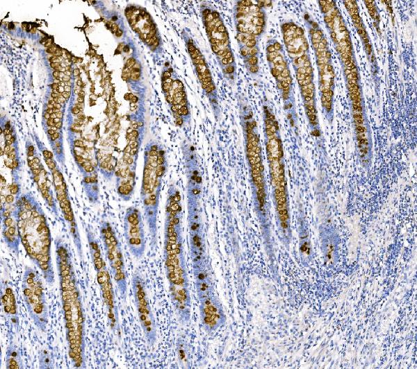
IHC analysis of MUC2 using anti-MUC2 antibody (A01212).
MUC2 was detected in a paraffin-embedded section of human colon cancer tissue. Heat mediated antigen retrieval was performed in EDTA buffer (pH 8.0, epitope retrieval solution). The tissue section was blocked with 10% goat serum. The tissue section was then incubated with 2 μg/ml rabbit anti-MUC2 Antibody (A01212) overnight at 4°C. Biotinylated goat anti-rabbit IgG was used as secondary antibody and incubated for 30 minutes at 37°C. The tissue section was developed using Strepavidin-Biotin-Complex (SABC) (Catalog # SA1022) with DAB as the chromogen.

Click image to see more details
IHC analysis of MUC2 using anti-MUC2 antibody (A01212).
MUC2 was detected in a paraffin-embedded section of mouse colon tissue. Heat mediated antigen retrieval was performed in EDTA buffer (pH 8.0, epitope retrieval solution). The tissue section was blocked with 10% goat serum. The tissue section was then incubated with 2 μg/ml rabbit anti-MUC2 Antibody (A01212) overnight at 4°C. Biotinylated goat anti-rabbit IgG was used as secondary antibody and incubated for 30 minutes at 37°C. The tissue section was developed using Strepavidin-Biotin-Complex (SABC) (Catalog # SA1022) with DAB as the chromogen.

Click image to see more details
IL-22 is the key cytokine underlying the effects of the human infant Th17-conditioned media. (A) Experimental design for enteroid stimulation. (B) Representative immunofluorescent staining of Ki67 (red), E-cadherin (green), and DAPI (blue) in infant enteroids cultured in control media (left), control media supplemented with infant Th17-conditioned media (middle), or control media plus Th17-conditioned media plus neutralizing anti-IL-22 (right) (Scale bars, 50 μm). (C, E) Fluorescence intensity of Ki67 and MUC2 per enteroid relative to DAPI following exposure to Th17-conditioned media. (technical replicates n=5; N= 4 individuals, statistical difference calculated by one-way ANOVA). Enteroids exposed to infant Th17-conditioned media showed more goblet cell (MUC2) staining as compared to control, and Th17-conditioned media plus neutralizing IL-22 groups (Scale bar, 50 μm). (D) Infant enteroids stimulated by neonatal Th17-conditioned media display increased goblet cell numbers/size as indicated by increased MUC2 intensity per enteroid. MUC2 intensity was normalized to DAPI (technical replicates n=5; N= 4 individuals, statistical differences calculated by t-test **P < 0.01, *** P <0.001, **** P <0.0001) (Scale bar, 50 μm). Enteroid samples used N001, N002, N011 and N012.
Index in PubMed under a CC BY license. PMID: 40375988

Click image to see more details
IHC analysis of MUC2 using anti-MUC2 antibody (A01212).
MUC2 was detected in a paraffin-embedded section of rat colon tissue. Heat mediated antigen retrieval was performed in EDTA buffer (pH 8.0, epitope retrieval solution). The tissue section was blocked with 10% goat serum. The tissue section was then incubated with 2 μg/ml rabbit anti-MUC2 Antibody (A01212) overnight at 4°C. Biotinylated goat anti-rabbit IgG was used as secondary antibody and incubated for 30 minutes at 37°C. The tissue section was developed using Strepavidin-Biotin-Complex (SABC) (Catalog # SA1022) with DAB as the chromogen.

Click image to see more details
IF analysis of MUC2 using anti-MUC2 antibody (A01212).
MUC2 was detected in paraffin-embedded section of human intestine cancer tissue. Heat mediated antigen retrieval was performed in citrate buffer (pH6, epitope retrieval solution) for 20 mins. The tissue section was blocked with 10% goat serum. The tissue section was then incubated with 5μg/mL rabbit anti-MUC2 Antibody (A01212) overnight at 4°C. DyLight®488 Conjugated Goat Anti-Rabbit IgG (BA1127) was used as secondary antibody at 1:100 dilution and incubated for 30 minutes at 37°C. The section was counterstained with DAPI. Visualize using a fluorescence microscope and filter sets appropriate for the label used.

Click image to see more details
IF analysis of MUC2 using anti-MUC2 antibody (A01212).
MUC2 was detected in paraffin-embedded section of human ileum tissue. Heat mediated antigen retrieval was performed in citrate buffer (pH6, epitope retrieval solution) for 20 mins. The tissue section was blocked with 10% goat serum. The tissue section was then incubated with 5μg/mL rabbit anti-MUC2 Antibody (A01212) overnight at 4°C. DyLight®488 Conjugated Goat Anti-Rabbit IgG (BA1127) was used as secondary antibody at 1:100 dilution and incubated for 30 minutes at 37°C. The section was counterstained with DAPI. Visualize using a fluorescence microscope and filter sets appropriate for the label used.

Click image to see more details
IF analysis of MUC2 using anti-MUC2 antibody (A01212).
MUC2 was detected in paraffin-embedded section of human colon organoid tissue. Heat mediated antigen retrieval was performed in citrate buffer (pH6, epitope retrieval solution) for 20 mins. The tissue section was blocked with 10% goat serum. The tissue section was then incubated with 5μg/mL rabbit anti-MUC2 Antibody (A01212) overnight at 4°C. DyLight®488 Conjugated Goat Anti-Rabbit IgG (BA1127) was used as secondary antibody at 1:100 dilution and incubated for 30 minutes at 37°C. The section was counterstained with DAPI. Visualize using a fluorescence microscope and filter sets appropriate for the label used.

Click image to see more details
IF analysis of MUC2 using anti-MUC2 antibody (A01212).
MUC2 was detected in paraffin-embedded section of mouse ileum tissue. Heat mediated antigen retrieval was performed in citrate buffer (pH6, epitope retrieval solution) for 20 mins. The tissue section was blocked with 10% goat serum. The tissue section was then incubated with 5μg/mL rabbit anti-MUC2 Antibody (A01212) overnight at 4°C. DyLight®488 Conjugated Goat Anti-Rabbit IgG (BA1127) was used as secondary antibody at 1:100 dilution and incubated for 30 minutes at 37°C. The section was counterstained with DAPI. Visualize using a fluorescence microscope and filter sets appropriate for the label used.

Click image to see more details
IF analysis of MUC2 using anti-MUC2 antibody (A01212).
MUC2 was detected in paraffin-embedded section of mouse ileum organoid tissue. Heat mediated antigen retrieval was performed in citrate buffer (pH6, epitope retrieval solution) for 20 mins. The tissue section was blocked with 10% goat serum. The tissue section was then incubated with 5μg/mL rabbit anti-MUC2 Antibody (A01212) overnight at 4°C. DyLight®488 Conjugated Goat Anti-Rabbit IgG (BA1127) was used as secondary antibody at 1:100 dilution and incubated for 30 minutes at 37°C. The section was counterstained with DAPI. Visualize using a fluorescence microscope and filter sets appropriate for the label used.

Click image to see more details
IF analysis of MUC2 using anti-MUC2 antibody (A01212).
MUC2 was detected in paraffin-embedded section of mouse intestine tissue. Heat mediated antigen retrieval was performed in citrate buffer (pH6, epitope retrieval solution) for 20 mins. The tissue section was blocked with 10% goat serum. The tissue section was then incubated with 5μg/mL rabbit anti-MUC2 Antibody (A01212) overnight at 4°C. DyLight®488 Conjugated Goat Anti-Rabbit IgG (BA1127) was used as secondary antibody at 1:100 dilution and incubated for 30 minutes at 37°C. The section was counterstained with DAPI. Visualize using a fluorescence microscope and filter sets appropriate for the label used.
Specific Publications For Anti-MUC2 Antibody (A01212)
Loading publications
Recommended Resources
Here are featured tools and databases that you might find useful.
- Boster's Pathways Library
- Protein Databases
- Bioscience Research Protocol Resources
- Data Processing & Analysis Software
- Photo Editing Software
- Scientific Literature Resources
- Research Paper Management Tools
- Molecular Biology Software
- Primer Design Tools
- Bioinformatics Tools
- Phylogenetic Tree Analysis
Customer Reviews
Have you used Anti-MUC2 Antibody?
Share your experimental results or join a short interview to earn up to $1,000 in product credits or other rewards.
0 Reviews For Anti-MUC2 Antibody
Customer Q&As
Have a question?
Find answers in Q&As, reviews.
Can't find your answer?
Submit your question
5 Customer Q&As for Anti-MUC2 Antibody
Question
We want using your anti-MUC2 antibody for dectin-2 family studies. Has this antibody been tested with western blotting on colon organoid tissue? We would like to see some validation images before ordering.
Verified Customer
Verified customer
Asked: 2020-04-10
Answer
I appreciate your inquiry. This A01212 anti-MUC2 antibody is validated on mouse ileum tissue, small intestine tissue, human ileum tissue, rectal cancer tissue, ileum organoid tissue, colon organoid tissue. It is guaranteed to work for IF, IHC in human, mouse, rat. Our Boster guarantee will cover your intended experiment even if the sample type has not been be directly tested.
Boster Scientific Support
Answered: 2020-04-10
Question
My team were happy with the WB result of your anti-MUC2 antibody. However we have seen positive staining in intestine secreted. note=in the intestine using this antibody. Is that expected? Could you tell me where is MUC2 supposed to be expressed?
Verified Customer
Verified customer
Asked: 2019-08-12
Answer
From literature, intestine does express MUC2. Generally MUC2 expresses in secreted. note=in the intestine, secreted. Regarding which tissues have MUC2 expression, here are a few articles citing expression in various tissues:
Cervix carcinoma, Pubmed ID: 18669648
Colon, Pubmed ID: 1400449, 1885763
Intestine, Pubmed ID: 8300571
Boster Scientific Support
Answered: 2019-08-12
Question
We are currently using anti-MUC2 antibody A01212 for mouse tissue, and we are content with the IF results. The species of reactivity given in the datasheet says human, mouse, rat. Is it possible that the antibody can work on dog tissues as well?
Verified Customer
Verified customer
Asked: 2019-06-18
Answer
The anti-MUC2 antibody (A01212) has not been tested for cross reactivity specifically with dog tissues, though there is a good chance of cross reactivity. We have an innovator award program that if you test this antibody and show it works in dog you can get your next antibody for free. Please contact me if I can help you with anything.
Boster Scientific Support
Answered: 2019-06-18
Question
We have seen staining in mouse cervix carcinoma. Are there any suggestions? Is anti-MUC2 antibody supposed to stain cervix carcinoma positively?
W. Parker
Verified customer
Asked: 2017-07-07
Answer
According to literature cervix carcinoma does express MUC2. According to Uniprot.org, MUC2 is expressed in intestine, colon, cervix carcinoma, among other tissues. Regarding which tissues have MUC2 expression, here are a few articles citing expression in various tissues:
Cervix carcinoma, Pubmed ID: 18669648
Colon, Pubmed ID: 1400449, 1885763
Intestine, Pubmed ID: 8300571
Boster Scientific Support
Answered: 2017-07-07
Question
Our lab used your anti-MUC2 antibody for IF on intestine in the past. I am using human, and I plan to use the antibody for IHC next. We want examining intestine as well as cervix carcinoma in our next experiment. Do you have any suggestion on which antibody would work the best for IHC?
B. Evans
Verified customer
Asked: 2016-11-08
Answer
I looked at the website and datasheets of our anti-MUC2 antibody and it seems that A01212 has been tested on human in both IF and IHC. Thus A01212 should work for your application. Our Boster satisfaction guarantee will cover this product for IHC in human even if the specific tissue type has not been validated. We do have a comprehensive range of products for IHC detection and you can check out our website bosterbio.com to find out more information about them.
Boster Scientific Support
Answered: 2016-11-08
