Click image to see more details
-

-
-
-
-
+1
Product Info Summary
| SKU: | M01212-1 |
|---|---|
| Size: | 100 μl |
| Reactive Species: | Human, Mouse, Rat |
| Host: | Rabbit |
| Application: | Flow Cytometry, IP, IF, IHC, ICC, WB |
Customers Who Bought This Also Bought

Product info
Product Name
Anti-MUC2 Rabbit Monoclonal Antibody
SKU/Catalog Number
M01212-1
BM5029 is an alternative SKU for this antibody, used in previous lots.
Size
100 μl
Form
Liquid
Description
Boster Bio Anti-MUC2 Rabbit Monoclonal Antibody catalog # M01212-1. Tested in WB, IHC, ICC/IF, IP, Flow Cytometry applications. This antibody reacts with Human, Mouse, Rat.
Storage & Handling
Store at -20°C for one year. For short term storage and frequent use, store at 4°C for up to one month. Avoid repeated freeze-thaw cycles.
Cite This Product
Anti-MUC2 Rabbit Monoclonal Antibody (Boster Biological Technology, Pleasanton CA, USA, Catalog # M01212-1)
Host
Rabbit
Contents
Rabbit IgG in stabilizing components, phosphate buffered saline, pH 7.4, 150mM NaCl, 0.02% sodium azide and 50% glycerol.
*This antibody is supplied in a stabilized formulation.
Compatibility with conjugation reactions depends on the chemistry of the conjugation method used.
For conjugation methods that are not compatible with the stabilizing components present in this formulation, a carrier-free antibody format is required.
Clonality
Monoclonal
Clone Number
AAEO-13
Isotype
Rabbit IgG
Immunogen
A synthesized peptide derived from human MUC2
Reactive Species
M01212-1 is reactive to MUC2 in Human, Mouse, Rat
Observed Molecular Weight
110 kDa
Calculated molecular weight
540.3 kDa
Antibody Validation
Boster validates all antibodies on WB, IHC, ICC, Immunofluorescence, and ELISA with known positive control and negative samples to ensure specificity and high affinity, including thorough antibody incubations.
Application & Images
Applications
M01212-1 is guaranteed for Flow Cytometry, IP, IF, IHC, ICC, WB Boster Guarantee
Recommend Dilution
WB 1:500-2000
IHC 1:50-200
ICC/IF 1:50-200
IP 1:20
FC 1:20
Tested application
Suggested blocking solution with 5% non-fat milk or BSA; (*)Recommended protein loading: 20-40 µg per lane
Use TE buffer pH 9.0 for antigen retrieval; (*) citrate buffer pH 6.0 is an alternative.
Validation Images & Assay Conditions
Click image to see more details
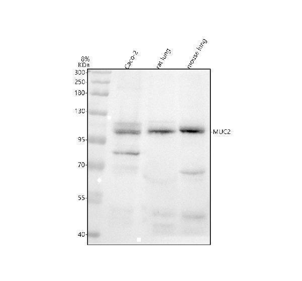
Western blot analysis of MUC2 using anti-MUC2 antibody (M01212-1).
Electrophoresis was performed on a 8% SDS-PAGE gel at 80V (Stacking gel) / 120V (Resolving gel) for 2 hours. The sample well of each lane was loaded with 30 ug of sample under reducing conditions.
Lane 1: human Caco-2 whole cell lysates,
Lane 2: rat lung tissue lysates,
Lane 3: mouse lung tissue lysates.
After electrophoresis, proteins were transferred to a nitrocellulose membrane at 150 mA for 50-90 minutes. Blocked the membrane with 5% non-fat milk/TBS for 1.5 hour at RT. The membrane was incubated with rabbit anti-MUC2 antigen affinity purified monoclonal antibody (M01212-1) at 1:500 overnight at 4°C, then washed with TBS-0.1%Tween 3 times with 5 minutes each and probed with a goat anti-rabbit IgG-HRP secondary antibody at a dilution of 1:5000 for 1.5 hour at RT. The signal is developed using an ECL Plus Western Blotting Substrate (Catalog # AR1196-200) with Tanon 5200 system. A specific band was detected for MUC2 at approximately 110 kDa. The expected band size for MUC2 is at 540 kDa.

Click image to see more details
IHC analysis of MUC2 using anti-MUC2 antibody (M01212-1).
MUC2 was detected in a paraffin-embedded section of human colon tissue. Heat mediated antigen retrieval was performed in EDTA buffer (pH 8.0, epitope retrieval solution). The tissue section was blocked with 10% goat serum. The tissue section was then incubated with 1:50 rabbit anti-MUC2 Antibody (M01212-1) overnight at 4°C. Peroxidase Conjugated Goat Anti-rabbit IgG was used as secondary antibody and incubated for 30 minutes at 37°C. The tissue section was developed using HRP Conjugated Rabbit IgG Super Vision Assay Kit (Catalog # SV0002) with DAB as the chromogen.

Click image to see more details
Polyamines enhance mucosal defense factors in rats with massive intestinal resection. (A-B) Fecal and serum secretory IgA was measured using ELISA. n = 5–7/group. (C) IgA in ileum tissue was assessed using western blotting. n = 4–6/group. (D) Fecal mucin was measured using a fluorometric assay. n = 4–6/group. (E) Representative images of ileal villus showing immunostaining by Muc2 (original magnification, ×400; scale bars = 200 μm). Left graphs show the number of goblet cells per unit villous area and the size of goblet cell secretion granule. (F) The expression of Claudin-3 in the ileum tissue was measured by western blot analysis. Representative images of ileal villus showing immunostaining by Claudin-3 (original magnification, ×200; scale bars = 100 μm). (G) Serum DAO was measured by ELISA. n = 5–7/group. (H) Serum GLP-2 was measured by ELISA. n = 5–7/group. (I) Fecal short-chain fatty acid (SCFA) content was measured using high-performance liquid chromatography. n = 3–6/group. Data are presented as mean ± SD. Results of one-way ANOVA are represented as follows: * P < 0.05; ** P < 0.01; *** P < 0.001; **** P < 0.0001.
Index in PubMed under a CC BY license. PMID: 38409241

Click image to see more details
The distribution of Lgr5 + ISCs in the intestinalmucosa and the subcellular localization and relative expression level detection of epithelial function proteins CDX2 and villin in the intestinal mucosa of IBD at 7 days after termination of DSS administration. (A) The Lgr5 + ISCs (brown) in the small intestinal mucosa: (A1) the normal group, the villi and the crypts were arranged compactly, and Lgr5 + ISCs were observed in the crypts; (A2) the DSS group, the villi and the crypts were scattered, with few Lgr5 + ISCs; (A3) the DSS + B. subtilis -fermented milk group, there were more Lgr5 + ISCs in villi and crypts compared with those in the DSS group. (B) The Lgr5 + ISCs (brown) in the colonic mucosa: (B1) the normal group, the glands were arranged compactly, and there were large amounts of Lgr5 + ISCs at the bottom of the glands; (B2) the DSS group: the ulcers were replaced by scars. No Lgr5 + ISCs were observed in the scars; (B3) the DSS + B. subtilis -fermented milk group: the colonic epithelium was integrated, with some regenerated glands. A number of Lgr5 + ISCs were observed at the bottom of the regenerated glands. (C) The CDX2 was localized in the epithelial cellular nuclei (brown) by immunohistochemistry staining in the small intestinal mucosa: (C1) the normal group: the villi and the crypts were arranged compactly, and CDX2 + epithelial cells were observed on the surface of the villi and the crypts; (C2) the DSS group: the villi and the crypts were scattered, and few CDX2 + epithelial cells were observed on the surface of the crypt and the villi; (C3) the DSS + B. subtilis -fermented milk group: more villi and crypts were observed in comparison with the DSS group, and there were more CDX2 + epithelial cells covering the villi and crypts. (D) The CDX2 was localized in the epithelial cellular nuclei (brown) by immunohistochemistry staining in the colonic mucosa: (D1) the normal group: the colonic glands were arranged compactly, and CDX2 + epithelial cells were observed on the surface of the glands; (D2) the DSS group: the glands were scattered, and few CDX2 + epithelial cells were observed in the scar; (D3) the DSS + B. subtilis -fermented milk group: more colonic glands were observed in comparison with the DSS group, and there were more CDX2 + epithelial cells in the glands. (E) The Mucin2 was localized in the cytoplasm of the goblet cells (brown) by immunohistochemistry staining in the small intestinal mucosa: (E1) the normal group, a number of Mucin2 + goblet cells observed in the epithelium; (E2) the DSS group: only few Mucin2 + goblet cells were observed in the remaining villi and crypts; (E3) the DSS + B. subtilis -fermented milk group: more Mucin2 + goblet cells were observed in the recovered mucosa. (F) The Mucin2 was localized in the cytoplasm of the goblet cells (brown) by immunohistochemistry staining in the colonic mucosa: (F1) the normal group, large amounts of Mucin2 + goblet cells were observed in the mucosa; (F2) the DSS group: only few Mucin2 + goblet cells were observed in the scars; (F3) the DSS + B. subtilis -fermented milk group: more Mucin2 + goblet cells were observed in the recovered colonic mucosa. (G,H) Western blotting was applied for detection of the relative expression level of Lgr5, CDX2, and Mucin2 in the samples containing equivalent ileum and colon. The expression level of Lgr5, CDX2, and Mucin2 in the DSS group was significantly lower than that of the normal (control) group. The expression level of Lgr5, CDX2, and Mucin2 in the DSS + B. subtilis -fermented milk (FM) group was significantly higher than that of the DSS group ( n = 5, ** represents p < 0.01).
Index in PubMed under a CC BY license. PMID: 33519783

Click image to see more details
IHC analysis of MUC2 using anti-MUC2 antibody (M01212-1).
MUC2 was detected in a paraffin-embedded section of human colon cancer tissue. Heat mediated antigen retrieval was performed in EDTA buffer (pH 8.0, epitope retrieval solution). The tissue section was blocked with 10% goat serum. The tissue section was then incubated with 1:50 rabbit anti-MUC2 Antibody (M01212-1) overnight at 4°C. Peroxidase Conjugated Goat Anti-rabbit IgG was used as secondary antibody and incubated for 30 minutes at 37°C. The tissue section was developed using HRP Conjugated Rabbit IgG Super Vision Assay Kit (Catalog # SV0002) with DAB as the chromogen.
Specific Publications For Anti-MUC2 Rabbit Monoclonal Antibody (M01212-1)
Loading publications
Recommended Resources
Here are featured tools and databases that you might find useful.
- Boster's Pathways Library
- Protein Databases
- Bioscience Research Protocol Resources
- Data Processing & Analysis Software
- Photo Editing Software
- Scientific Literature Resources
- Research Paper Management Tools
- Molecular Biology Software
- Primer Design Tools
- Bioinformatics Tools
- Phylogenetic Tree Analysis
Customer Reviews
Have you used Anti-MUC2 Rabbit Monoclonal Antibody?
Share your experimental results or join a short interview to earn up to $1,000 in product credits or other rewards.
0 Reviews For Anti-MUC2 Rabbit Monoclonal Antibody
Customer Q&As
Have a question?
Find answers in Q&As, reviews.
Can't find your answer?
Submit your question
15 Customer Q&As for Anti-MUC2 Rabbit Monoclonal Antibody
Question
Is a blocking peptide available for product anti-MUC2 Rabbit Monoclonal antibody (M01212-1)?
Verified Customer
Verified customer
Asked: 2020-04-16
Answer
We do provide the blocking peptide for product anti-MUC2 Rabbit Monoclonal antibody (M01212-1). If you would like to place an order for it please contact support@bosterbio.com and make a special request.
Boster Scientific Support
Answered: 2020-04-16
Question
Do you have a BSA free version of anti-MUC2 Rabbit Monoclonal antibody M01212-1 available?
Verified Customer
Verified customer
Asked: 2020-02-11
Answer
Thank you for your recent telephone inquiry. I can confirm that some lots of this anti-MUC2 Rabbit Monoclonal antibody M01212-1 are BSA free. For now, these lots are available and we can make a BSA free formula for you free of charge. It will take 3 extra days to prepare. If you require this antibody BSA free again in future, please do not hesitate to contact me and I will be pleased to check which lots we have in stock that are BSA free.
Boster Scientific Support
Answered: 2020-02-11
Question
I am looking for to test anti-MUC2 Rabbit Monoclonal antibody M01212-1 on rat colon for research purposes, then I may be interested in using anti-MUC2 Rabbit Monoclonal antibody M01212-1 for diagnostic purposes as well. Is the antibody suitable for diagnostic purposes?
Verified Customer
Verified customer
Asked: 2020-02-04
Answer
The products we sell, including anti-MUC2 Rabbit Monoclonal antibody M01212-1, are only intended for research use. They would not be suitable for use in diagnostic work. If you have the means to develop a product into diagnostic use, and are interested in collaborating with us and develop our product into an IVD product, please contact us for more discussions.
Boster Scientific Support
Answered: 2020-02-04
Question
Does M01212-1 anti-MUC2 Rabbit Monoclonal antibody work on parafin embedded sections? If so, which fixation method do you recommend we use (PFA, paraformaldehyde, other)?
Verified Customer
Verified customer
Asked: 2020-01-17
Answer
You can see on the product datasheet, M01212-1 anti-MUC2 Rabbit Monoclonal antibody as been tested on WB. It is best to use PFA for fixation because it has better tissue penetration ability. PFA needs to be prepared fresh before use. Long term stored PFA turns into formalin, as the PFA molecules congregate and become formalin.
Boster Scientific Support
Answered: 2020-01-17
Question
My boss were well pleased with the WB result of your anti-MUC2 Rabbit Monoclonal antibody. However we have seen positive staining in cervix carcinoma secreted. note=in the intestine using this antibody. Is that expected? Could you tell me where is MUC2 supposed to be expressed?
Verified Customer
Verified customer
Asked: 2019-12-26
Answer
Based on literature, cervix carcinoma does express MUC2. Generally MUC2 expresses in secreted. note=in the intestine, secreted. Regarding which tissues have MUC2 expression, here are a few articles citing expression in various tissues:
Cervix carcinoma, Pubmed ID: 18669648
Colon, Pubmed ID: 1400449, 1885763
Intestine, Pubmed ID: 8300571
Boster Scientific Support
Answered: 2019-12-26
Question
My question regarding product M01212-1, anti-MUC2 Rabbit Monoclonal antibody. I was wondering if it would be possible to conjugate this antibody with biotin. I would need it to be without BSA or sodium azide. I am planning on using a buffer exchange of sodium azide with PBS only. Would there be problems for me to conjugate the antibody and store it in -20 degrees in small aliquots?
J. Collins
Verified customer
Asked: 2019-06-17
Answer
We suggest not storing this antibody with PBS buffer only in -20 degrees. If you want to store it in -20 degrees it is best to add some cryoprotectant like glycerol. If you want carrier free M01212-1 anti-MUC2 Rabbit Monoclonal antibody, we can provide it to you in a special formula with trehalose and/or glycerol. These molecules will not interfere with conjugation chemistry and provide a good level of protection for the antibody from degradation. Please be sure to specify this in your purchase order.
Boster Scientific Support
Answered: 2019-06-17
Question
We have tried in the past anti-MUC2 Rabbit Monoclonal antibody for IF on intestine last year. I am using human, and We intend to use the antibody for IP next. I am interested in examining intestine as well as cervix carcinoma in our next experiment. Could give a recommendation on which antibody would work the best for IP?
Verified Customer
Verified customer
Asked: 2019-06-04
Answer
I have checked the website and datasheets of our anti-MUC2 Rabbit Monoclonal antibody and it appears that M01212-1 has been validated on human in both IF and IP. Thus M01212-1 should work for your application. Our Boster satisfaction guarantee will cover this product for IP in human even if the specific tissue type has not been validated. We do have a comprehensive range of products for IP detection and you can check out our website bosterbio.com to find out more information about them.
Boster Scientific Support
Answered: 2019-06-04
Question
Would anti-MUC2 Rabbit Monoclonal antibody M01212-1 work for WB with colon?
Verified Customer
Verified customer
Asked: 2019-04-16
Answer
According to the expression profile of colon, MUC2 is highly expressed in colon. So, it is likely that anti-MUC2 Rabbit Monoclonal antibody M01212-1 will work for WB with colon.
Boster Scientific Support
Answered: 2019-04-16
Question
We have seen staining in rat colon. Are there any suggestions? Is anti-MUC2 Rabbit Monoclonal antibody supposed to stain colon positively?
Verified Customer
Verified customer
Asked: 2018-09-21
Answer
According to literature colon does express MUC2. According to Uniprot.org, MUC2 is expressed in intestine, colon, cervix carcinoma, among other tissues. Regarding which tissues have MUC2 expression, here are a few articles citing expression in various tissues:
Cervix carcinoma, Pubmed ID: 18669648
Colon, Pubmed ID: 1400449, 1885763
Intestine, Pubmed ID: 8300571
Boster Scientific Support
Answered: 2018-09-21
Question
I was wanting to use your anti-MUC2 Rabbit Monoclonal antibody for WB for rat colon on frozen tissues, but I want to know if it has been tested for this particular application. Has this antibody been tested and is this antibody a good choice for rat colon identification?
Z. Kulkarni
Verified customer
Asked: 2017-02-28
Answer
It shows on the product datasheet, M01212-1 anti-MUC2 Rabbit Monoclonal antibody has been tested for IP, IF, WB on human, rat tissues. We have an innovator award program that if you test this antibody and show it works in rat colon in IHC-frozen, you can get your next antibody for free.
Boster Scientific Support
Answered: 2017-02-28
Question
Is this M01212-1 anti-MUC2 Rabbit Monoclonal antibody reactive to the isotypes of MUC2?
S. Edwards
Verified customer
Asked: 2015-11-17
Answer
The immunogen of M01212-1 anti-MUC2 Rabbit Monoclonal antibody is A synthesized peptide derived from human MUC2. Could you tell me which isotype you are interested in so I can help see if the immunogen is part of this isotype?
Boster Scientific Support
Answered: 2015-11-17
Question
I see that the anti-MUC2 Rabbit Monoclonal antibody M01212-1 works with WB, what is the protocol used to produce the result images on the product page?
C. Collins
Verified customer
Asked: 2014-03-24
Answer
You can find protocols for WB on the "support/technical resources" section of our navigation menu. If you have any further questions, please send an email to support@bosterbio.com
Boster Scientific Support
Answered: 2014-03-24
Question
Please see the WB image, lot number and protocol we used for colon using anti-MUC2 Rabbit Monoclonal antibody M01212-1. Please let me know if you require anything else.
S. Dhar
Verified customer
Asked: 2013-12-26
Answer
Thank you very much for the data. Our lab team are working to resolve this as quickly as possible, and we appreciate your patience and understanding! You have provided everything we needed. Please let me know if there is anything you need in the meantime.
Boster Scientific Support
Answered: 2013-12-26
Question
We are currently using anti-MUC2 Rabbit Monoclonal antibody M01212-1 for rat tissue, and we are satisfied with the IF results. The species of reactivity given in the datasheet says human, rat. Is it true that the antibody can work on monkey tissues as well?
R. Bhatt
Verified customer
Asked: 2013-07-12
Answer
The anti-MUC2 Rabbit Monoclonal antibody (M01212-1) has not been validated for cross reactivity specifically with monkey tissues, though there is a good chance of cross reactivity. We have an innovator award program that if you test this antibody and show it works in monkey you can get your next antibody for free. Please contact me if I can help you with anything.
Boster Scientific Support
Answered: 2013-07-12
Question
Thanks for helping with my inquiry over the phone. Here are the WB image, lot number and protocol we used for colon using anti-MUC2 Rabbit Monoclonal antibody M01212-1. Let me know if you need anything else.
J. Krishna
Verified customer
Asked: 2013-05-02
Answer
I appreciate the data. You have provided everything we needed. Our lab team are working to resolve your inquiry as quickly as possible, and we appreciate your patience and understanding! Please let me know if there is anything you need in the meantime.
Boster Scientific Support
Answered: 2013-05-02
