MUC5B / Mucin-5B · IHC design guide

Design Immunohistochemistry for MUC5B

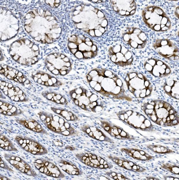
Plan chromogenic IHC on paraffin sections with A00719-2 at 2–5 μg/ml (datasheet A00719-2). Use rectal glandular cells as a positive reference (HPA tissue IHC), and account for MUC5B secretion when interpreting staining beyond producing cells (UniProt).

Evidence assembled Oct 2026 · For research use; verify linked source records and product datasheet before use
Immunohistochemistry protocol sheet for MUC5B (IHC for MUC5B): expected localisation Secreted; no transmembrane segment (UniProt), antibody A00719-2, validated IHC image, and IHC protocol steps
Printable MUC5B IHC protocol sheet — expected localisation Secreted; no transmembrane segment (UniProt), antibody A00719-2, controls and protocol steps. Open the full MUC5B IHC guide →

MUC5B Immunohistochemistry Experimental Design Guide

Expected localisation, validated protocols, controls and antibodies — the at-a-glance facts below, then the full design guide.

Must know before staining
Expected localisation Secreted; no transmembrane segment (UniProt)
Staining pattern Selective staining in mucus-producing cells; compartment unspecified (HPA tissue IHC)
Antigen retrieval EDTA pH 8.0 HIER, heat-mediated (datasheet A00719-2)
Positive control ⓘ Appendix+4 more · see all
Negative control ⓘ Adipose tissue+4 more · see all
Important caveats
Reasons your staining may differ from the expected pattern.
Fixation Keep fixation consistent across sections. (standard IHC practice; not target-specific)
Caveat Secreted MUC5B may stain beyond its producing cells (UniProt)
Regulation No specific expression regulator reported (UniProt)
Isoform / epitope No isoforms listed; mature chain spans aa 26–5762 (UniProt)
Section 1

Recommended MUC5B IHC & IF Protocols

The catalog antibody has a datasheet IHC-P protocol (datasheet A00719-2). The published IHC protocols below cover airway biopsies, pediatric lung, ferret tissues, and rat lung (PMC11678853; PMC4596737; PMC10286488; PMC6376022).

Recommended immunohistochemistry (IHC-P) protocol parameters
SampleParaffin-embedded human rectal cancer tissue; fixative not specified (datasheet A00719-2)
FixationImage fixative and duration unreported (datasheet A00719-2); verify before use.
Sectioning4–5 µm sections on charged slides (standard)
DeparaffinisationXylene, graded ethanol series to water (standard)
Antigen retrievalHeat retrieval: EDTA pH 8.0 (datasheet A00719-2); 20 min, 95–100 °C (standard)
Peroxidase block3% H2O2, 10 min, room temperature (standard)
Blocking10% goat serum (datasheet A00719-2)
Primary antibodyRabbit anti-MUC5B, 2-5 μg/ml (datasheet A00719-2)
Primary incubationOvernight at 4 °C (datasheet A00719-2)
DetectionHRP-conjugated secondary, DAB chromogen (datasheet A00719-2)
CounterstainHematoxylin, blue, dehydrate and mount (standard)
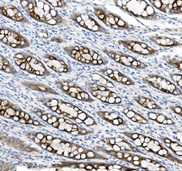
Expected resultMUC5B-positive staining in glandular cells of appendix (HPA tissue IHC: High). HPA tissue profile: Selective expression in mucus producing cells. No signal in the no-primary control.
💡Decision noteStart with heat-mediated EDTA retrieval at pH 8.0 for the catalog antibody (datasheet A00719-2). Citrate at pH 6.0 is a published alternative (PMC11678853; PMC10286488).
Section 2

What Is the Expected MUC5B Staining Pattern?

MUC5B is a secreted mucin with no transmembrane segment (UniProt Q9HC84 topology). In paraffin-section IHC, expect selective staining in mucus-producing glandular or respiratory epithelial cells; secreted material may also be visible near those cells (HPA tissue IHC; UniProt Q9HC84 subcellular location). HPA rates its tissue pattern Enhanced, while cautioning that secreted protein and tissue RNA need not occupy the same location (HPA tissue IHC).

What am I looking at on my slide?
Strong staining in bronchial or nasopharyngeal respiratory epithelium, or in cervical or gallbladder glandular cells (HPA: High in each listed cell population).This fits the observed tissue pattern. Assess which cells stain and whether adjacent secretions also contain signal; extracellular staining alone needs context because MUC5B is secreted (HPA tissue IHC; UniProt Q9HC84 subcellular location).
Vesicle-like intracellular signal in a mucus-producing cell, with possible signal in material it secretes (HPA subcellular ICC-IF: supported vesicles; UniProt Q9HC84: Secreted).A secretory distribution is plausible. The vesicle assignment comes from ICC-IF and should guide interpretation cautiously; it does not establish a required vesicle pattern in paraffin-section IHC (HPA subcellular ICC-IF; HPA tissue IHC).
Predominant nuclear or uniform membrane-rim staining, without a matching mucus-producing cell pattern (UniProt Q9HC84 topology; HPA tissue IHC profile).Treat this as a localization mismatch requiring review, rather than a confirmed MUC5B result: MUC5B is secreted and has no transmembrane segment (UniProt Q9HC84 topology; general IHC interpretation practice).
Strong staining across unrelated cells or broad, even chromogen throughout the section (HPA tissue IHC: selective expression in mucus-producing cells).This conflicts with HPA's selective profile. Consider nonspecific binding, uneven background, or endogenous detection activity; compare matched negative and detection-only controls before assigning cellular positivity (HPA tissue IHC; general chromogenic IHC practice).
No staining in a known-positive region, such as bronchial respiratory epithelium or cervical glandular cells (HPA: High in those cell populations).An absent control signal makes a negative study section hard to interpret. Check that the expected cells are present, then review the primary antibody and detection workflow using the same run (HPA tissue IHC; general IHC control practice).
💡Expected MUC5B appearanceCall positive when mucus-producing glandular or respiratory epithelial cells show a selective, clearly visible secretory pattern, with high staining possible in several listed tissues; isolated nuclear, uniform membrane-rim, or indiscriminate staining warrants control review (HPA tissue IHC: selective profile and High examples; UniProt Q9HC84 topology; general IHC interpretation practice).
How each factor affects the staining
Positive-control tissue and cell selection (HPA tissue IHC).Bronchus respiratory epithelium and cervix glandular cells are High examples; salivary gland glandular cells are Medium. Score the listed cells, rather than treating every cell in a positive tissue as positive (HPA tissue IHC).
Secreted protein versus cellular RNA location (HPA reliability description; UniProt Q9HC84: Secreted).Signal in secretions can extend beyond the producing cell. A mismatch between protein location and tissue RNA is therefore possible and does not, by itself, overturn an IHC result (HPA reliability description; UniProt Q9HC84 subcellular location).
Topology and processing (UniProt Q9HC84).MUC5B has a signal peptide at residues 1–25, a chain at 26–5762, and no transmembrane segment. Interpret a dominant membrane outline cautiously; these facts do not define an antibody epitope (UniProt Q9HC84 topology and processing).
Antibody evidence (HPA antibodies; HPA tissue IHC).HPA008246 and CAB009396 each have Enhanced IHC validation; HPA describes Enhanced tissue reliability as consistent antibody staining and RNA evidence, with added complexity for secreted proteins. This supports the reported pattern, not every signal in a new section (HPA antibodies; HPA tissue IHC reliability).
IF/ICC Q: What localization is reported? (HPA subcellular ICC-IF).A: Vesicles are the supported main location in ICC-IF; images are listed for A-549, PC-3, and U2OS. This is context for interpreting localization, not an IHC protocol or proof that every paraffin section shows discrete vesicles (HPA subcellular ICC-IF).
Why is my staining missing, weak or wrong?
SituationLikely causeNext action
Known-positive bronchial or cervical cells have no signal (HPA: High in the specified cells).The expected cells may be absent from the sampled region, or the run may have failed (HPA tissue IHC; general IHC control practice).Confirm the cell population on the counterstained section and inspect a known-positive control processed in the same run (HPA tissue IHC; general IHC control practice).
The entire section shows diffuse chromogen (general chromogenic IHC observation).Nonspecific background or endogenous detection activity can obscure selective staining (general chromogenic IHC practice).Compare detection-only and negative controls; review blocking, washes, and detection conditions before scoring cells (general chromogenic IHC practice).
Adipocytes stain strongly in an adipose-tissue control (HPA: Not detected in adipocytes).The result conflicts with the reported cell pattern and may reflect nonspecific or detection-related signal (HPA tissue IHC; general IHC interpretation practice).Review control staining and cellular morphology, then reassess antibody and detection conditions before calling it MUC5B (HPA tissue IHC; general IHC control practice).
Only luminal material stains while producing cells are unclear (UniProt Q9HC84: Secreted).Secreted MUC5B may lie outside its source cells; extracellular signal alone leaves cellular attribution uncertain (UniProt Q9HC84 subcellular location; HPA reliability description).Inspect adjacent epithelium and glands on the counterstain and compare the known-positive control before assigning the source (HPA tissue IHC; general IHC interpretation practice).
A sharp nuclear or continuous membrane pattern dominates (UniProt Q9HC84 topology).That distribution does not follow the reported secreted, nontransmembrane topology (UniProt Q9HC84 topology).Check morphology and negative controls, and seek concordance with selective mucus-producing cells before accepting specificity (HPA tissue IHC; general IHC control practice).
The salivary-gland control looks weaker than a bronchial control (HPA: Medium versus High in the specified cells).HPA reports different staining levels for those cell populations; equal intensity is not an expected control criterion (HPA tissue IHC).Evaluate each listed cell population against its reported level and the controls in that run; avoid scoring whole-tissue intensity alone (HPA tissue IHC; general IHC control practice).

Sample controls for MUC5B IHC & IF

🧪Run bronchus first: respiratory epithelial cells should stain strongly (HPA: High in bronchus respiratory epithelial cells). Use adipose tissue as the negative tissue, where adipocytes are not detected (HPA: Not detected in adipose adipocytes); on the bronchus slide, adjacent stromal cells should lack specific cellular staining and serve as an internal background comparator (standard IHC practice).
Positive control tissue: Appendix (Glandular cells, HPA High)
Negative control tissue: Adipose tissue (HPA Not detected)
ICC-IF cell lines (HPA subcellular resource): HPA ICC-IF images show MUC5B in A-549, PC-3, U2OS, with annotated localisation: Vesicles (supported) (HPA subcellular).
Technical controls: Include no-primary and secondary-only controls, a rabbit IgG isotype control matched to the primary antibody’s clonality, and MUC5B knockout material if available as a biological negative (selected-SKU caption: rabbit primary; standard IHC practice). Quench endogenous peroxidase in bronchus sections before HRP/DAB detection (selected-SKU caption: HRP/DAB; standard IHC practice).
⚠️Feasibility: A target-specific fixation window or fixation effect is unreported, and the selected-SKU paraffin-section caption does not state the fixative (selected-SKU caption: fixative not stated). The caption uses heat retrieval in EDTA at pH 8.0, but does not establish that retrieval is required (selected-SKU caption: EDTA retrieval). Paraffin IHC is documented here, while the supplied evidence does not establish whether frozen sections or IF are easier; interpret luminal mucus staining cautiously because MUC5B is secreted (selected-SKU caption: paraffin IHC; UniProt Q9HC84: secreted).

HPA tissue IHC evidence for MUC5B

Comprehensive Human Protein Atlas IHC scoring per tissue (reliability: Enhanced — High consistency between antibody staining and RNA expression data. At least one protein variant secreted, tissue location of RNA and protein might differ and correlation is complex.). Rows are taken directly from the HPA tissue chart — click any row's HPA link to view the source.

Positive expression · recommended positive controls

TissueCell typeLevelEvidenceSource
Appendix Glandular cells High Protein (IHC) HPA →
Bronchus Respiratory epithelial cells High Protein (IHC) HPA →
Cervix Glandular cells High Protein (IHC) HPA →
Colon Glandular cells High Protein (IHC) HPA →
Gallbladder Glandular cells High Protein (IHC) HPA →

Undetected expression · recommended negative controls

TissueCell typeLevelEvidenceSource
Adipose tissue Adipocytes Not detected Protein (IHC) HPA →
Adrenal gland Glandular cells Not detected Protein (IHC) HPA →
Bone marrow Hematopoietic cells Not detected Protein (IHC) HPA →
Breast Adipocytes Not detected Protein (IHC) HPA →
Caudate Glial cells Not detected Protein (IHC) HPA →
Section 3

Advanced MUC5B IHC Tips

Troubleshoot MUC5B chromogenic IHC in paraffin sections by checking retrieval, tissue context, secreted mucus staining, and controls before scoring.

Which retrieval condition should I start with for MUC5B paraffin sections?
Start with heat-mediated retrieval in EDTA at pH 8.0 for paraffin sections stained with A00719-2 (datasheet A00719-2). The documented image used 2 μg/ml primary antibody overnight at 4°C, so keep antibody exposure constant while evaluating retrieval (caption A00719-2). If staining is weak, compare a modest increase in heating time on matched sections, recording both signal and tissue damage; no retrieval duration is supplied for this antibody (caption A00719-2; standard IHC practice). Include a known positive glandular region, such as rectum, and a no-primary control so improved staining is judged against background rather than intensity alone (HPA: rectal glandular cells High; standard IHC practice).
Could fixation explain weak or inconsistent MUC5B staining?
The A00719-2 image describes a paraffin section but does not state its fixative, so MUC5B-specific sensitivity to fixation is unknown (caption A00719-2). Record the fixative, fixation interval, and processing history for each block, then compare similarly processed sections before attributing a staining difference to biology (standard IHC practice). On paired sections, hold EDTA retrieval at pH 8.0 and the documented 2 μg/ml primary incubation overnight at 4°C constant (datasheet A00719-2; caption A00719-2). If those comparisons implicate processing, optimize fixation prospectively with matched specimens and assess morphology alongside signal; the available evidence does not establish a target-specific fixation effect (standard IHC practice; caption A00719-2).
Where should convincing MUC5B staining appear in a section?
Expect staining associated with mucus-producing epithelium and secreted material, since MUC5B is secreted and has no transmembrane segment (UniProt Q9HC84: subcellular location and topology; HPA: selective expression in mucus-producing cells). Airway submucosal gland mucous cells are a documented source, while HPA reports High staining in bronchial respiratory epithelial cells and rectal glandular cells (UniProt Q9HC84: tissue specificity; HPA: bronchus and rectum). Evaluate intracellular glandular staining and extracellular mucus separately, because secretion can place protein away from the cells that produced it (UniProt Q9HC84: secreted; HPA: reliability description). Predominantly nuclear staining would conflict with the reported localisation and warrants review of morphology, detection controls, and antibody background (UniProt Q9HC84: secreted; standard IHC practice).
How do glycosylation and an unmapped epitope affect interpretation?
UniProt lists 0 isoforms and 38 glycosylation sites for MUC5B, but the supplied A00719-2 caption does not identify the antibody epitope (UniProt Q9HC84: isoforms and glycosylation; caption A00719-2). Therefore, a negative section cannot by itself establish absence of protein or prove that a particular glycan masked the epitope (standard IHC interpretation). Compare serial sections using the documented EDTA pH 8.0 retrieval before changing one processing variable at a time (datasheet A00719-2; standard IHC practice). Score cell-associated and extracellular staining separately, and report epitope accessibility as unresolved if morphology and a positive control are satisfactory but the test region remains negative (UniProt Q9HC84: secreted; standard IHC practice).
How should I plan an IF follow-up to the chromogenic MUC5B result?
Treat IF as a separate assay: the supplied A00719-2 evidence is a paraffin-section chromogenic IHC image, with no IF conditions reported (caption A00719-2). Multiplex MUC5B with an independently validated marker of the expected mucus-producing epithelial cells, and inspect each single-marker control for bleed-through before interpreting overlap (HPA: selective expression in mucus-producing cells; standard IF practice). Choose a fluorophore channel after measuring tissue autofluorescence, with a far-red channel as an option when shorter wavelengths are noisy (standard IF practice). Because MUC5B is secreted and the antibody epitope is unspecified, assess intracellular vesicles with permeabilisation only when testing an intracellularly accessible epitope, and assess deposited extracellular mucus without assuming permeabilisation is necessary (UniProt Q9HC84: secreted; HPA: vesicles; caption A00719-2).
What should I check when DAB appears throughout the section?
First compare a no-primary slide and a matched positive section to distinguish detection background from expected mucus-associated staining (standard IHC practice; UniProt Q9HC84: secreted). The A00719-2 image used 10% goat serum blocking, a peroxidase-conjugated secondary for 30 minutes at 37°C, and DAB development (caption A00719-2). Include an endogenous peroxidase block as a general chromogenic IHC step, then check whether diffuse brown signal persists without primary antibody (standard IHC practice). If background remains, review secondary specificity, blocking, washes, primary concentration, and DAB development time while retaining a positive glandular control; the documented primary concentration was 2 μg/ml (standard IHC practice; caption A00719-2).
How should I quantify patchy MUC5B staining in glandular tissue? ⚠ ANSWER MARKED FOR VERIFICATION
Define glandular epithelium and luminal or extracellular mucus as separate regions before scoring, because MUC5B is secreted and the protein may lie beyond its producer cell (UniProt Q9HC84: secreted; HPA: reliability description). For cell-associated staining, report the percentage of positive glandular cells and, if intensity is reproducible, an H-score from 0–300 using prespecified intensity categories (standard IHC practice). For deposited mucus, measure positive area divided by the sampled mucus or luminal area, rather than including unrelated stroma in the denominator (standard image-analysis practice). Keep retrieval, DAB development, exposure, and region selection consistent across slides, and inspect a positive rectal glandular control in each staining run (datasheet A00719-2; HPA: rectal glandular cells High; standard IHC practice).
How can I distinguish true MUC5B signal from artefact?
Give greatest weight to staining in mucus-producing glandular or respiratory epithelium and associated secretions, consistent with the reported tissue pattern and secreted localisation (HPA: selective expression in mucus-producing cells; UniProt Q9HC84: secreted). Strong nuclear staining, staining confined to cut edges or necrotic areas, and uniform colour across unrelated compartments should prompt a morphology and control review (UniProt Q9HC84: secreted; standard IHC practice). Compare a no-primary slide and an endogenous peroxidase-blocked slide when DAB signal could reflect detection chemistry (standard IHC practice). Use the rectal glandular High pattern as a positive reference, while remembering that secreted protein and tissue RNA need not occupy the same location (HPA: rectal glandular cells High; HPA: reliability description).
Boster reagents

Best MUC5B / Mucin-5B IHC Antibodies

A00719-2 has human paraffin-section MUC5B IHC data (A00719-2 image caption). M00719-1 has human, mouse and rat stomach IHC images, but targets Mucin 5AC (M00719-1 title and image captions).

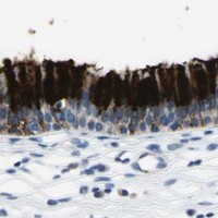
Real IHC data IHC analysis of MUC5B using anti-MUC5B antibody (A00719-2). MUC5B was detected in a paraffin-embedded section of human rectal cancer tissue. Heat mediated antigen retrieval was performed in EDTA buffer (pH 8.0, epitope retrieval solution). The tissue section was blocked with 10% goat serum. The tissue section was then incubated with 2 μg/ml rabbit anti-MUC5B Antibody (A00719-2) overnight at 4°C. Peroxidase Conjugated Goat Anti-rabbit IgG was used as secondary antibody and incubated for 30 minutes at 37°C. The tissue section was developed using HRP Conjugated Rabbit IgG Super Vision Assay Kit (Catalog # SV0002) with DAB as the chromogen.
Anti-MUC5B Antibody
Cat # A00719-2
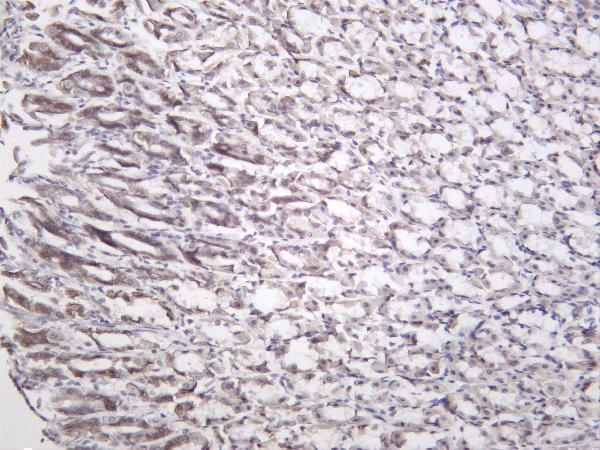
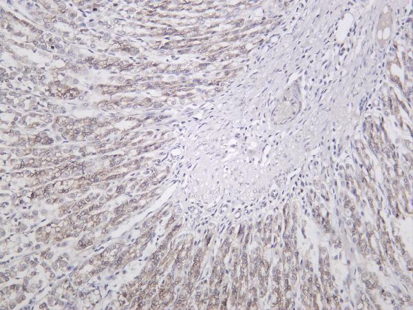
Real IHC data Mouse stomach was stained with anti-Mucin 5AC rabbit antibody
Anti-Mucin 5AC Rabbit Monoclonal Antibody
Cat # M00719-1

A00719-2 will render with human rectal cancer paraffin-section IHC data (A00719-2 image caption). M00719-1 will render with a mouse stomach IHC image and lists human, mouse and rat reactivity, but its stated target is Mucin 5AC (M00719-1 title, image captions and reactivity list).

Which to pick: Choose A00719-2 for human MUC5B paraffin-section IHC; its caption reports EDTA retrieval at pH 8.0 and antibody at 2 μg/ml, but does not report the fixative (A00719-2 image caption). M00719-1 lists IF and human, mouse and rat reactivity, but it targets Mucin 5AC, so it is unsuitable as a MUC5B IF/ICC or cross-species reagent (M00719-1 title, applications and reactivity list). No MUC5B IF/ICC or cross-species validation is provided for A00719-2 (A00719-2 applications and reactivity list).

Each figure is that product's own IHC / IF validation image from its datasheet.

References

  1. UniProt Consortium. UniProt entry Q9HC84 (MUC5B_HUMAN, Mucin-5B).
  2. Human Protein Atlas. MUC5B tissue IHC expression (reliability: Enhanced).
  3. Human Protein Atlas. MUC5B subcellular location (ICC-IF): Localized to vesicles..
  4. Human Protein Atlas. MUC5B antibody validation summary (2 antibodies).
  5. Expression Levels of MUC5AC and MUC5B in Airway Goblet Cells Are Associated with Traits of COPD and Progression of Chronic Airflow Limitation. International journal of molecular sciences 2024 — PMC11678853.
  6. MUC5B expression and location in surfactant protein C mutations in children. Pediatric pulmonology 2015 — PMC4596737.
  7. Immunohistochemical detection of MUC5AC and MUC5B mucins in ferrets. BMC research notes 2023 — PMC10286488.
  8. Increase in secreted airway mucins and partial Muc5b STAT6/FoxA2 regulation during Pneumocystis primary infection. Scientific reports 2019 — PMC6376022.
  9. PubMed PMID:11713095 — UniProt-cited evidence.
  10. PubMed PMID:9790959 — UniProt-cited evidence.
  11. PubMed PMID:9804771 — UniProt-cited evidence.