MYB / Transcriptional activator Myb · IHC design guide

Design Immunohistochemistry for MYB

Plan chromogenic MYB IHC around its mainly nuclear tissue pattern (HPA tissue IHC). Compare positive hematopoietic cells in bone marrow with cardiomyocytes, where staining was not detected (HPA tissue IHC).

Evidence assembled Oct 2026 · For research use; verify linked source records and product datasheet before use
Immunohistochemistry protocol sheet for MYB (IHC for MYB): expected localisation Mainly nuclear tissue staining (HPA tissue IHC), antibody PB9288, validated IHC image, and IHC protocol steps
Printable MYB IHC protocol sheet — expected localisation Mainly nuclear tissue staining (HPA tissue IHC), antibody PB9288, controls and protocol steps. Open the full MYB IHC guide →

MYB Immunohistochemistry Experimental Design Guide

Expected localisation, validated protocols, controls and antibodies — the at-a-glance facts below, then the full design guide.

Must know before staining
Expected localisation Mainly nuclear tissue staining (HPA tissue IHC)
Staining pattern Nuclear staining in gastrointestinal and lymphoid tissues (HPA tissue IHC)
Antigen retrieval EDTA pH 8.0 HIER, heat-mediated (datasheet PB9288)
Positive control ⓘ Appendix+4 more · see all
Negative control ⓘ Adipose tissue+4 more · see all
Important caveats
Reasons your staining may differ from the expected pattern.
Fixation Keep fixation consistent across sections (standard IHC practice; not target-specific); Matched tissue-IHC evidence does not establish the fixation claim. Validate the specimen-specific method before use. (selected-SKU IHC image PB9288)
Caveat Presumed off-target binding has been observed (HPA tissue IHC)
Regulation Expression regulation is unreported (UniProt)
Isoform / epitope 12 isoforms; check antibody epitope coverage (UniProt)
Section 1

Recommended MYB IHC & IF Protocols

The catalog antibody uses EDTA pH 8.0 heat retrieval (datasheet PB9288). The published MYB IHC protocols below provide additional staining conditions (PMC5597477; PMC9197148).

Recommended immunohistochemistry (IHC-P) protocol parameters
SampleParaffin-embedded human breast cancer tissue; fixative not specified (datasheet PB9288)
FixationImage fixative and duration unreported (datasheet PB9288); verify before use.
Sectioning4–5 µm sections on charged slides (standard)
DeparaffinisationXylene, graded ethanol series to water (standard)
Antigen retrievalHeat retrieval: EDTA pH 8.0 (datasheet PB9288); 20 min, 95–100 °C (standard)
Peroxidase block3% H2O2, 10 min, room temperature (standard)
Blocking10% goat serum (datasheet PB9288)
Primary antibodyRabbit anti-MYB, 2-5μg/ml (datasheet PB9288)
Primary incubationOvernight at 4 °C (datasheet PB9288)
DetectionHRP-conjugated secondary, DAB chromogen (datasheet PB9288)
CounterstainHematoxylin, blue, dehydrate and mount (standard)
Expected resultMYB-positive staining in glandular cells of appendix (HPA tissue IHC: High). HPA tissue profile: Mainly nuclear expression in several different tissue types, including gastrointestinal tract and lymphoid tissues. No signal in the no-primary control.
💡Decision noteStart with EDTA pH 8.0 retrieval for the catalog antibody (datasheet PB9288). Use CC1 retrieval when following the published platform protocol (PMC9197148).
Section 2

What Is the Expected MYB Staining Pattern?

MYB is a nuclear, DNA-binding transcriptional activator with no transmembrane segment (UniProt P10242). In paraffin-section IHC, expect mainly nuclear staining in selected cell populations, including bone marrow hematopoietic cells, lymph-node germinal-center cells, and appendix or rectum glandular cells (HPA: High; mainly nuclear expression). HPA rates tissue IHC reliability Approved, while noting presumed off-target binding was disregarded and external verification remains pending (HPA: tissue IHC).

What am I looking at on my slide?
Distinct nuclear staining in hematopoietic cells, germinal-center cells, or appendix or rectum glandular cells.This fits the reported compartment and cell populations (UniProt P10242: nucleus; HPA: mainly nuclear; High in these cells). Compare cell types within each section; a tissue-level positive call should not make every cell positive. HPA's Approved rating still carries an off-target caveat (HPA: tissue IHC).
Predominantly cytoplasmic staining, with little nuclear signal, in a putative positive cell population.Treat this as discordant with the expected IHC pattern (UniProt P10242: nucleus; HPA: mainly nuclear tissue expression). HPA reports uncertain additional cytosolic localisation by ICC-IF, but that observation alone does not validate predominantly cytoplasmic chromogenic IHC (HPA: subcellular).
Strong staining in adipocytes, cardiomyocytes, or skeletal-muscle myocytes.These cell types are reported Not detected (HPA: tissue IHC). Check for nonspecific binding or endogenous detection activity using appropriate controls (general IHC practice). The HPA reliability note specifically acknowledges presumed off-target binding, so unexpected cells need independent scrutiny (HPA: tissue IHC).
Diffuse chromogen across cells or tissue, without clear nuclear boundaries.This cannot be confidently scored as the mainly nuclear MYB pattern (HPA: tissue IHC; UniProt P10242: nucleus). Review background with a no-primary control, then assess blocking, antibody concentration, washes, and endogenous enzyme activity as appropriate to the detection system (general IHC practice).
No staining in bone marrow hematopoietic cells or lymph-node germinal-center cells.These are reported High and can serve as positive tissue references (HPA: tissue IHC). First check that cells and nuclei are preserved and that the detection controls worked; then review antibody dilution and retrieval against the IHC-validated conditions available for the antibody (general IHC practice). A negative slide alone does not establish absence of MYB.
💡Expected MYB appearanceCall a convincing positive result when selected cells show clear, predominantly nuclear chromogen, with strong signal possible in HPA High populations; diffuse staining or strong signal in HPA Not detected cell types warrants control-based review (UniProt P10242: nucleus; HPA: tissue IHC).
How each factor affects the staining
Cell selection and reliabilityHPA reports High staining in several distinct populations, including bone marrow hematopoietic cells and lymph-node germinal-center cells, while adipocytes and cardiomyocytes are Not detected (HPA: tissue IHC). Its Approved rating includes a pending external-verification caveat (HPA: tissue IHC).
Isoforms and antibody epitopeUniProt lists 12 MYB isoforms and three HTH myb-type domains (UniProt P10242). No antibody epitope is supplied here, so isoform recognition or a domain-specific staining difference cannot be assigned from this record.
Processing and modificationsUniProt lists a single chain spanning residues 1–640, no signal peptide or propeptide, no transmembrane segment, and acetylated or phosphorylated residues (UniProt P10242). These annotations do not establish an IHC retrieval requirement or a fixation effect.
Antibody validationCAB017704 has Approved IHC status; HPA071605 has Supported ICC status, with no IHC status listed (HPA: antibody validation). Use the application-specific status when judging a discrepant result; an ICC designation does not establish paraffin-section performance.
IF/ICC Q&A: Is cytosolic MYB expected?HPA supports nucleoplasmic ICC-IF localisation and labels additional cytosolic localisation uncertain (HPA: subcellular). Score nucleoplasmic signal as the stronger localisation evidence; assess cytosolic signal against controls before interpreting it.
Why is my staining missing, weak or wrong?
SituationLikely causeNext action
The positive reference tissue has no nuclear signal.The assay may have failed, or the selected area may lack the reported positive cells (HPA: High in bone marrow hematopoietic cells and lymph-node germinal-center cells; general IHC practice).Confirm the relevant cells on the section, check detection controls, and review the IHC-validated antibody conditions, including dilution and retrieval where specified (general IHC practice).
A specimen is called negative because most cells are unstained.MYB expression is cell-type dependent; HPA reports High staining in specific populations rather than every cell in a tissue (HPA: tissue IHC).Score the relevant cell population and its nuclei separately, using a reported positive population as a reference (HPA: tissue IHC; general IHC practice).
Brown signal is widespread but nuclear detail is weak.Background from nonspecific binding or detection chemistry may obscure localisation (general IHC practice).Inspect a no-primary control and review blocking, washes, antibody concentration, and endogenous enzyme blocking where applicable (general IHC practice). Do not score diffuse chromogen as the mainly nuclear pattern (HPA: tissue IHC).
Adipocytes or cardiomyocytes stain strongly.Those cell types are Not detected in HPA tissue IHC; off-target binding or endogenous detection activity is possible (HPA: tissue IHC; general IHC practice).Check no-primary and tissue controls, verify that the signal belongs to the named cell type, and interpret it cautiously in light of HPA's off-target warning (HPA: tissue IHC; general IHC practice).
Cytoplasmic signal dominates an otherwise positive slide.That pattern conflicts with the nuclear IHC expectation; HPA's additional cytosolic ICC-IF localisation is uncertain (UniProt P10242: nucleus; HPA: tissue IHC and subcellular).Compare nuclear and cytoplasmic staining in the same cells, inspect controls, and require corroboration before treating cytoplasmic IHC as MYB-specific (general IHC practice).
Staining changes after retrieval conditions are adjusted.Retrieval can change staining in paraffin-section IHC generally, but no MYB-specific fixation sensitivity or retrieval effect is established by these payloads (general IHC practice; HPA: tissue IHC).Compare conditions on the same reported positive and negative cell populations, retaining the condition that gives interpretable nuclear signal with acceptable background (HPA: tissue IHC; general IHC practice).

Sample controls for MYB IHC & IF

🧪Run bone marrow first: hematopoietic cells should show nuclear staining (HPA: High in hematopoietic cells; UniProt P10242: nucleus). Use heart muscle as a negative tissue: cardiomyocytes should lack staining (HPA: Not detected in cardiomyocytes); on the bone marrow slide, assess morphologically identified non-target cells as internal negatives and expect their nuclei to lack specific signal (standard IHC practice).
Positive control tissue: Appendix (Glandular cells, HPA High)
Negative control tissue: Adipose tissue (HPA Not detected)
ICC-IF cell lines (HPA subcellular resource): HPA ICC-IF images show MYB in HEL, REH, U2OS, RPTEC/TERT1, hTERT-RPE1 (serum starved), with annotated localisation: Nucleoplasm (supported) (HPA subcellular).
Technical controls: Include a no-primary, secondary-only control; a host- and clonality-matched rabbit IgG isotype control; and MYB-knockout material or a validated peptide-block control (selected tissue-IHC caption: rabbit primary; standard IHC practice). Quench endogenous peroxidase in bone marrow before HRP/DAB detection because hematopoietic cells can produce background signal (selected tissue-IHC caption: HRP/DAB; standard IHC practice).
⚠️Feasibility: A target-specific fixation window and fixation effect are unreported in the supplied evidence; the selected PB9288 paraffin-section caption also leaves the fixative unreported (selected tissue-IHC caption). Use its heat-mediated EDTA retrieval at pH 8.0 as an IHC-P starting point, then verify nuclear signal and background in bone marrow; the caption does not establish retrieval dependence for that tissue (selected tissue-IHC caption; HPA: High in bone marrow hematopoietic cells). The evidence does not establish whether frozen sections or IF are easier, while endogenous peroxidase is a practical bone marrow artefact to control in chromogenic IHC (standard IHC practice).

HPA tissue IHC evidence for MYB

Comprehensive Human Protein Atlas IHC scoring per tissue (reliability: Approved — Presumed off target binding observed and disregarded. Pending external verification.). Rows are taken directly from the HPA tissue chart — click any row's HPA link to view the source.

Positive expression · recommended positive controls

TissueCell typeLevelEvidenceSource
Appendix Glandular cells High Protein (IHC) HPA →
Bone marrow Hematopoietic cells High Protein (IHC) HPA →
Colon Endothelial cells High Protein (IHC) HPA →
Fallopian tube Ciliated cells (cell body) High Protein (IHC) HPA →
Lymph node Germinal center cells High Protein (IHC) HPA →

Undetected expression · recommended negative controls

TissueCell typeLevelEvidenceSource
Adipose tissue Adipocytes Not detected Protein (IHC) HPA →
Adrenal gland Glandular cells Not detected Protein (IHC) HPA →
Heart muscle Cardiomyocytes Not detected Protein (IHC) HPA →
Liver Cholangiocytes Not detected Protein (IHC) HPA →
Ovary Ovarian stroma cells Not detected Protein (IHC) HPA →
Section 3

Advanced MYB IHC Tips

These MYB troubleshooting answers focus on nuclear chromogenic staining in paraffin sections, with one entry for the separate IF/ICC workflow.

How should I retrieve MYB in paraffin sections when nuclear staining is weak?
Start with heat-mediated retrieval in EDTA at pH 8.0 for paraffin sections (datasheet PB9288). The catalog image used that retrieval before an overnight incubation at 4°C with 2 μg/ml antibody, so change one variable at a time when troubleshooting weak nuclei (datasheet PB9288; standard IHC practice). Compare retrieved and unretrieved sections from the same block, keeping detection and DAB development matched (standard IHC practice). If staining remains weak, test another retrieval condition as a documented fallback, checking whether stronger treatment damages morphology or raises background (standard IHC practice). Judge improvement by distinct nuclear staining in an appropriate cell population, rather than darker tissue overall (UniProt P10242 nucleus; standard IHC practice).
Could fixation explain weak or uneven MYB staining?
The PB9288 tissue caption describes a paraffin section but does not state its fixative, so MYB-specific fixation sensitivity is unknown (datasheet PB9288). Record the actual fixative, fixation interval and tissue thickness for each specimen before comparing staining across blocks (standard IHC practice). Process a reference section alongside the problem section, using the same EDTA pH 8.0 retrieval and detection conditions (datasheet PB9288; standard IHC practice). If staining differs, review preservation and morphology, then vary retrieval in a controlled comparison rather than assigning the difference to MYB biology (standard IHC practice). Do not infer a fixation effect from tissue-expression patterns or MYB modifications (HPA tissue IHC; UniProt P10242 modified residues).
What should I do when MYB staining looks mainly cytoplasmic?
Prioritise a nuclear staining pattern when assessing chromogenic MYB IHC: UniProt places MYB in the nucleus, and HPA describes mainly nuclear tissue expression (UniProt P10242 subcellular location; HPA tissue IHC). Use a counterstain to decide whether DAB overlaps nuclei rather than adjacent cytoplasm, and inspect cells at high magnification (standard IHC practice). HPA reports supported nucleoplasmic localisation by ICC/IF, while its additional cytosolic location is uncertain (HPA subcellular). Compare suspect cytoplasmic signal with a no-primary control and an appropriate nuclear-positive area under identical detection conditions (standard IHC practice). Treat cytoplasmic staining alone as insufficient evidence for MYB-positive cells, especially when nuclear staining is absent (UniProt P10242 subcellular location; standard IHC practice).
Why might an antibody miss MYB-positive nuclei despite adequate retrieval?
MYB has 12 annotated isoforms, so establish which sequence the antibody immunogen represents before interpreting a negative nuclear stain (UniProt P10242 isoforms; standard IHC practice). The record places three myb-type DNA-binding domains at residues 35–193 and lists modified residues including acetyllysine 471 and phosphoserine 532 (UniProt P10242 domains and modified residues). Those annotations identify regions to check against the stated immunogen; they do not establish that a particular modification blocks binding (UniProt P10242; standard IHC practice). Compare a second antibody with a documented, distinct epitope if available, using matched paraffin sections and scoring only convincing nuclear signal (standard IHC practice). Keep the PB9288 EDTA pH 8.0 retrieval constant during that comparison (datasheet PB9288; standard IHC practice).
How can I check MYB localisation by multiplex IF/ICC?
For the separate IF/ICC workflow, pair MYB with a validated marker of the expected cell population; HPA reports high MYB staining in bone-marrow hematopoietic cells (HPA tissue IHC; standard IF practice). Select fluorophores after inspecting unstained specimen autofluorescence, and favour a well-separated, longer-wavelength channel when background is strong (standard IF practice). MYB is nuclear and has no transmembrane segment, so permeabilise sufficiently to expose nuclear epitopes rather than planning access to a membrane-facing domain (UniProt P10242 topology and subcellular location; standard IF practice). Include single-colour and secondary-only controls to assess spectral bleed-through and nonspecific signal (standard IF practice). Interpret nucleoplasmic staining as the stronger localisation evidence; HPA labels additional cytosolic localisation uncertain (HPA subcellular).
How do I separate MYB signal from diffuse DAB background?
The PB9288 example used 10% goat-serum blocking, 2 μg/ml primary antibody overnight at 4°C, and HRP/DAB detection (datasheet PB9288). If DAB appears diffuse, compare a no-primary section and shorten development or titrate primary antibody while keeping retrieval and section handling matched (standard IHC practice). Include a peroxidase-blocking step and inspect the no-primary control for endogenous enzyme activity; that block is a general chromogenic IHC measure (standard IHC practice). Check section edges and damaged areas separately because concentrated or trapped reagent can complicate interpretation there (standard IHC practice). Score MYB only where discrete nuclear staining rises above the local background in intact cells (UniProt P10242 nucleus; standard IHC practice).
What is a defensible way to quantify MYB in heterogeneous sections? ⚠ ANSWER MARKED FOR VERIFICATION
Define the cell population and analysis area before measuring MYB, then count intact nuclei with signal above a threshold set from matched controls (UniProt P10242 nucleus; standard IHC practice). Report the percentage of positive nuclei or an H-score that combines nuclear intensity and positive-cell fraction, using the same scoring rules for every section (standard IHC practice). For dispersed positive cells, density per mm² can complement the percentage, provided the analysed area excludes folds and necrosis (standard IHC practice). Normalise positive counts to eligible nuclei in the same cell population, and report area when using density (standard IHC practice). Analyse relevant cell types separately because HPA reports differing MYB patterns across tissues and cell populations (HPA tissue IHC).
When is a positive MYB IHC result convincing rather than artefactual?
A convincing result shows DAB in nuclei of intact cells, consistent with MYB's nuclear annotation and HPA's mainly nuclear tissue pattern (UniProt P10242 subcellular location; HPA tissue IHC). Interpret staining by cell type: HPA reports high staining in bone-marrow hematopoietic cells and lymph-node germinal-center cells, but no detected staining in adipocytes (HPA tissue IHC). Review section edges, necrotic regions and the no-primary control before calling isolated dark deposits positive (standard IHC practice). Suspect endogenous peroxidase when DAB persists without primary antibody, and review the peroxidase block (standard IHC practice). HPA marks its tissue IHC assessment Approved while noting presumed off-target binding and pending external verification, so corroborate unexpected patterns independently (HPA tissue IHC reliability).
Boster reagents

Best MYB / Transcriptional activator Myb IHC Antibodies

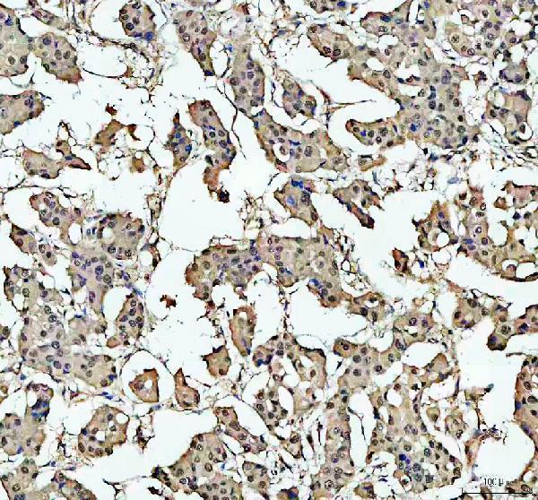
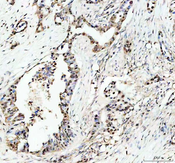
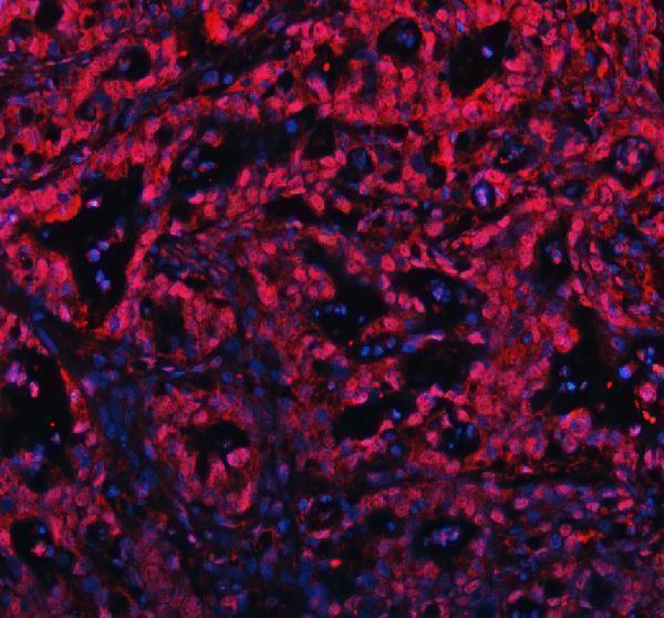
PB9288 has IHC data from a human breast cancer paraffin section and IF data from a human pancreas cancer paraffin section (PB9288 IHC and IF captions).

Real IHC data IHC analysis of c-Myb using anti-c-Myb antibody (PB9288). c-Myb was detected in a paraffin-embedded section of human breast cancer tissue. Heat mediated antigen retrieval was performed in EDTA buffer (pH 8.0, epitope retrieval solution). The tissue section was blocked with 10% goat serum. The tissue section was then incubated with 2 μg/ml rabbit anti-c-Myb Antibody (PB9288) overnight at 4°C. Peroxidase Conjugated Goat Anti-rabbit IgG was used as secondary antibody and incubated for 30 minutes at 37°C. The tissue section was developed using HRP Conjugated Rabbit IgG Super Vision Assay Kit (Catalog # SV0002) with DAB as the chromogen.
Anti-c-Myb Antibody ®
Cat # PB9288

PB9288 is the only SKU that will render (cards: PB9288). Its IHC image shows human breast cancer tissue, and its IF image shows human pancreas cancer tissue, both in paraffin sections (PB9288 IHC and IF captions).

Which to pick: For tissue IHC, choose PB9288: its own image documents chromogenic staining of a human breast cancer paraffin section; the fixative is unreported (PB9288 IHC caption). For IF, PB9288 has a human pancreas cancer paraffin-section image (PB9288 IF caption); for ICC, the rabbit monoclonal P00157 lists ICC/IF but has no captioned ICC example (catalog: P00157). No listed SKU has documented nonhuman reactivity, so there is no supported cross-species choice (catalog: M00157, P00157 and PB9288 reactivity).

Each figure is that product's own IHC / IF validation image from its datasheet.

References

  1. UniProt Consortium. UniProt entry P10242 (MYB_HUMAN, Transcriptional activator Myb).
  2. Human Protein Atlas. MYB tissue IHC expression (reliability: Approved).
  3. Human Protein Atlas. MYB subcellular location (ICC-IF): Mainly localized to the nucleoplasm. In addition localized to the cytosol..
  4. Human Protein Atlas. MYB antibody validation summary (2 antibodies).
  5. Myb Immunohistochemical Staining and Fluorescence in situ Hybridization in Salivary Rare Basaloid Lesions. Frontiers in oncology 2020 — PMC7339910.
  6. Predictors of Outcome in Adenoid Cystic Carcinoma of Salivary Glands: A Clinicopathologic Study With Correlation Between MYB Fusion and Protein Expression. The American journal of surgical pathology 2017 — PMC5597477.
  7. The clinical behavior and genomic features of the so-called adenoid cystic carcinomas of the solid variant with basaloid features. Modern pathology : an official journal of the United States and Canadian Academy of Pathology, Inc 2022 — PMC9197148.
  8. MYB expression and translocation in adenoid cystic carcinomas and other salivary gland tumors with clinicopathologic correlation. The American journal of surgical pathology 2011 — PMC3127258.
  9. PubMed PMID:3540945 — UniProt-cited evidence.
  10. PubMed PMID:2202948 — UniProt-cited evidence.
  11. PubMed PMID:15169551 — UniProt-cited evidence.