This website uses cookies to ensure you get the best experience on our website.


- Table of Contents
Plan chromogenic NANOS1 IHC on paraffin sections with human-reactive antibody A07276, starting at 2.5 μg/mL (datasheet). Assess cytoplasmic and membranous staining against the tissue profile, allowing for its low consistency with RNA expression (HPA tissue IHC).

Expected localisation, validated protocols, controls and antibodies — the at-a-glance facts below, then the full design guide.
| Expected localisation | Cytoplasmic and membranous tissue staining (HPA tissue IHC) | |
| Staining pattern | Cytoplasmic and membranous signal in several tissues (HPA tissue IHC) | |
| Antigen retrieval | Citrate pH 6.0 HIER, 95–98 °C, 20 min (rule: cytoplasmic / membrane antigen) | |
| Positive control | Adrenal gland+4 more · see all | |
| Negative control | None in HPA (detected in all 45 tissues); use no-primary + isotype controls |
| Fixation | Keep fixation consistent across paraffin sections (standard IHC practice; not target-specific) | |
| Caveat | Staining shows low consistency with RNA expression (HPA tissue IHC) | |
| Regulation | Varies by germ-cell stage (UniProt) | |
| Isoform / epitope | No annotated isoforms; one 1–292 chain (UniProt) |
Compare the catalog antibody’s IHC-P protocol with published NANOS1 staining in Nile tilapia testis (PMC13412013) and dogfish gonads (PMC5932020).
| Sample | Tissue sections; selected-image fixative not specified (standard IHC workflow) |
| Fixation | Image fixative and duration unreported (datasheet A07276); verify before use. |
| Sectioning | 4–5 µm sections on charged slides (standard) |
| Deparaffinisation | Xylene, graded ethanol series to water (standard) |
| Antigen retrieval | Heat-induced epitope retrieval in citrate buffer, pH 6.0, 20 min at 95–98 °C (standard rule: cytoplasmic / membrane antigen) |
| Peroxidase block | 3% H2O2, 10 min, room temperature (standard) |
| Blocking | 10% normal serum of the secondary host, 30 min, room temperature (standard) |
| Primary antibody | Rabbit anti-NANOS1, 2.5 μg/mL (datasheet A07276) |
| Primary incubation | Overnight at 4 °C (standard) |
| Detection | HRP-polymer secondary, DAB chromogen 5–10 min (standard) |
| Counterstain | Hematoxylin, blue, dehydrate and mount (standard) |
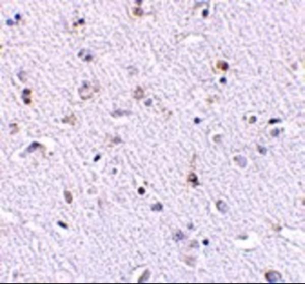
| Expected result | NANOS1-positive staining in glandular cells of adrenal gland (HPA tissue IHC: High). HPA tissue profile: Cytoplasmic and membranous expression in several tissues. No signal in the no-primary control. |
NANOS1 is cytoplasmic, with perinuclear enrichment reported in germ cells, and has no transmembrane segment (UniProt Q8WY41). Look for staining in spermatogonia and spermatocytes (UniProt Q8WY41), while recognizing that HPA reports cytoplasmic and membranous staining across several tissues (HPA tissue IHC). The HPA IHC result is Approved but has low consistency with RNA expression, so tissue distribution alone warrants cautious interpretation (HPA tissue IHC).
| Cytoplasmic staining in spermatogonia or spermatocytes, possibly enriched near the nucleus. | This fits the reported germ-cell distribution and perinuclear localization (UniProt Q8WY41). Assess the signal within identifiable cells and compare it with a matched control; color alone cannot establish antibody specificity (general IHC practice). |
| Strong staining confined to nuclei, with little cytoplasmic signal. | Treat this as a compartment mismatch for tissue IHC because UniProt places NANOS1 in the cytoplasm (UniProt Q8WY41). HPA reports uncertain nucleoplasmic localization in ICC-IF, so nuclear signal is a prompt to check controls and assay context, rather than proof of an artefact (HPA subcellular ICC-IF). |
| Staining appears mainly in cells outside the expected germ-cell population. | Check cell identity before calling it nonspecific: HPA reports high IHC staining in several somatic cell populations, including adrenal glandular cells and bone-marrow hematopoietic cells (HPA tissue IHC). If staining also appears in a no-primary control, investigate endogenous detection activity; otherwise, assess possible cross-reactivity with an independent specificity control (general IHC practice). |
| Diffuse color covers tissue and spaces between cells, obscuring cell boundaries. | This does not give an interpretable cytoplasmic pattern (UniProt Q8WY41; general IHC practice). Compare a no-primary control and review blocking, washing, antibody concentration, and chromogen development before assigning the signal to NANOS1 (general IHC practice). |
| No signal appears in an expected positive cell population. | First verify that the expected cells are present: UniProt places adult expression mainly in spermatogonia and also in spermatocytes, but not late postmeiotic germ cells (UniProt Q8WY41). A blank result in the intended positive cells calls for a detection and retrieval check; it does not establish absence of NANOS1 by itself (general IHC practice). |
| Cell population and tissue | UniProt emphasizes germline development and adult spermatogonia, with expression also in spermatocytes (UniProt Q8WY41). HPA records high IHC staining in multiple somatic populations and low staining in soft-tissue fibroblasts (HPA tissue IHC). Use the cells visible on the section when interpreting a result; neither profile alone proves specificity. |
| Compartment and membrane-associated staining | UniProt places NANOS1 in the cytoplasm and reports no transmembrane segment (UniProt Q8WY41). HPA describes cytoplasmic and membranous tissue staining (HPA tissue IHC). Membranous color may match that observation, but topology alone cannot confirm that a membrane outline represents specific NANOS1 staining. |
| Strength of tissue IHC evidence | The listed antibody CAB012219 has Approved IHC status, while the HPA tissue profile notes low consistency between antibody staining and RNA expression (HPA antibodies; HPA tissue IHC). Interpret unexpected cell populations with matched controls and morphology; Approved status does not resolve that reported inconsistency. |
| Processing and isoform annotation | UniProt lists a single 1–292 chain, no signal peptide or propeptide, and no annotated isoforms or glycosylation sites (UniProt Q8WY41). These annotations give no basis here for predicting a cleaved, shed, or glycosylated staining pattern; the antibody epitope is not supplied. |
| IF/ICC: should its localization set the paraffin IHC call? | HPA ICC-IF supports cytosol localization and labels nucleoplasm uncertain; images are listed for A-431, U-251MG, and U2OS (HPA subcellular ICC-IF). Use that as a compartment cross-check, while scoring this paraffin IHC section against its tissue morphology and controls (general IHC practice). |
| Antigen retrieval | Retrieval is a general variable in paraffin IHC (general IHC practice). No NANOS1-specific retrieval condition or fixation sensitivity is supplied, so optimize against a suitable positive section and its controls without attributing a weak result to a documented NANOS1 fixation effect. |
| Situation | Likely cause | Next action |
|---|---|---|
| The intended positive germ cells are blank. | The sampled area may lack spermatogonia or spermatocytes (UniProt Q8WY41), or the IHC detection workflow may have failed (general IHC practice). | Confirm cell identity on the counterstained section, then review the positive control, retrieval, primary-antibody step, and detection reagents (general IHC practice). Avoid treating late postmeiotic cells as the expected positive population (UniProt Q8WY41). |
| Color persists when the primary antibody is omitted. | Endogenous enzyme activity, endogenous biotin where a biotin-based system is used, or detection-reagent background can produce color (general IHC practice). | Identify the detection chemistry and apply its appropriate endogenous-activity block; compare the adjusted no-primary control with the test section (general IHC practice). Do not score persistent control color as NANOS1. |
| Many unrelated cells stain intensely. | Some somatic staining is reported by HPA, while its tissue IHC profile also has low consistency with RNA data (HPA tissue IHC). Excess primary antibody or cross-reactivity remains possible (general IHC practice). | Map staining to cell types and tissue compartments, compare controls, and titrate the IHC-validated antibody under the same detection conditions (general IHC practice). Seek independent specificity evidence before making a new cell-type claim. |
| The slide has widespread haze or granular deposits. | Inadequate blocking or washing, concentrated antibody, or overdevelopment can obscure a cell-bound signal (general IHC practice). | Check the no-primary control, improve blocking and washes, and adjust primary-antibody concentration or chromogen development one variable at a time (general IHC practice). Reassess whether clear cytoplasmic cell boundaries emerge. |
| Staining is exclusively nuclear. | That conflicts with UniProt's cytoplasmic assignment, although HPA calls nucleoplasmic ICC-IF localization uncertain (UniProt Q8WY41; HPA subcellular ICC-IF). | Recheck cell boundaries and counterstain, compare negative and positive controls, and verify the pattern with independent specificity evidence (general IHC practice). Do not promote uncertain ICC-IF nuclear localization to an established tissue IHC pattern. |
| The section shows a sharp membrane outline without convincing cytoplasmic color. | HPA reports membranous tissue staining, but NANOS1 has no transmembrane segment (HPA tissue IHC; UniProt Q8WY41). The outline alone cannot settle specificity. | Review adjacent cells and matched controls for a reproducible cell-associated pattern, then corroborate an isolated membrane result before scoring it as NANOS1 (general IHC practice). |
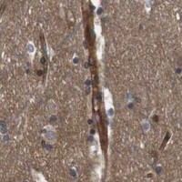

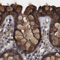


Comprehensive Human Protein Atlas IHC scoring per tissue (reliability: Approved — Low consistency between antibody staining and RNA expression data.). Rows are taken directly from the HPA tissue chart — click any row's HPA link to view the source.
| Tissue | Cell type | Level | Evidence | Source |
|---|---|---|---|---|
| Adrenal gland | Glandular cells | High | Protein (IHC) | HPA → |
| Appendix | Glandular cells | High | Protein (IHC) | HPA → |
| Bone marrow | Hematopoietic cells | High | Protein (IHC) | HPA → |
| Bronchus | Respiratory epithelial cells | High | Protein (IHC) | HPA → |
| Cervix | Glandular cells | High | Protein (IHC) | HPA → |
| Tissue | Cell type | Level | Evidence | Source |
|---|---|---|---|---|
| None in HPA: NANOS1 is detected in all 45 scored tissues. Use a no-primary (secondary-only) and an isotype control instead. | ||||
Troubleshoot NANOS1 staining in paraffin sections by checking retrieval, compartment, cell identity, and controls before interpreting chromogenic signal.
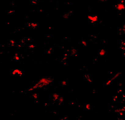
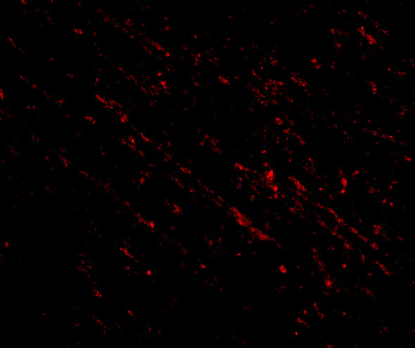
The catalog shows human brain IHC and IF images for anti-NANOS1 antibodies (A07276 IHC image caption; A07276-1 IF image caption). Listed reactivity includes human, mouse, and rat (catalog reactivity).
A07276 renders with a human brain IHC image at 2.5 μg/mL and lists human IHC-P and IF (A07276 IHC image caption; A07276 applications/reactivity). A07276-1 renders with a human brain IF image at 20 μg/mL and lists IF plus human, mouse, and rat reactivity (A07276-1 IF image caption; A07276-1 applications/reactivity).
Which to pick: Choose A07276 for human paraffin-section chromogenic IHC: it lists IHC-P, and its own IHC image shows human brain tissue; the fixative is unreported (A07276 applications; A07276 IHC image caption). For IF, A07276-1 has a human brain IF image, but ICC is not listed for either SKU (A07276-1 IF image caption; catalog applications). For work involving mouse or rat, A07276-1 lists those species as reactive, though its pictured IF sample is human; both antibodies have rabbit hosts and no reported clone, so clonality cannot guide the choice (A07276-1 reactivity; A07276-1 IF image caption; catalog host/clone).