This website uses cookies to ensure you get the best experience on our website.


- Table of Contents
Plan chromogenic NANOS3 IHC on paraffin sections using testis spermatogonia as a positive reference (HPA tissue IHC). Start the catalog antibody at 2.5 μg/mL (datasheet) and assess nuclear and cytoplasmic staining as molecular expectations, since the tissue-IHC compartment is unconfirmed (UniProt; HPA tissue IHC).

Expected localisation, validated protocols, controls and antibodies — the at-a-glance facts below, then the full design guide.
| Expected localisation | Nucleus and cytoplasm expected; IHC compartment unconfirmed (UniProt) | |
| Staining pattern | Medium testis spermatogonia; low ovarian follicle cells; site unknown (HPA tissue IHC) | |
| Antigen retrieval | Tris-EDTA pH 9.0 HIER, 95–98 °C, 20 min (rule: nuclear antigen) | |
| Positive control | Testis | |
| Negative control | Adipose tissue+4 more · see all |
| Fixation | Keep paraffin-section fixation consistent across samples (standard IHC practice; not target-specific); Matched tissue-IHC evidence does not establish the fixation claim. Validate the specimen-specific method before use. (selected-SKU IHC image A07449) | |
| Caveat | Oocyte staining cannot be correlated with the RNA-seq data (HPA tissue IHC) | |
| Regulation | Highest in oocytes across oogenesis (UniProt) | |
| Isoform / epitope | Two isoforms; no cleavage annotated; epitope coverage unknown (UniProt) |
The catalog antibody’s IHC-P protocol is accompanied by published NANOS3 staining protocols for human tissue, bovine ovary, and macaque testis (PMC3090199; PMC4846992; PMC13079449).
| Sample | Tissue sections; selected-image fixative not specified (standard IHC workflow) |
| Fixation | Image fixative and duration unreported (datasheet A07449); verify before use. |
| Sectioning | 4–5 µm sections on charged slides (standard) |
| Deparaffinisation | Xylene, graded ethanol series to water (standard) |
| Antigen retrieval | Heat-induced epitope retrieval in Tris-EDTA buffer, pH 9.0, 20 min at 95–98 °C (standard rule: nuclear antigen) |
| Peroxidase block | 3% H2O2, 10 min, room temperature (standard) |
| Blocking | 10% normal serum of the secondary host, 30 min, room temperature (standard) |
| Primary antibody | Rabbit anti-NANOS3, 2.5 μg/mL (datasheet A07449) |
| Primary incubation | Overnight at 4 °C (standard) |
| Detection | HRP-polymer secondary, DAB chromogen 5–10 min (standard) |
| Counterstain | Hematoxylin, blue, dehydrate and mount (standard) |
| Expected result | NANOS3-positive staining in spermatogonia cells of testis (HPA tissue IHC: Medium). HPA tissue profile: Expression in spermatogonia in testis and ovarian follicles. No signal in the no-primary control. |
NANOS3 is a non-membrane protein reported in the nucleus and cytoplasm (UniProt P60323: localization, topology). In tissue IHC, expect expression in testicular spermatogonia and ovarian follicles (HPA tissue IHC: Enhanced). HPA reports medium staining in spermatogonia and low staining in ovarian follicle cells; UniProt also reports expression in oocytes and several germ-cell stages (HPA tissue IHC; UniProt P60323: tissue specificity).
| Spermatogonia stain at medium intensity in testis; ovarian follicle cells show weaker staining. | This matches the cell-specific HPA tissue IHC observations (HPA: medium in spermatogonia; low in ovarian follicle cells). Assess cell identity alongside intensity: UniProt reports other germ-cell stages, but the supplied HPA positive-cell entry specifically identifies spermatogonia (UniProt P60323: tissue specificity; HPA tissue IHC). |
| Signal is confined to cell borders or forms a membrane-like rim. | Treat a membrane-only pattern as suspect: NANOS3 has no transmembrane segment and is assigned to the nucleus and cytoplasm (UniProt P60323: topology, localization). Recheck morphology and the detection controls before scoring it as NANOS3; HPA ICC-IF supports nucleoplasm and cytosol, though that does not prescribe an identical IHC appearance (HPA subcellular). |
| Strong staining appears in cells listed as negative, such as adipocytes in adipose tissue. | Investigate specificity or detection background before calling those cells positive (HPA tissue IHC: adipocytes not detected; general IHC practice). A negative HPA observation is a comparison point, not proof that every specimen must be blank. Check whether staining follows tissue structures or also appears in a no-primary control (general IHC practice). |
| Brown signal spreads across most cells, extracellular areas, or the entire section. | Diffuse signal obscures the selective spermatogonial pattern reported in testis (HPA tissue IHC: medium in spermatogonia). Review blocking, washes, antibody concentration, and detection controls as general IHC background checks (general IHC practice). Distribution alone cannot identify which step caused the background. |
| No staining is visible in testicular spermatogonia expected to be positive. | First verify that spermatogonia are present in the section; HPA reports medium staining in those cells (HPA tissue IHC). If they are present, review the catalog antibody’s IHC-P instructions and run the recommended controls (general IHC practice). An unstained section alone cannot distinguish technical failure from a specimen-specific result. |
| Cell type and tissue | HPA identifies medium staining in testicular spermatogonia and low staining in ovarian follicle cells (HPA tissue IHC). UniProt reports strongest ovarian expression in oocytes, but HPA says oocyte protein expression cannot be correlated with its RNA samples because that cell type is absent from them (UniProt P60323: tissue specificity; HPA: reliability description). |
| Subcellular comparison | UniProt assigns NANOS3 to nucleus, cytoplasm, stress granules, and P-bodies (UniProt P60323: localization). HPA ICC-IF supports a mainly nucleoplasmic location with additional cytosol (HPA subcellular). Use those assignments to assess plausibility, while recognizing that an ICC-IF image does not establish the visibility of small cytoplasmic structures in chromogenic IHC. |
| Antibody validation | HPA lists HPA062989 as IHC Enhanced and ICC Supported, and CAB080164 as IHC Supported with no ICC status supplied (HPA antibodies). The tissue profile has Enhanced reliability (HPA tissue IHC). These labels support comparison with the reported pattern; they do not establish the performance of an unspecified catalog antibody. |
| Isoforms and epitope coverage | UniProt lists two NANOS3 isoforms, while the supplied record gives no antibody epitope or isoform-specific validation (UniProt P60323: isoforms; HPA antibodies). Do not assign a staining difference to one isoform without antibody-specific evidence. NANOS3 is annotated as a 1–173 chain with no signal peptide or propeptide (UniProt P60323: processing). |
| Detection background | For chromogenic IHC, endogenous detection activity and incomplete blocking can produce signal independent of primary-antibody binding (general IHC practice). Use a no-primary control to assess that possibility. This is a general workflow factor, not an HPA or UniProt finding about NANOS3. |
| Situation | Likely cause | Next action |
|---|---|---|
| Testis shows no signal where spermatogonia are visible. | The expected comparison is medium spermatogonial staining (HPA tissue IHC); the image alone does not identify the failed step. | Confirm cell identity, check the catalog antibody’s IHC-P instructions, and compare with processing and detection controls (general IHC practice). Do not infer NANOS3 absence from one negative section. |
| Ovary appears weak beside testis. | HPA reports low staining in ovarian follicle cells versus medium staining in testicular spermatogonia (HPA tissue IHC). | Score the relevant cell populations separately (general IHC practice). Do not use the HPA RNA comparison to discount oocyte protein expression: oocytes were absent from its RNA samples (HPA: reliability description). |
| A membrane rim is the dominant pattern. | That distribution conflicts with the reported nuclear and cytoplasmic localization and lack of a transmembrane segment (UniProt P60323: localization, topology). | Inspect compartment boundaries at higher magnification and compare detection controls (general IHC practice). Treat the rim as unconfirmed until a cell-associated pattern is resolved. |
| Many unrelated cells or tissue spaces are brown. | Broad staining differs from the selective HPA tissue pattern (HPA tissue IHC); background has several possible technical causes (general IHC practice). | Compare a no-primary control, then review blocking, washes, and antibody concentration under the catalog IHC-P instructions (general IHC practice). Change one condition at a time. |
| Adipocytes in adipose tissue show strong staining. | HPA lists those adipocytes as not detected, so the result conflicts with that reference observation (HPA tissue IHC). | Verify cell identity and assess a no-primary control before interpreting the signal as NANOS3 (general IHC practice). Record any persistent difference as specimen and antibody dependent. |
| IF/ICC: where should cellular signal appear? | HPA reports mainly nucleoplasmic signal with additional cytosol in ICC-IF; UniProt also lists nuclear and cytoplasmic localization (HPA subcellular; UniProt P60323: localization). | Use those compartments as the IF/ICC interpretation reference, and consult the separate IF/ICC guide for that application (HPA subcellular; general IHC/IF practice). |


Comprehensive Human Protein Atlas IHC scoring per tissue (reliability: Enhanced — Protein expression in oocytes cannot be correlated with RNA expression data due to cell type not present in RNA-seq samples.). Rows are taken directly from the HPA tissue chart — click any row's HPA link to view the source.
| Tissue | Cell type | Level | Evidence | Source |
|---|---|---|---|---|
| Testis | Spermatogonia cells | Medium | Protein (IHC) | HPA → |
| Tissue | Cell type | Level | Evidence | Source |
|---|---|---|---|---|
| Adipose tissue | Adipocytes | Not detected | Protein (IHC) | HPA → |
| Adrenal gland | Glandular cells | Not detected | Protein (IHC) | HPA → |
| Appendix | Glandular cells | Not detected | Protein (IHC) | HPA → |
| Bone marrow | Hematopoietic cells | Not detected | Protein (IHC) | HPA → |
| Breast | Adipocytes | Not detected | Protein (IHC) | HPA → |
Troubleshoot NANOS3 staining in paraffin sections using the catalog antibody’s brain tissue image, the page retrieval setting, and independent expression and localisation evidence.
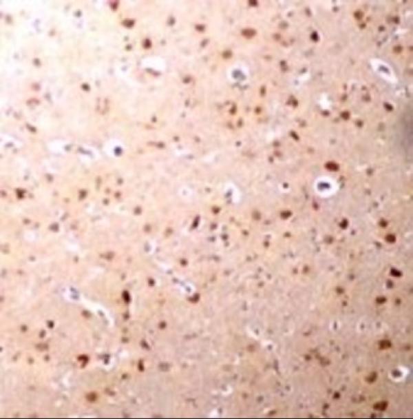
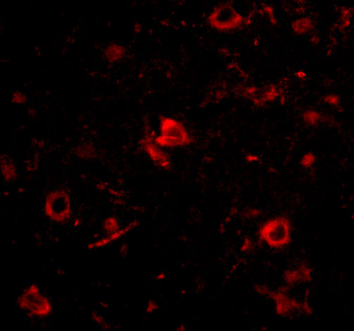
A07449 supports IHC-P and IF (catalog: applications), with images of human brain tissue for each (catalog: IHC and IF captions); listed reactivity includes human, mouse, and rat (catalog: reactivity).
A07449 will render with a human brain tissue IHC image at 2.5 μg/mL (A07449 IHC caption). A07449 also has a human brain tissue IF image at 20 μg/mL (A07449 IF caption).
Which to pick: For paraffin-section tissue IHC, choose A07449 because IHC-P is listed (catalog: applications); its IHC caption shows human brain tissue at 2.5 μg/mL but does not report the fixative (A07449 IHC caption). For IF, choose A07449 based on its human brain tissue IF image at 20 μg/mL (A07449 IF caption); ICC validation is unreported (catalog: applications). For mouse or rat studies, A07449 lists reactivity with both species, although the supplied IHC and IF images show human tissue (catalog: reactivity; A07449 IHC and IF captions); its host is rabbit and clonality is unreported (catalog: host and clone).