This website uses cookies to ensure you get the best experience on our website.


- Table of Contents
Plan chromogenic IHC-P for NAP1L5 using its mainly nuclear tissue pattern, most abundant in CNS (HPA tissue IHC). Start the catalog antibody at 5 μg/mL (datasheet: A14492) and interpret staining in light of the reported antibody–RNA discordance (HPA tissue IHC).

Expected localisation, validated protocols, controls and antibodies — the at-a-glance facts below, then the full design guide.
| Expected localisation | Mainly nuclear in tissues (HPA tissue IHC) | |
| Staining pattern | Mainly nuclear across tissues; most abundant in CNS (HPA tissue IHC) | |
| Antigen retrieval | Tris-EDTA pH 9.0 HIER, 95–98 °C, 20 min (rule: nuclear antigen) | |
| Positive control | Appendix+4 more · see all | |
| Negative control | Adipose tissue+4 more · see all |
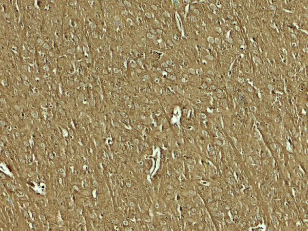
| Fixation | Keep fixation consistent across sections (standard IHC practice; not target-specific); Matched tissue-IHC evidence does not establish the fixation claim. Validate the specimen-specific method before use. (selected-SKU IHC image A14492) | |
| Caveat | Low antibody–RNA concordance may complicate interpretation (HPA tissue IHC) | |
| Regulation | Brain-predominant expression (UniProt) | |
| Isoform / epitope | No isoforms or processing annotated; epitope map unresolved (UniProt) |
Compare the catalog antibody’s IHC-P protocol with the published NAP1L5 staining protocol for carcinoma and tumor transplantation tissues (PMC9740361).
| Sample | Tissue sections; selected-image fixative not specified (standard IHC workflow) |
| Fixation | Image fixative and duration unreported (datasheet A14492); verify before use. |
| Sectioning | 4–5 µm sections on charged slides (standard) |
| Deparaffinisation | Xylene, graded ethanol series to water (standard) |
| Antigen retrieval | Heat-induced epitope retrieval in Tris-EDTA buffer, pH 9.0, 20 min at 95–98 °C (standard rule: nuclear antigen) |
| Peroxidase block | 3% H2O2, 10 min, room temperature (standard) |
| Blocking | 10% normal serum of the secondary host, 30 min, room temperature (standard) |
| Primary antibody | Rabbit anti-NAP1L5, 5 μg/mL (datasheet A14492) |
| Primary incubation | Overnight at 4 °C (standard) |
| Detection | HRP-polymer secondary, DAB chromogen 5–10 min (standard) |
| Counterstain | Hematoxylin, blue, dehydrate and mount (standard) |
| Expected result | NAP1L5-positive staining in glandular cells of appendix (HPA tissue IHC: Medium). HPA tissue profile: Mainly nuclear expression in several tissues, most abundant CNS. No signal in the no-primary control. |
NAP1L5 is a nuclear protein without a transmembrane segment (UniProt Q96NT1: subcellular location and topology). In paraffin-section IHC, expect mainly nuclear staining, most abundant in CNS tissues; HPA reports medium staining in cortical and caudate neuronal cells and cerebellar molecular-layer cells (HPA: tissue IHC). HPA rates its tissue IHC profile Approved but reports low consistency between antibody staining and RNA expression, so interpret isolated positive cells cautiously (HPA: tissue IHC reliability).
| Distinct nuclear staining in cortical or caudate neuronal cells. | This matches HPA's medium staining in those cells and its mainly nuclear tissue profile (HPA: tissue IHC). Compare the signal with adjacent morphology and controls before scoring it as target-associated staining (general IHC practice). |
| Strong membrane staining, or cytoplasmic staining with no discernible nuclear component. | A dominant membrane pattern conflicts with the nuclear location and lack of transmembrane segment (UniProt Q96NT1: location and topology). Cytoplasmic signal alone is insufficient to establish the expected tissue IHC pattern; examine background and detection controls (HPA: tissue IHC; general IHC practice). |
| Prominent staining of adipocytes or liver cholangiocytes. | HPA reports these cells as not detected in tissue IHC (HPA: adipose tissue and liver IHC). Treat unexpected staining as a specificity or endogenous-detection question; inspect the negative control and tissue morphology before interpreting it as NAP1L5 (general IHC practice). |
| Diffuse color across nuclei, cytoplasm, and surrounding tissue. | A widespread haze does not resolve the mainly nuclear pattern reported by HPA (HPA: tissue IHC). Uneven blocking, excessive detection signal, or incomplete washing can cause nonspecific background; compare with a no-primary control (general IHC practice). |
| No staining in cortical or caudate neuronal cells. | HPA reports medium staining in these cells, so an absent result warrants a run check (HPA: tissue IHC). Confirm tissue preservation and morphology, then review the catalog antibody's IHC-P conditions and the run controls (general IHC practice). |
| Cell type and tissue | Brain is the predominant expression site (UniProt Q96NT1: tissue specificity). HPA finds medium neuronal staining in cortex and caudate, but low staining in hippocampal neuronal cells; choose and score reference areas by cell type (HPA: tissue IHC). |
| Compartment | UniProt places NAP1L5 in the nucleus and reports no transmembrane segment (UniProt Q96NT1: location and topology). HPA describes tissue staining as mainly nuclear; membrane-dominant staining needs scrutiny (HPA: tissue IHC). |
| Evidence strength | HPA labels the tissue IHC profile Approved while noting low consistency with RNA expression (HPA: tissue IHC reliability). Its listed antibody HPA058227 is Approved for IHC, which does not resolve every unexpected cell-level result (HPA: antibody validation; HPA: tissue IHC reliability). |
| Protein processing | UniProt lists one 1–182 chain, no signal peptide or propeptide, and no annotated glycosylation sites or modified residues (UniProt Q96NT1: processing and modifications). These annotations give no basis to expect a secreted or membrane-associated staining pattern (UniProt Q96NT1: processing and topology). |
| IHC-P workflow variables | Antigen retrieval, blocking, primary-antibody concentration, washing, and chromogenic detection can affect contrast in routine paraffin-section IHC (general IHC practice). Target-specific fixation sensitivity and an optimal retrieval condition are unreported in the supplied UniProt and HPA records. |
| Situation | Likely cause | Next action |
|---|---|---|
| Nuclear signal is weak in an HPA-positive reference area. | Cortical and caudate neuronal cells have medium HPA staining, so weak signal may reflect the selected cells or the run conditions (HPA: tissue IHC; general IHC practice). | Check neuronal morphology and run controls, then review the catalog antibody's IHC-P retrieval and detection instructions before adjusting routine conditions (general IHC practice). |
| Most cells show brown haze. | Widespread color obscures HPA's mainly nuclear tissue pattern and may reflect nonspecific detection or inadequate washing (HPA: tissue IHC; general IHC practice). | Compare a no-primary control, inspect blocking and washes, and reduce detection intensity if the control also develops color (general IHC practice). |
| Signal appears mainly at cell membranes. | Membrane dominance conflicts with NAP1L5's nuclear location and absence of a transmembrane segment (UniProt Q96NT1: location and topology). | Check morphology and control sections; do not score membrane-only color as the expected NAP1L5 pattern (HPA: tissue IHC; general IHC practice). |
| A nominally negative cell population stains strongly. | HPA reports adipocytes and liver cholangiocytes as not detected; cross-reactivity or endogenous detection activity may explain unexpected color (HPA: tissue IHC; general IHC practice). | Check the no-primary control and localize color to specific cell types before assigning target expression (general IHC practice). |
| Cortex is positive but hippocampal neuronal cells are faint. | This difference can match HPA's medium cortical and low hippocampal neuronal staining (HPA: tissue IHC). | Score the regions separately and compare like cell types; faint hippocampal signal alone does not establish a failed run (HPA: tissue IHC; general IHC practice). |
| What should a separate IF/ICC experiment show? | HPA places NAP1L5 mainly in the nucleoplasm, with an additional cytosolic location in ICC-IF (HPA: subcellular ICC-IF). | Use the separate IF/ICC guide for that application; compare nuclear and cytosolic fluorescence with appropriate controls rather than applying the tissue IHC pattern as an exclusive ICC-IF rule (HPA: subcellular ICC-IF; general IF practice). |


![HPA ICC-IF BJ [Human fibroblast] — NAP1L5 (HPA058227)](https://images.proteinatlas.org/58227/1599_B11_3_blue_red_green.jpg)
![HPA ICC-IF BJ [Human fibroblast] — NAP1L5 (HPA058227)](https://images.proteinatlas.org/58227/1599_B11_5_blue_red_green.jpg)
Comprehensive Human Protein Atlas IHC scoring per tissue (reliability: Approved — Low consistency between antibody staining and RNA expression data.). Rows are taken directly from the HPA tissue chart — click any row's HPA link to view the source.
| Tissue | Cell type | Level | Evidence | Source |
|---|---|---|---|---|
| Appendix | Glandular cells | Medium | Protein (IHC) | HPA → |
| Bronchus | Basal cells | Medium | Protein (IHC) | HPA → |
| Caudate | Neuronal cells | Medium | Protein (IHC) | HPA → |
| Cerebellum | Cells in molecular layer | Medium | Protein (IHC) | HPA → |
| Cerebral cortex | Neuronal cells | Medium | Protein (IHC) | HPA → |
| Tissue | Cell type | Level | Evidence | Source |
|---|---|---|---|---|
| Adipose tissue | Adipocytes | Not detected | Protein (IHC) | HPA → |
| Bone marrow | Hematopoietic cells | Not detected | Protein (IHC) | HPA → |
| Breast | Adipocytes | Not detected | Protein (IHC) | HPA → |
| Liver | Cholangiocytes | Not detected | Protein (IHC) | HPA → |
| Lung | Alveolar cells | Not detected | Protein (IHC) | HPA → |
Troubleshoot NAP1L5 staining in paraffin section IHC using nuclear localisation and brain enriched expression as reference points (UniProt Q96NT1; HPA tissue IHC).
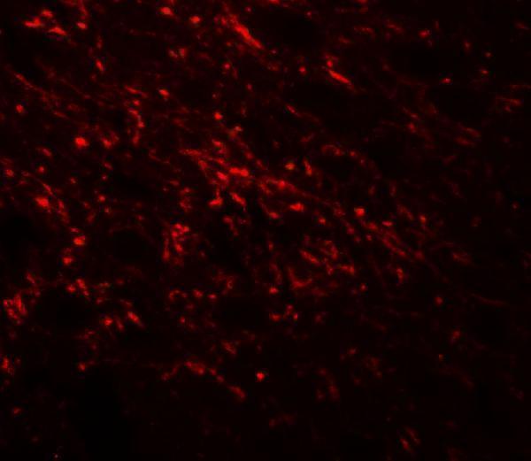
Two anti-NAP1L5 antibodies have mouse brain tissue IHC/IF images and a human HeLa cell IF/ICC image (catalog image captions). Both list human, mouse, and rat reactivity (catalog reactivity).
A14492 lists IHC-P and IF, with mouse brain tissue images at 5 μg/mL for IHC and 20 μg/mL for IF (catalog applications and A14492 image captions). A14492-1 lists IF/ICC and has a HeLa cell IF image at 5 μg/mL (catalog applications and A14492-1 IF caption).
Which to pick: For paraffin-section tissue IHC, choose A14492: IHC-P is listed, and its own IHC caption shows mouse brain tissue at 5 μg/mL; the fixative is unreported (catalog applications and A14492 IHC caption). For cell IF/ICC, choose A14492-1, whose IF caption shows HeLa cells at 5 μg/mL (catalog applications and A14492-1 IF caption). Both list human, mouse, and rat reactivity; the pictured IHC evidence is mouse tissue for A14492, while the pictured cell IF evidence is human HeLa cells for A14492-1 (catalog reactivity and image captions).