NCL / Nucleolin · IHC design guide

Design Immunohistochemistry for NCL

Plan NCL paraffin section IHC around nuclear staining across tissues (HPA tissue IHC). The catalog antibody was used at 2 μg/mL overnight at 4°C, followed by biotinylated secondary antibody and DAB detection (datasheet A00228-1).

Evidence assembled Oct 2026 · For research use; verify linked source records and product datasheet before use
Immunohistochemistry protocol sheet for NCL (IHC for NCL): expected localisation Nuclear staining in tissue (HPA tissue IHC); cytoplasm annotated (UniProt), antibody A00228-1, validated IHC image, and IHC protocol steps
Printable NCL IHC protocol sheet — expected localisation Nuclear staining in tissue (HPA tissue IHC); cytoplasm annotated (UniProt), antibody A00228-1, controls and protocol steps. Open the full NCL IHC guide →

NCL Immunohistochemistry Experimental Design Guide

Expected localisation, validated protocols, controls and antibodies — the at-a-glance facts below, then the full design guide.

Must know before staining
Expected localisation Nuclear staining in tissue (HPA tissue IHC); cytoplasm annotated (UniProt)
Staining pattern Nuclear staining across all tissues (HPA tissue IHC)
Antigen retrieval EDTA pH 8.0 HIER, heat-mediated (datasheet A00228-1)
Positive control ⓘ Adipose tissue+4 more · see all
Negative control ⓘ None in HPA (detected in all 45 tissues); use no-primary + isotype controls
Important caveats
Reasons your staining may differ from the expected pattern.
Fixation Matched tissue-IHC evidence does not establish the fixation claim. Validate the specimen-specific method before use. (selected-SKU IHC image A00228-1)
Caveat Antibody staining has medium consistency with RNA (HPA tissue IHC)
Regulation Major nucleolar protein in growing cells (UniProt)
Isoform / epitope No isoforms annotated; mature chain spans residues 2–710 (UniProt)
Section 1

Recommended NCL IHC & IF Protocols

The catalog antibody protocol and two published IHC protocols provide starting conditions for paraffin sections (datasheet A00228-1; PMC12898300; PMC7920503).

Recommended immunohistochemistry (IHC-P) protocol parameters
SampleParaffin-embedded human metaplasia of squamous cells of the renal pelvis tissue; fixative not specified (datasheet A00228-1)
FixationImage fixative and duration unreported (datasheet A00228-1); verify before use.
Sectioning4–5 µm sections on charged slides (standard)
DeparaffinisationXylene, graded ethanol series to water (standard)
Antigen retrievalHeat retrieval: EDTA pH 8.0 (datasheet A00228-1); 20 min, 95–100 °C (standard)
Peroxidase block3% H2O2, 10 min, room temperature (standard)
Blocking10% goat serum (datasheet A00228-1)
Primary antibodyRabbit anti-NCL, 1-2μg/ml (datasheet A00228-1)
Primary incubationOvernight at 4 °C (datasheet A00228-1)
DetectionStreptavidin-biotin complex (SABC), DAB chromogen (datasheet A00228-1)
CounterstainHematoxylin, blue, dehydrate and mount (standard)
Expected resultNCL-positive staining in adipocytes of adipose tissue (HPA tissue IHC: High). HPA tissue profile: Nuclear expression in all tissues. No signal in the no-primary control.
💡Decision noteStart with heat-mediated EDTA, pH 8.0 (datasheet A00228-1); citrate, pH 6.0, is a published NCL alternative (PMC12898300).
Section 2

What Is the Expected NCL Staining Pattern?

NCL is chiefly nuclear and nucleolar, with a documented cytoplasmic mRNP-granule pool (UniProt P19338). In paraffin-section IHC, expect nuclear staining across tissues, including high staining in the cell populations listed by HPA (HPA: nuclear expression in all tissues; listed cells High). HPA rates its tissue pattern Supported, with medium consistency against RNA data (HPA: reliability). NCL has no transmembrane segment (UniProt P19338 topology).

What am I looking at on my slide?
Distinct nuclear staining in tissue cells, sometimes concentrated within nucleoli.This fits HPA's broad nuclear IHC pattern and UniProt's nucleolar location (HPA: nuclear expression in all tissues; UniProt P19338). Assess staining in recognizable cells, using the counterstain to distinguish nuclei from pigment or deposits (general IHC practice).
Cytoplasmic staining dominates while nuclei remain weak or blank.Question the result because the expected tissue pattern is nuclear (HPA: tissue IHC). A discrete cytoplasmic pool is biologically plausible in mRNP granules (UniProt P19338); diffuse cytoplasm alone does not establish that pool. Compare with a nuclear positive control and detection controls (general IHC practice).
Strong staining appears in an unexpected cell population.HPA reports nuclear expression across tissues and lists specific High populations, including bone-marrow hematopoietic cells and bronchial respiratory epithelial cells (HPA: tissue IHC). Check cellular identity and nuclear localization before calling the result nonspecific; cross-reactivity or endogenous chromogen activity are possibilities, not diagnoses (general IHC practice).
Colour spreads through stroma, empty spaces, or many cells without clear nuclear boundaries.This lacks the cell-associated nuclear pattern reported for NCL (HPA: tissue IHC). Diffuse background can reflect nonspecific antibody binding or detection chemistry (general IHC practice). A matched negative detection control helps identify background without assuming which reagent caused it.
No signal is visible in a known-positive section.Bone-marrow hematopoietic cells and adipocytes in adipose tissue are reported High by HPA (HPA: tissue IHC), so absent staining there warrants a run-level check. Confirm tissue morphology, control performance, antibody suitability for IHC-P, and detection reagents before interpreting the sample as NCL-negative (general IHC practice).
💡Expected NCL appearanceCall a result positive when identifiable cells show predominantly nuclear, often nucleolar, staining, with strong signal plausible in HPA-listed High populations; diffuse extracellular colour or cytoplasm-only staining is suspect and needs controls (HPA: tissue IHC; UniProt P19338; general IHC practice).
How each factor affects the staining
Tissue and cell choiceHPA reports nuclear expression in all tissues and High staining in its listed cell populations, with low tissue RNA specificity (HPA: tissue IHC). These observations support positive-control selection, but HPA supplies no negative tissue or universal intensity threshold.
Compartment and topologyUniProt places NCL in nucleoli and a cytoplasmic mRNP-granule pool, and reports no transmembrane segment (UniProt P19338). HPA tissue IHC emphasizes nuclear signal (HPA: tissue IHC); do not score a cell outline alone as the expected positive pattern.
Antibody evidenceHPA023981 and CAB004210 are Supported for IHC; HPA071110 has ICC support but no IHC rating in the supplied record (HPA: antibody validation). The tissue profile itself is Supported, with medium RNA–staining consistency (HPA: reliability), so evaluate the observed pattern alongside controls.
IF/ICC Q: Where should NCL appear?A: HPA reports enhanced nucleoplasm, nucleoli, and nucleoli-rim localization, plus supported mitotic-chromosome localization (HPA: subcellular ICC-IF). This answers the localization question; the paraffin-section IHC pattern and validation remain the basis for this guide's slide interpretation (HPA: tissue IHC).
Why is my staining missing, weak or wrong?
SituationLikely causeNext action
Known-positive tissue has no nuclear signal.The staining run or antibody application may have failed; the observation conflicts with reported High cells (HPA: tissue IHC).Inspect the positive control and morphology, then check the antibody's documented IHC-P use and detection steps; adjust retrieval only within the validated workflow (general IHC practice).
Signal is present only in cytoplasm.A cytoplasmic pool is possible, but it does not explain loss of the expected nuclear pattern by itself (UniProt P19338; HPA: tissue IHC).Recheck the counterstain and focal plane, compare a nuclear positive control, and examine the negative detection control (general IHC practice).
Broad brown haze obscures nuclei.Background from nonspecific binding or detection chemistry is possible (general IHC practice).Compare a no-primary control, inspect blocking and washing, and assess whether signal resolves into nuclei before scoring NCL (general IHC practice; HPA: tissue IHC).
Unexpected cells show strong chromogen.Cell identification may be uncertain, or cross-reactivity or endogenous detection activity may contribute (general IHC practice).Check morphology and nuclear placement against HPA's listed High populations; use an appropriate negative detection control to investigate background (HPA: tissue IHC; general IHC practice).
A cell-surface-like rim is the only visible signal.That pattern does not match HPA's tissue nuclear profile; UniProt reports no transmembrane segment (HPA: tissue IHC; UniProt P19338 topology).Do not score the rim alone as a positive nucleus. Check focus, counterstain, and control sections, then reassess localization (general IHC practice).
Staining varies between sections or runs.Run conditions may differ; HPA's Supported tissue profile does not establish a target-specific fixation or retrieval effect (HPA: reliability).Compare positive controls processed with each run and review documented staining conditions before attributing the difference to NCL biology (general IHC practice).

Sample controls for NCL IHC & IF

🧪Run bone marrow first and expect nucleolar staining in hematopoietic cells (HPA: High in hematopoietic cells; UniProt P19338: nucleolar localization). HPA detects NCL in all 45 scored tissues, so use no-primary and isotype controls instead of a negative tissue; mature erythrocytes on the positive slide should lack nucleolar staining because they are anucleate, but they are not a validated NCL-negative cell population (HPA: no negative tissue; standard histology).
Positive control tissue: Adipose tissue (Adipocytes, HPA High)
Negative control tissue: None in HPA: NCL is detected in all 45 scored tissues. Use a no-primary (secondary-only) and an isotype control instead.
ICC-IF cell lines (HPA subcellular resource): HPA ICC-IF images show NCL in A-431, U-251MG, U2OS, HAP1, HEL, with annotated localisation: Nucleoplasm (enhanced), Nucleoli (enhanced), Nucleoli rim (enhanced) (HPA subcellular).
Technical controls: Include a no-primary, secondary-only section and an isotype IgG matched to the primary antibody’s host species and clonality; use an NCL knockout specimen as a biological negative where available (standard IHC controls). Quench endogenous peroxidase in bone marrow, particularly in myeloid cells, and block endogenous biotin if using the caption’s streptavidin–biotin detection system (standard IHC practice; selected A00228-1 caption: SABC/DAB).
⚠️Feasibility: No target-specific fixation window or fixation effect is reported, and the selected A00228-1 paraffin-section caption does not state a fixative (selected A00228-1 caption). That example uses heat retrieval in EDTA at pH 8.0, but retrieval dependence is unreported (selected A00228-1 caption). ICC-IF images support assessing nucleolar localization in cultured cells, while relative ease versus frozen sections is unreported; in bone marrow, endogenous peroxidase can produce misleading chromogenic signal (HPA: ICC-IF subcellular images; standard IHC practice).

HPA tissue IHC evidence for NCL

Comprehensive Human Protein Atlas IHC scoring per tissue (reliability: Supported — Medium consistency between antibody staining and RNA expression data.). Rows are taken directly from the HPA tissue chart — click any row's HPA link to view the source.

Positive expression · recommended positive controls

TissueCell typeLevelEvidenceSource
Adipose tissue Adipocytes High Protein (IHC) HPA →
Adrenal gland Glandular cells High Protein (IHC) HPA →
Appendix Endocrine cells High Protein (IHC) HPA →
Bone marrow Hematopoietic cells High Protein (IHC) HPA →
Breast Adipocytes High Protein (IHC) HPA →

Undetected expression · recommended negative controls

TissueCell typeLevelEvidenceSource
None in HPA: NCL is detected in all 45 scored tissues. Use a no-primary (secondary-only) and an isotype control instead.
Section 3

Advanced NCL IHC Tips

Troubleshoot NCL staining in paraffin sections by checking retrieval, compartment, controls and scoring before interpreting changes in signal.

What retrieval should I use when NCL staining is weak?
Start with heat-mediated retrieval in EDTA at pH 8.0 for this paraffin-section assay (datasheet A00228-1). The selected tissue image used this retrieval before incubation with 2 μg/ml primary antibody overnight at 4°C, so keep those conditions together when first reproducing its result (datasheet A00228-1). If staining remains weak, check that heating was consistent across slides and that sections stayed covered by buffer, then compare a known positive tissue on the same run (standard IHC practice; HPA: nuclear expression in all tissues). Record any retrieval changes alongside nuclear signal and tissue damage, because stronger brown staining alone does not establish improved NCL detection (standard IHC practice; HPA: nuclear expression in all tissues).
Could fixation explain inconsistent NCL staining between paraffin blocks?
The selected IHC caption identifies a paraffin-embedded section but does not state its fixative; target-specific NCL sensitivity to fixation is therefore unknown (datasheet A00228-1: fixative not stated). For block comparisons, record the actual fixative, fixation duration and processing history, then stain sections together with the same EDTA pH 8.0 retrieval and 2 μg/ml primary concentration (standard IHC practice; datasheet A00228-1). Compare staining in intact nuclei within comparable regions before attributing a difference to biology (standard IHC practice; HPA: nuclear expression in all tissues). Do not infer a fixation effect from NCL localisation, modifications or the HPA tissue pattern, which do not test fixation sensitivity (UniProt P19338: localisation and modified residues; HPA: tissue IHC profile).
Where should convincing NCL staining appear in tissue sections?
Look first for nuclear staining, with nucleolar enrichment where morphology permits, because NCL is a major nucleolar protein and HPA reports nuclear expression across tissues (UniProt P19338: function and localisation; HPA: tissue IHC profile). HPA also reports nucleoplasm and nucleoli-rim localisation, so a nucleus need not appear as one uniformly stained compartment (HPA: subcellular profile). Cytoplasmic signal warrants careful review rather than automatic rejection: NCL can occur in cytoplasmic mRNP granules containing untranslated mRNAs (UniProt P19338: subcellular localisation). Compare the signal with the counterstain and adjacent intact cells, and treat diffuse staining outside cells or concentrated along section edges as possible artefact (standard IHC practice).
How should epitope uncertainty affect interpretation of NCL staining?
The supplied record lists 0 isoforms and places 4 RNA recognition motifs between residues 307–647, but it does not identify the catalog antibody’s epitope (UniProt P19338: isoforms and domains; datasheet A00228-1: epitope not supplied). Consequently, a change in staining cannot be assigned to a particular domain or modified residue from this IHC result alone (UniProt P19338: domains and modified residues; standard IHC interpretation). NCL has numerous annotated modified residues, so avoid claiming that this antibody distinguishes a modification state without direct validation (UniProt P19338: modified residues). Compare localisation and controls across identically processed sections, and request epitope information before drawing domain-specific conclusions (standard IHC practice).
How can IF help assess an ambiguous chromogenic NCL pattern?
On a separate IF/ICC workflow, assess NCL alongside a marker for the expected cell type and a nuclear counterstain, then compare compartments within the same cells (standard IF practice; HPA: nuclear expression in all tissues). Select fluorophores after checking the tissue’s autofluorescence, and include single-channel controls when signals overlap (standard IF practice). Because NCL lacks a transmembrane segment and its principal reported locations are nuclear, permeabilisation must allow antibody access to intracellular epitopes; the antibody’s exact epitope is not supplied (UniProt P19338: topology and localisation; datasheet A00228-1: epitope not supplied). Treat IF localisation as supporting evidence while interpreting the paraffin-section chromogenic result with its own EDTA pH 8.0 retrieval conditions (datasheet A00228-1; standard IHC interpretation).
How do I distinguish NCL staining from chromogenic background?
Check a no-primary control and inspect whether brown signal follows intact nuclear structures rather than tissue edges, folds or damaged regions (standard IHC practice; HPA: nuclear expression in all tissues). The selected procedure used 10% goat serum, a biotinylated goat anti-rabbit secondary and DAB detection, so reproduce its blocking and detection sequence when investigating excess signal (datasheet A00228-1). Use an appropriate peroxidase block and assess background from the secondary and detection reagents as general chromogenic IHC controls (standard IHC practice). Shorten chromogen development or adjust antibody exposure only after checking controls, and judge improvement by preserved nuclear contrast rather than a uniformly paler section (standard IHC practice; HPA: tissue IHC profile).
What should I score when comparing NCL across sections? ⚠ ANSWER MARKED FOR VERIFICATION
Define comparable viable regions before scoring, then report the percentage of positive cells and a nuclear or nucleolar intensity score; an H-score can combine the percentage at each intensity (standard IHC quantification; UniProt P19338: nucleolar localisation). Keep the scoring compartment explicit because NCL can also appear in cytoplasmic mRNP granules (UniProt P19338: subcellular localisation). Normalise positive counts to the number of eligible cells, or report density per mm² of assessed tissue when cell counting is impractical (standard IHC quantification). Apply the same EDTA pH 8.0 retrieval, detection settings and scoring thresholds across sections, and exclude folds, edges and necrotic areas consistently (datasheet A00228-1; standard IHC practice).
When is an apparent NCL-positive cell likely to be artefactual?
Give greatest weight to signal within intact nuclei, especially nucleolar structures, because that matches the reported NCL distribution and the broad nuclear tissue pattern (UniProt P19338: localisation; HPA: tissue IHC profile). A cytoplasmic pattern alone is less decisive: cytoplasmic mRNP granules are reported, but diffuse brown deposit at an edge or in necrosis calls for control review (UniProt P19338: subcellular localisation; standard IHC practice). Check cell identity against morphology and the counterstain before treating staining in an unexpected cell population as a biological finding (standard IHC practice). If a no-primary control shows similar deposit, investigate endogenous enzyme activity or detection background before calling cells NCL-positive (standard IHC practice).
Boster reagents

Best NCL / Nucleolin IHC Antibodies

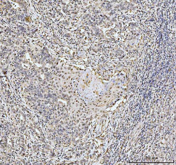
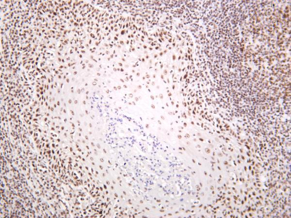
The catalog includes NCL antibodies with IHC images from human renal pelvis metaplasia, human tonsil, mouse pancreas, and rat pancreas; one also has IF data from A431 cells (catalog image captions).

Real IHC data IHC analysis of NCL using anti-NCL antibody (A00228-1). NCL was detected in a paraffin-embedded section of human metaplasia of squamous cells of the renal pelvis tissue. Heat mediated antigen retrieval was performed in EDTA buffer (pH 8.0, epitope retrieval solution). The tissue section was blocked with 10% goat serum. The tissue section was then incubated with 2 μg/ml rabbit anti-NCL Antibody (A00228-1) overnight at 4°C. Biotinylated goat anti-rabbit IgG was used as secondary antibody and incubated for 30 minutes at 37°C. The tissue section was developed using Strepavidin-Biotin-Complex (SABC) (Catalog # SA1022) with DAB as the chromogen.
Anti-Nucleolin/NCL Antibody ®
Cat # A00228-1
Real IHC data Human tonsil was stained with anti-Nucleolin rabbit antibody
Anti-Nucleolin Rabbit Monoclonal Antibody
Cat # M00228-4

A00228-1 has paraffin-section IHC images from human renal pelvis metaplasia, mouse pancreas, and rat pancreas, plus an IF image from A431 cells (A00228-1 image captions). M00228-4 has a human tonsil IHC image and lists human and mouse reactivity and IF as an application (M00228-4 catalog).

Which to pick: For tissue IHC, choose A00228-1 when its documented paraffin-section procedure fits your sample; its caption specifies EDTA retrieval at pH 8.0 and 2 μg/ml primary antibody, but does not report the fixative (A00228-1 IHC caption). For IF/ICC, A00228-1 has an A431 cell IF image and lists both applications; M00228-4 lists IF but provides no IF image (catalog applications and image captions). For work across species, A00228-1 lists human, monkey, mouse, and rat reactivity, with IHC images for human, mouse, and rat; M00228-4 is a rabbit monoclonal with a human tonsil IHC image whose processing and fixative are unreported (catalog reactivity and IHC captions).

Each figure is that product's own IHC / IF validation image from its datasheet.

References

  1. UniProt Consortium. UniProt entry P19338 (NUCL_HUMAN, Nucleolin).
  2. Human Protein Atlas. NCL tissue IHC expression (reliability: Supported).
  3. Human Protein Atlas. NCL subcellular location (ICC-IF): Mainly localized to the nucleoplasm, nucleoli and nucleoli rim. In addition localized to the mitotic chromosome..
  4. Human Protein Atlas. NCL antibody validation summary (3 antibodies).
  5. Expression Patterns of LALBA and Nucleolin and Their Clinical, Prognostic, and Immune Relevance in Breast Cancer Tissues of Mexican Patients. International journal of molecular sciences 2026 — PMC12898300.
  6. Helicobacter pylori in colorectal neoplasms: is there an aetiological relationship?. World journal of surgical oncology 2007 — PMC1885433.
  7. EIF4EBP1 overexpression is associated with poor survival and disease progression in patients with hepatocellular carcinoma. PloS one 2015 — PMC4319970.
  8. Overexpression of Nucleolin is a Potential Prognostic Marker in Endometrial Carcinoma. Cancer management and research 2021 — PMC7920503.
  9. PubMed PMID:2737305 — UniProt-cited evidence.
  10. PubMed PMID:2394707 — UniProt-cited evidence.
  11. PubMed PMID:14702039 — UniProt-cited evidence.