This website uses cookies to ensure you get the best experience on our website.


- Table of Contents
Use cervical squamous epithelium as a high-staining reference for NCOA7 paraffin-section IHC (HPA tissue IHC). Compare nuclear and cytoplasmic staining (HPA tissue IHC) with matched controls (standard IHC practice), and check epitope coverage across 7 isoforms (UniProt).

Expected localisation, validated protocols, controls and antibodies — the at-a-glance facts below, then the full design guide.
| Expected localisation | Nuclear and cytoplasmic staining (HPA tissue IHC) | |
| Staining pattern | Nuclear and cytoplasmic signal in CNS, squamous cells and nerves (HPA tissue IHC) | |
| Antigen retrieval | Tris-EDTA pH 8.0 HIER, heat-mediated (datasheet A05814-1) | |
| Positive control | Cervix+4 more · see all | |
| Negative control | Adipose tissue+4 more · see all |
| Fixation | Matched tissue-IHC evidence does not establish the fixation claim. Validate the specimen-specific method before use. (selected-SKU IHC image A05814-1) | |
| Caveat | Staining and RNA show low consistency; validate specificity (HPA tissue IHC) | |
| Regulation | Estradiol promotes nuclear localization (UniProt) | |
| Isoform / epitope | 7 isoforms; epitope coverage is unspecified (UniProt) |
The catalog antibody uses heat-mediated Tris-EDTA retrieval at pH 8.0 (datasheet A05814-1). The published paraffin-section IHC protocols below cover breast and oral cancer tissue (PMC11063752; PMC12819905; PMC5312364).
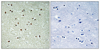
| Sample | Paraffin-embedded Human brain tissue; fixative not specified (datasheet A05814-1) |
| Fixation | Image fixative and duration unreported (datasheet A05814-1); verify before use. |
| Sectioning | 4–5 µm sections on charged slides (standard) |
| Deparaffinisation | Xylene, graded ethanol series to water (standard) |
| Antigen retrieval | Heat retrieval: Tris-EDTA pH 8.0 (datasheet A05814-1); 20 min, 95–100 °C (standard) |
| Peroxidase block | 3% H2O2, 10 min, room temperature (standard) |
| Blocking | 10% normal serum of the secondary host, 30 min, room temperature (standard) |
| Primary antibody | Rabbit anti-NCOA7, 1:100-1:300 (datasheet A05814-1) |
| Primary incubation | Overnight at 4 °C (standard) |
| Detection | HRP-polymer secondary, DAB chromogen 5–10 min (standard) |
| Counterstain | Hematoxylin, blue, dehydrate and mount (standard) |
| Expected result | NCOA7-positive staining in squamous epithelial cells of cervix (HPA tissue IHC: High). HPA tissue profile: Nuclear and cytoplasmic expression in several cell types, mainly in the CNS, squamous epithelia and peripheral nerves. No signal in the no-primary control. |
NCOA7 should show nuclear and cytoplasmic IHC staining in selected cell types, especially CNS cells and squamous epithelia (HPA tissue IHC). Nucleoplasm and nuclear bodies are the main ICC/IF locations, with additional Golgi staining (HPA subcellular ICC/IF). NCOA7 has no transmembrane segment (UniProt Q8NI08 topology). Interpret tissue staining cautiously: HPA rates it Approved but reports low consistency with RNA data and pending external verification (HPA tissue IHC).
| Nuclear and cytoplasmic signal in cervical squamous epithelial cells, with stronger staining than nearby unstained cells. | This fits a positive IHC result: HPA reports High staining in cervical squamous epithelial cells and a nuclear and cytoplasmic tissue profile (HPA tissue IHC). Judge the cell type and compartment together; intensity alone does not establish specificity (general IHC practice). |
| Signal is confined to cell borders or extracellular material, with no convincing nuclear or cytoplasmic staining. | This is a compartment mismatch with the reported tissue profile (HPA tissue IHC). NCOA7 has no transmembrane segment (UniProt Q8NI08 topology). Check morphology and controls before calling it positive; the pattern may reflect staining artefact (general IHC practice). |
| Strong staining appears chiefly in adipocytes or bone marrow hematopoietic cells. | HPA reports NCOA7 as Not detected in those cell populations (HPA tissue IHC). Consider antibody cross-reactivity or endogenous detection activity, and compare an appropriate negative control before assigning the signal to NCOA7 (general IHC practice). |
| Weak colour spreads across cells and tissue spaces without clear cellular boundaries. | Diffuse background does not establish NCOA7 localisation (general IHC practice). Assess blocking, washes and detection controls; a true call should resolve to plausible cells and nuclear or cytoplasmic compartments (HPA tissue IHC; general IHC practice). |
| Cervical squamous epithelial cells show no detectable signal. | That conflicts with HPA's High staining report for those cells (HPA tissue IHC). Check the antibody's IHC-P instructions and control performance before interpreting the specimen as negative; HPA also flags low RNA–staining consistency (HPA tissue IHC; general IHC practice). |
| Cell type and tissue | HPA reports High cervical squamous staining; Medium Purkinje, cortical neuronal, esophageal squamous, gallbladder glandular and skin keratinocyte staining; and Low soft-tissue fibroblast staining (HPA tissue IHC). Select and score the actual cell population, rather than averaging a whole section (general IHC practice). |
| Nuclear distribution | NCOA7 is nuclear and cytoplasmic without estradiol and predominantly nuclear after estradiol stimulation (UniProt Q8NI08, PubMed:17391516). Hormone context may therefore inform a compartment comparison; the supplied HPA tissue data do not establish treatment-specific IHC intensity (HPA tissue IHC). |
| Golgi localisation | UniProt lists Rab-dependent recruitment to cis- and trans-Golgi network membranes by similarity (UniProt Q8NI08). HPA additionally approves Golgi localisation in ICC/IF (HPA subcellular ICC/IF). A Golgi-like pattern can be plausible, but it is not a required tissue IHC finding. |
| Isoforms and antibody coverage | UniProt lists 7 NCOA7 isoforms (UniProt Q8NI08). The payload supplies no antibody epitope or isoform coverage, so a negative stain cannot identify which isoform is absent; consult the selected antibody's documented specificity before making that inference (general IHC practice). |
| Evidence strength | HPA calls its tissue IHC reliability Approved while noting low agreement with RNA expression and pending external verification (HPA tissue IHC). Four listed antibodies have Approved IHC status; the supplied status alone does not validate every specimen or unexpected cell pattern (HPA antibodies; general IHC practice). |
| ICC/IF Q&A | What should ICC/IF show? Mainly nucleoplasm and nuclear bodies, with additional Golgi staining (HPA subcellular ICC/IF). That compartment evidence helps interpret localisation; it does not specify an IHC-P staining protocol (HPA subcellular ICC/IF). |
| Situation | Likely cause | Next action |
|---|---|---|
| No stain in a cervical squamous epithelial positive control. | The expected High cell-level signal is absent (HPA tissue IHC); the failed step cannot be identified from the supplied evidence. | Confirm the correct cells are present, then review the IHC-validated antibody's documented IHC-P conditions, retrieval, dilution and detection controls (general IHC practice). Do not infer NCOA7-specific fixation sensitivity; none is supplied. |
| Only a few cells stain in a tissue expected to be positive. | NCOA7 staining is cell-type dependent; HPA reports different levels even among listed positive populations (HPA tissue IHC). | Identify and score the named cell population—for example, Purkinje cells in cerebellum—against its reported Medium level, rather than treating the whole tissue as uniformly positive (HPA tissue IHC; general IHC practice). |
| Adipocytes or marrow hematopoietic cells stain strongly. | Both are reported Not detected (HPA tissue IHC); cross-reactivity or endogenous chromogenic detection activity is possible (general IHC practice). | Compare a detection-only negative control, check endogenous activity blocking and inspect the cell morphology; require concordant localisation before reporting NCOA7 (general IHC practice; HPA tissue IHC). |
| Background obscures nuclei and cell boundaries. | Non-specific antibody binding or detection background can obscure a cellular IHC pattern (general IHC practice). | Review blocking, antibody dilution, washes and the negative control using the antibody's documented IHC-P conditions; score only resolved cellular staining (general IHC practice). |
| Signal appears exclusively along cell membranes. | A border-only pattern conflicts with HPA's nuclear and cytoplasmic tissue profile and is not explained by a transmembrane segment (HPA tissue IHC; UniProt Q8NI08 topology). | Recheck the compartment under adequate magnification and compare controls; treat the border-only result as unconfirmed rather than assigning it to NCOA7 (general IHC practice). |
| A novel tissue pattern differs from the HPA example. | HPA reports low RNA–staining consistency and pending external verification despite an Approved tissue rating (HPA tissue IHC). | Record cell type, compartment and intensity, and seek independent antibody or orthogonal evidence before making a firm biological claim (general IHC practice). |


Comprehensive Human Protein Atlas IHC scoring per tissue (reliability: Approved — Low consistency between antibody staining and RNA expression data. Pending external verification.). Rows are taken directly from the HPA tissue chart — click any row's HPA link to view the source.
| Tissue | Cell type | Level | Evidence | Source |
|---|---|---|---|---|
| Cervix | Squamous epithelial cells | High | Protein (IHC) | HPA → |
| Cerebellum | Purkinje cells | Medium | Protein (IHC) | HPA → |
| Cerebral cortex | Neuronal cells | Medium | Protein (IHC) | HPA → |
| Esophagus | Squamous epithelial cells | Medium | Protein (IHC) | HPA → |
| Gallbladder | Glandular cells | Medium | Protein (IHC) | HPA → |
| Tissue | Cell type | Level | Evidence | Source |
|---|---|---|---|---|
| Adipose tissue | Adipocytes | Not detected | Protein (IHC) | HPA → |
| Appendix | Glandular cells | Not detected | Protein (IHC) | HPA → |
| Bone marrow | Hematopoietic cells | Not detected | Protein (IHC) | HPA → |
| Caudate | Glial cells | Not detected | Protein (IHC) | HPA → |
| Duodenum | Glandular cells | Not detected | Protein (IHC) | HPA → |
Troubleshoot NCOA7 staining in paraffin sections by checking retrieval, compartment, cell identity, and controls before interpreting chromogenic signal.
Two rabbit polyclonal anti-NCOA7 antibodies have human brain IHC images (catalog captions); both list human and mouse reactivity (catalog). One also lists IF, without an IF image (catalog: A05814-1).
A05814-1 has a human brain paraffin-section IHC image and lists IF, IHC, and human/mouse reactivity (catalog: A05814-1 caption and applications). A05814-2 has a formalin-fixed, paraffin-embedded human brain IHC image and lists IHC-P and human/mouse reactivity (catalog: A05814-2 caption and applications).
Which to pick: For tissue IHC, choose A05814-2 when paraffin-section paraffin sections match your sample; its own image documents that preparation and DAB detection (catalog: A05814-2 caption). For IF, choose A05814-1 because IF is listed at 1:50, while A05814-2 does not list IF; ICC performance is unreported (catalog: applications and dilutions). Both are rabbit polyclonals with listed human/mouse reactivity, but their IHC images show human brain only; A05814-1’s paraffin-section caption does not report the fixative (catalog: dilution_raw, reactivity, and captions). The selected A05814-1 tissue-IHC caption documents paraffin sections, but does not specify the fixative (selected-SKU IHC image A05814-1).