This website uses cookies to ensure you get the best experience on our website.


- Table of Contents
Plan chromogenic IHC for NDFIP1 in paraffin sections using the catalog antibody at a starting concentration of 5 μg/mL (datasheet). Compare cytoplasmic staining in lung macrophages with an appropriate negative control (HPA tissue IHC).

Expected localisation, validated protocols, controls and antibodies — the at-a-glance facts below, then the full design guide.
| Expected localisation | Cytoplasmic staining observed; endosome and Golgi membranes annotated (HPA tissue IHC; UniProt) | |
| Staining pattern | Cytoplasmic staining, including in lung macrophages (HPA tissue IHC) | |
| Antigen retrieval | Citrate pH 6.0 HIER, 95–98 °C, 20 min (rule: cytoplasmic / membrane antigen) | |
| Positive control | Adrenal gland+4 more · see all | |
| Negative control | Adipose tissue+4 more · see all |
| Fixation | Keep formalin fixation consistent across paraffin sections. (standard IHC practice; not target-specific) | |
| Caveat | Endogenous peroxidase in lung macrophages may confound staining (HPA tissue IHC; standard IHC practice) | |
| Regulation | Stimulus-linked expression change not specified (UniProt) | |
| Isoform / epitope | 2 isoforms; check epitope against membrane topology (UniProt) |
Compare the catalog antibody protocol with three published NDFIP1 IHC protocols for cell line arrays, substantia nigra sections, and HNSCC patient sections (PMC8320194; PMC3901732; PMC8486716).
| Sample | Tissue sections; selected-image fixative not specified (standard IHC workflow) |
| Fixation | Image fixative and duration unreported (datasheet A04644); verify before use. |
| Sectioning | 4–5 µm sections on charged slides (standard) |
| Deparaffinisation | Xylene, graded ethanol series to water (standard) |
| Antigen retrieval | Heat-induced epitope retrieval in citrate buffer, pH 6.0, 20 min at 95–98 °C (standard rule: cytoplasmic / membrane antigen) |
| Peroxidase block | 3% H2O2, 10 min, room temperature (standard) |
| Blocking | 10% normal serum of the secondary host, 30 min, room temperature (standard) |
| Primary antibody | Rabbit anti-NDFIP1, 5 μg/mL (datasheet A04644) |
| Primary incubation | Overnight at 4 °C (standard) |
| Detection | HRP-polymer secondary, DAB chromogen 5–10 min (standard) |
| Counterstain | Hematoxylin, blue, dehydrate and mount (standard) |
| Expected result | NDFIP1-positive staining in glandular cells of adrenal gland (HPA tissue IHC: High). HPA tissue profile: General cytoplasmic expression. No signal in the no-primary control. |
NDFIP1 is a three-pass membrane protein associated with endosome and Golgi membranes (UniProt Q9BT67 topology and subcellular location). In paraffin-section IHC, expect a predominantly cytoplasmic pattern, with strong staining possible in neuronal cells, lung macrophages, testicular Leydig cells, and several glandular cell populations (HPA: general cytoplasmic expression; High in those cells). Treat the pattern as a guide rather than an absolute rule: HPA rates its tissue IHC evidence Approved, with medium consistency against RNA data (HPA: reliability).
| Cytoplasmic staining in cerebral cortex neurons or lung macrophages, with recognizable cell boundaries and a clean counterstain. | This fits the reported compartment and high-staining cell types (HPA: general cytoplasmic expression; High in cerebral cortex neuronal cells and lung macrophages). A granular or perinuclear accent can be compatible with endosome and Golgi membranes, but HPA does not specify either appearance for tissue IHC (UniProt Q9BT67 subcellular location; HPA: tissue profile). |
| Strong nuclear-only staining dominates cells while their cytoplasm is unstained. | A nuclear-only result does not match the reported general cytoplasmic tissue profile or the annotated endosome and Golgi membrane locations (HPA: tissue profile; UniProt Q9BT67 subcellular location). Check staining controls and repeat before interpreting the nuclear signal as NDFIP1; these sources do not establish nuclear-only tissue staining. |
| Adipocytes or cardiomyocytes stain strongly, especially when expected positive cells stain weakly. | These cell types were reported as not detected in the HPA tissue survey (HPA: Not detected in adipocytes and cardiomyocytes). Consider antibody cross-reactivity or chromogenic detection background, then compare a no-primary control and an expected positive section (general IHC practice). A discordant cell alone cannot identify the cause. |
| Color spreads through stroma, lumens, or empty areas instead of outlining stained cells. | Noncellular color cannot be assigned to NDFIP1 expression from the reported cellular pattern (HPA: general cytoplasmic expression). Investigate nonspecific reagent binding, residual endogenous enzyme activity, and counterstain or chromogen deposits using process controls (general chromogenic IHC practice). |
| No staining appears in a section containing a reported high-staining cell population. | An absent signal in an expected positive cell type makes the run inconclusive before it establishes biological absence (HPA: High in lung macrophages, cerebral cortex neurons, and testicular Leydig cells). Review the section, antibody application, retrieval, detection reagents, and positive control together (general IHC practice). |
| Compartment and topology | NDFIP1 has three transmembrane segments and annotated endosome and Golgi membrane locations (UniProt Q9BT67 topology and subcellular location). These support a cytoplasmic membrane-associated interpretation of the tissue pattern (HPA: general cytoplasmic expression). The supplied record does not locate the catalog antibody's epitope, so topology alone cannot prescribe retrieval conditions. |
| Cell-type variation | High staining is reported in selected neuronal, macrophage, Leydig, myoepithelial, respiratory epithelial, and glandular cells; other populations are low or not detected (HPA: tissue IHC). Choose controls by cell type within the section, since whole-tissue names can hide which cells supply a valid positive or negative reference (general IHC practice). |
| Strength of the tissue evidence | The tissue antibody is rated Approved, with medium consistency between staining and RNA expression; its IHC validation is not listed as Enhanced (HPA: reliability; HPA: HPA009682 IHC Approved). Interpret isolated, unexpected staining conservatively and compare morphology and controls before assigning it to NDFIP1 (general IHC practice). |
| Isoforms and extracellular detection | Two isoforms are listed, and NDFIP1 has been detected in exosomes released through an exosomal pathway (UniProt Q9BT67 isoforms and subcellular location). The payload does not establish isoform-specific tissue patterns or show that chromogenic extracellular deposits identify exosomal NDFIP1; avoid either inference from staining alone. |
| IF/ICC cross-check: what can it add? | HPA summarizes subcellular localization as Membrane but provides no ICC-IF image cell lines or main location (HPA: subcellular summary). That entry cannot confirm a particular punctate IF pattern or substitute for the paraffin-section IHC tissue observations; use the separate IF/ICC guide for its experimental workflow. |
| Situation | Likely cause | Next action |
|---|---|---|
| A reported high-staining population is blank. | The run may have failed, or the sampled section may lack the relevant cells; absence is not interpretable without a working positive control (HPA: High in lung macrophages and cerebral cortex neuronal cells; general IHC practice). | Confirm the cell population on the counterstained section and run an expected positive section alongside it; verify antibody application, antigen retrieval, and detection reagents against the chosen assay instructions (general IHC practice). Do not infer target-specific fixation sensitivity from this result. |
| Nuclei stain strongly while cytoplasm remains clear. | This conflicts with the reported general cytoplasmic tissue pattern and annotated membrane locations (HPA: tissue profile; UniProt Q9BT67 subcellular location). The observation alone does not distinguish off-target binding from detection background. | Compare a no-primary control, inspect cell morphology, and repeat with controlled reagent and detection settings (general IHC practice). Score nuclear-only color separately from the expected cytoplasmic signal until specificity is supported. |
| Adipocytes, cardiomyocytes, or squamous epithelium stain unexpectedly. | HPA reports no detection in adipocytes, cardiomyocytes, and esophageal or vaginal squamous epithelial cells (HPA: tissue IHC). Strong staining there is discordant, although HPA's Approved rating does not make every individual cell an absolute negative (HPA: reliability). | Compare a no-primary control and a high-staining cell population in the same run; assess morphology and whether the signal remains cell-associated (general IHC practice). Record a persistent discordance instead of treating it as established NDFIP1 expression. |
| Broad brown haze obscures cell boundaries. | Diffuse color is difficult to reconcile with the reported cellular cytoplasmic profile (HPA: tissue profile). Nonspecific reagent binding or residual endogenous peroxidase can contribute to chromogenic background (general IHC practice). | Use a no-primary control, verify the blocking and endogenous-enzyme steps appropriate to the detection system, and adjust development to preserve cell boundaries (general chromogenic IHC practice). Reassess intensity only after background is controlled. |
| Only a few cells stain in a tissue described as NDFIP1 positive. | Expression is cell-type dependent: HPA names specific high-staining cells rather than uniformly high staining across each tissue (HPA: tissue IHC). Its overall profile is general cytoplasmic expression with low tissue specificity at the RNA level (HPA: tissue profile and RNA specificity). | Identify the reported cell population on the section and score it separately from neighboring cells (general IHC practice). Compare with a matched positive control before increasing antibody concentration or calling the tissue negative. |
| IF/ICC shows a membrane signal that differs from the paraffin-section pattern. | HPA's IF entry says Membrane but supplies no ICC-IF image cell lines or detailed main location; its tissue IHC entry reports general cytoplasmic expression (HPA: subcellular summary; HPA: tissue profile). The two entries do not establish identical appearances across applications. | Evaluate the IHC section against its own cell-type and cytoplasmic references, and assess IF/ICC in its separate guide with application-specific controls (HPA: tissue IHC and subcellular summary; general assay practice). Do not change the IHC interpretation solely to match an unsupported detailed IF pattern. |
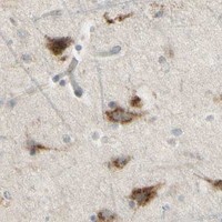
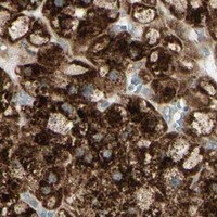
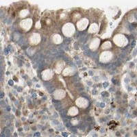
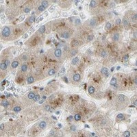
Comprehensive Human Protein Atlas IHC scoring per tissue (reliability: Approved — Medium consistency between antibody staining and RNA expression data.). Rows are taken directly from the HPA tissue chart — click any row's HPA link to view the source.
| Tissue | Cell type | Level | Evidence | Source |
|---|---|---|---|---|
| Adrenal gland | Glandular cells | High | Protein (IHC) | HPA → |
| Breast | Myoepithelial cells | High | Protein (IHC) | HPA → |
| Cerebral cortex | Neuronal cells | High | Protein (IHC) | HPA → |
| Gallbladder | Glandular cells | High | Protein (IHC) | HPA → |
| Lung | Macrophages | High | Protein (IHC) | HPA → |
| Tissue | Cell type | Level | Evidence | Source |
|---|---|---|---|---|
| Adipose tissue | Adipocytes | Not detected | Protein (IHC) | HPA → |
| Esophagus | Squamous epithelial cells | Not detected | Protein (IHC) | HPA → |
| Heart muscle | Cardiomyocytes | Not detected | Protein (IHC) | HPA → |
| Smooth muscle | Smooth muscle cells | Not detected | Protein (IHC) | HPA → |
| Spleen | Cells in red pulp | Not detected | Protein (IHC) | HPA → |
Troubleshoot NDFIP1 chromogenic IHC in paraffin sections using the reported tissue staining, protein topology, and matched controls.
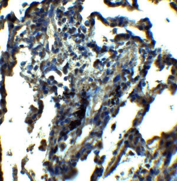
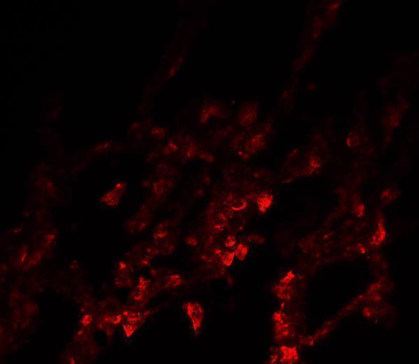
A04644 has IHC and IF images from human lung tissue (catalog image captions) and lists Human and Mouse reactivity (catalog: reactivity).
A04644 has a human lung tissue IHC image at 5 μg/mL (catalog IHC image caption). Its IF image shows human lung tissue at 20 μg/mL (catalog IF image caption).
Which to pick: Choose A04644 for paraffin section IHC: IHC-P is listed, and its own IHC image shows human lung tissue at 5 μg/mL; the fixative is unreported (catalog: applications; catalog IHC image caption). For IF, A04644 has a human lung tissue image at 20 μg/mL; ICC validation is unreported (catalog: applications; catalog IF image caption). For cross-species work, A04644 lists Human and Mouse reactivity, although the supplied images show human tissue only; clonality is unreported.