This website uses cookies to ensure you get the best experience on our website.


- Table of Contents
Plan NDUFS2 chromogenic IHC in paraffin sections using its granular cytoplasmic tissue profile and an IHC-validated antibody (HPA tissue IHC; datasheet A05618-3). Compare stained cell types with documented positive and undetected populations while keeping fixation and scoring consistent (HPA tissue IHC; standard IHC practice).

Expected localisation, validated protocols, controls and antibodies — the at-a-glance facts below, then the full design guide.
| Expected localisation | Granular cytoplasm in tissue sections (HPA tissue IHC) | |
| Staining pattern | Many cell types show granular cytoplasmic staining (HPA tissue IHC) | |
| Antigen retrieval | EDTA pH 8.0 HIER, heat-mediated (datasheet A05618-3) | |
| Positive control | Appendix+4 more · see all | |
| Negative control | Adipose tissue+4 more · see all |
| Fixation | Matched tissue-IHC evidence does not establish the fixation claim. Validate the specimen-specific method before use. (selected-SKU IHC image A05618-3) | |
| Caveat | Staining and RNA expression show medium consistency (HPA tissue IHC) | |
| Regulation | No specific regulator annotated (UniProt) | |
| Isoform / epitope | 2 isoforms; epitope differences unreported (UniProt) |
The catalog antibody uses EDTA pH 8.0 heat retrieval (datasheet: A05618-3). Published NDUFS2 IHC protocols cover tissue, mouse xenograft tumors, and rat brain (PMC7446242; PMC11039712; PMC8374556).
| Sample | Paraffin-embedded human liver cancer tissue; fixative not specified (datasheet A05618-3) |
| Fixation | Image fixative and duration unreported (datasheet A05618-3); verify before use. |
| Sectioning | 4–5 µm sections on charged slides (standard) |
| Deparaffinisation | Xylene, graded ethanol series to water (standard) |
| Antigen retrieval | Heat retrieval: EDTA pH 8.0 (datasheet A05618-3); 20 min, 95–100 °C (standard) |
| Peroxidase block | 3% H2O2, 10 min, room temperature (standard) |
| Blocking | 10% goat serum (datasheet A05618-3) |
| Primary antibody | Rabbit anti-NDUFS2, 2-5 μg/ml (datasheet A05618-3) |
| Primary incubation | Overnight at 4 °C (datasheet A05618-3) |
| Detection | HRP-conjugated secondary, DAB chromogen (datasheet A05618-3) |
| Counterstain | Hematoxylin, blue, dehydrate and mount (standard) |
| Expected result | NDUFS2-positive staining in glandular cells of appendix (HPA tissue IHC: High). HPA tissue profile: Ubiquitous cytoplasmic expression with a granular pattern. No signal in the no-primary control. |
NDUFS2 is a Complex I subunit at the mitochondrial inner membrane, with no annotated transmembrane segment (UniProt O75306 topology). In paraffin-section IHC, expect granular cytoplasmic staining, including in cardiomyocytes, neurons and several glandular or epithelial cell types reported as High (HPA tissue IHC). HPA describes the overall tissue pattern as ubiquitous, but rates its antibody staining versus RNA consistency as medium (HPA tissue IHC: Enhanced reliability).
| Granular cytoplasmic signal in heart-muscle cardiomyocytes or cerebellar Purkinje cells. | This matches the expected tissue and compartment pattern: both cell types are High by HPA tissue IHC, and NDUFS2 belongs to mitochondrial Complex I (HPA tissue IHC; UniProt O75306 function). Judge the cellular pattern alongside a no-primary control, since chromogen deposits alone do not establish target specificity (general chromogenic IHC practice). |
| Strong, predominantly nuclear or cell-surface staining with little granular cytoplasmic signal. | That compartment pattern conflicts with the HPA granular cytoplasmic IHC profile and UniProt mitochondrial inner-membrane assignment (HPA tissue IHC; UniProt O75306 subcellular location). Treat it as suspect staining and review controls and antibody specificity before scoring it as NDUFS2 (general IHC practice). |
| Prominent staining in adipocytes or bone-marrow hematopoietic cells while expected positive cells are weak. | HPA reports NDUFS2 as Not detected in those cell types, so this pattern raises concern for cross-reactivity or detection-system activity (HPA tissue IHC; general chromogenic IHC practice). A Not detected HPA result is an assay observation, however, and does not prove absolute protein absence. |
| Uniform brown haze across cells, stroma or the section edge, without resolvable cytoplasmic granules. | This does not reproduce HPA's granular cytoplasmic profile (HPA tissue IHC). Excess background can arise from nonspecific antibody binding, incomplete blocking or detection chemistry; compare a no-primary control and assess whether the haze tracks tissue features (general chromogenic IHC practice). |
| No signal in heart-muscle cardiomyocytes or another HPA High reference cell type. | An entirely blank expected-positive area makes the IHC run or antibody conditions questionable, although HPA's High rating does not guarantee every specimen will stain (HPA tissue IHC; general IHC practice). Check the reference tissue, detection controls and documented antibody conditions before interpreting a study specimen as negative. |
| Compartment and topology | NDUFS2 is assigned to the mitochondrial inner membrane, with no annotated transmembrane segment (UniProt O75306 topology). HPA IHC describes granules in cytoplasm; its microscopy profile does not resolve the inner membrane itself (HPA tissue IHC). |
| Cell-type selection | Heart-muscle cardiomyocytes, cortical neurons and bronchial respiratory epithelial cells are High; adipocytes in adipose tissue are Not detected (HPA tissue IHC). Compare named cell populations, since a whole-section average can obscure the reported contrast (general IHC interpretation). |
| Antibody evidence | HPA lists HPA055140 as IHC Enhanced and HPA061953 as IHC Supported; the tissue profile has Enhanced reliability with medium staining-versus-RNA consistency (HPA antibody validation; HPA tissue IHC). These labels support a starting expectation, not a guarantee for another antibody or specimen. |
| Processing and isoforms | UniProt annotates a chain spanning residues 34–463 and two isoforms, but the payload gives no epitope for the chosen antibody (UniProt O75306 processing and isoforms). Do not predict which isoform or processed form its stain distinguishes without antibody-specific evidence. |
| IF/ICC Q&A | Q: Should IF/ICC show the same location? A: HPA's approved main ICC-IF location is mitochondria; it also reports calyx and connecting-piece staining (HPA subcellular). Those IF observations do not establish a paraffin IHC protocol or expected staining in every tissue. |
| Situation | Likely cause | Next action |
|---|---|---|
| Expected-positive tissue has no detectable chromogen. | The run may have a reagent, detection or antibody-condition problem; HPA reports High cardiomyocyte and Purkinje-cell staining but does not establish a universal signal threshold (HPA tissue IHC; general IHC practice). | Inspect a known-positive section and detection control, then verify the chosen IHC-validated antibody's documented retrieval and dilution conditions before judging the study tissue (general IHC practice). |
| Signal is mainly nuclear or along cell borders. | The distribution conflicts with mitochondrial localization and the granular cytoplasmic IHC profile (UniProt O75306 subcellular location; HPA tissue IHC). | Compare with a no-primary control and an HPA High cell type; reassess antibody specificity if the compartment mismatch persists (general IHC practice; HPA tissue IHC). |
| Brown deposits appear broadly, including where HPA reports Not detected cells. | Nonspecific binding or endogenous detection activity can produce apparent positives in chromogenic IHC; HPA lists adipose-tissue adipocytes as Not detected (general IHC practice; HPA tissue IHC). | Review blocking and the detection-system control; score distinct cells against their HPA reference pattern rather than assigning every brown deposit to NDUFS2 (general IHC practice; HPA tissue IHC). |
| Granules are present, but a diffuse brown veil makes them hard to score. | Background can obscure a granular pattern through nonspecific binding or overly strong detection conditions (general chromogenic IHC practice; HPA tissue IHC profile). | Compare no-primary and positive-reference sections, then optimize the IHC-validated antibody and detection conditions using their documented starting settings (general IHC practice). |
| A study section stains less strongly than an HPA High example. | HPA High is a reported cell-type observation, while its overall tissue-IHC reliability description notes medium agreement between staining and RNA (HPA tissue IHC). Intensity alone cannot establish loss of NDUFS2. | Compare the same cell type with a reference section processed in the same run, and assess pattern, controls and morphology together (general IHC practice). |
| An IF image seems to disagree with the paraffin IHC slide. | HPA assigns mitochondria as the approved main ICC-IF location, whereas its tissue-IHC description is granular cytoplasmic staining; these are different readouts (HPA subcellular; HPA tissue IHC). | Evaluate each image against its application-specific HPA pattern and controls; do not transfer an ICC-IF localization observation into an IHC protocol decision (HPA subcellular; general IHC/IF practice). |
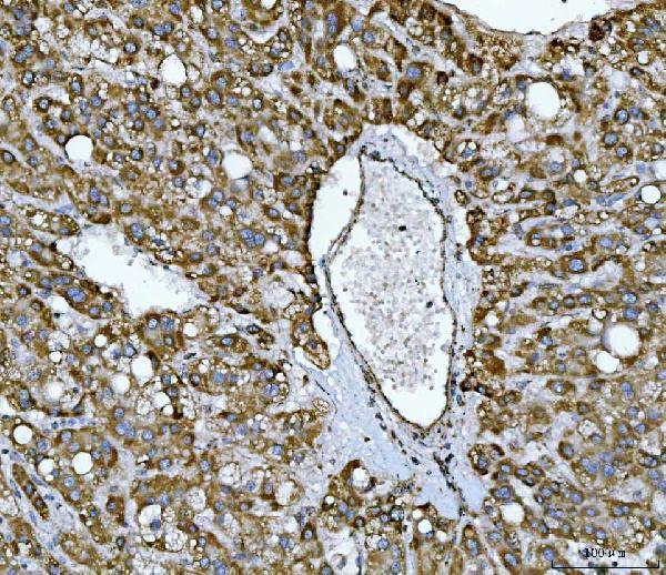
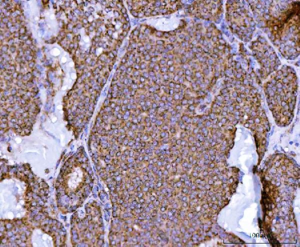
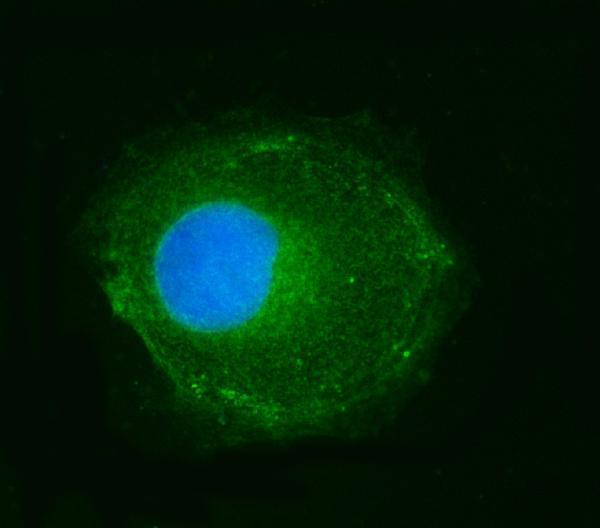
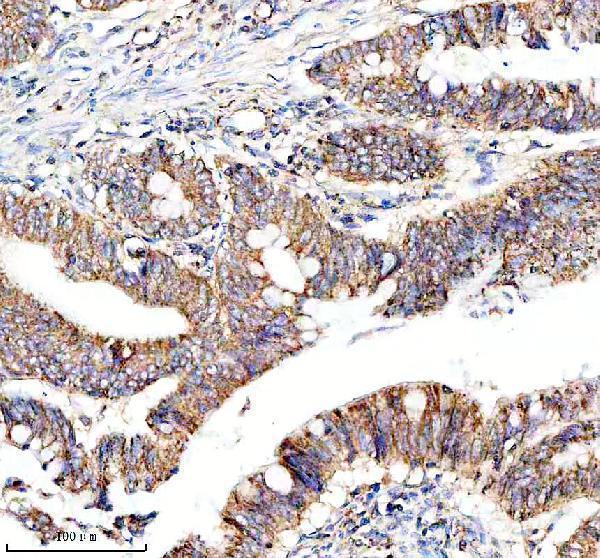


Comprehensive Human Protein Atlas IHC scoring per tissue (reliability: Enhanced — Medium consistency between antibody staining and RNA expression data.). Rows are taken directly from the HPA tissue chart — click any row's HPA link to view the source.
| Tissue | Cell type | Level | Evidence | Source |
|---|---|---|---|---|
| Appendix | Glandular cells | High | Protein (IHC) | HPA → |
| Bronchus | Respiratory epithelial cells | High | Protein (IHC) | HPA → |
| Cerebellum | Purkinje cells | High | Protein (IHC) | HPA → |
| Cerebral cortex | Neuronal cells | High | Protein (IHC) | HPA → |
| Colon | Glandular cells | High | Protein (IHC) | HPA → |
| Tissue | Cell type | Level | Evidence | Source |
|---|---|---|---|---|
| Adipose tissue | Adipocytes | Not detected | Protein (IHC) | HPA → |
| Bone marrow | Hematopoietic cells | Not detected | Protein (IHC) | HPA → |
| Breast | Adipocytes | Not detected | Protein (IHC) | HPA → |
| Cervix | Glandular cells | Not detected | Protein (IHC) | HPA → |
| Epididymis | Glandular cells | Not detected | Protein (IHC) | HPA → |
Troubleshoot NDUFS2 staining in paraffin sections by checking retrieval, tissue processing, mitochondrial localisation, controls, and scoring before interpreting chromogenic signal.
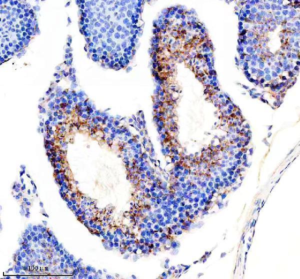
The catalog shows NDUFS2 IHC in human cancer sections and mouse and rat testis sections (catalog IHC captions), plus IF in CACO-2 cells and rat cardiac muscle (A05618-3 IF captions).
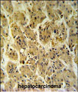
A05618-3 has paraffin-section IHC images from human liver, breast, colorectal and endometrial cancers, plus IF images from CACO-2 cells, human liver cancer and rat cardiac muscle (A05618-3 image captions); M05618-1 has paraffin-section IHC images from human colon cancer and mouse and rat testis (M05618-1 image captions). A05618-1 has a formalin-fixed, paraffin-embedded hepatocarcinoma IHC image; the caption does not report the species (A05618-1 IHC caption).
Which to pick: For tissue IHC, A05618-3 has several human paraffin-section examples with EDTA retrieval, although their fixative is unreported (A05618-3 IHC captions); choose A05618-1 when a documented paraffin-section, paraffin-embedded example is useful (A05618-1 IHC caption). For IF/ICC, choose A05618-3: both applications are listed, with a 5 μg/ml recommendation and IF images from cells and tissue (A05618-3 catalog and IF captions). For cross-species tissue IHC, M05618-1 is a rabbit monoclonal with human, mouse and rat reactivity and paraffin-section images in all three species after EDTA retrieval; the captions do not report the fixative (M05618-1 catalog and IHC captions). The selected A05618-3 tissue-IHC caption documents paraffin sections, but does not specify the fixative (selected-SKU IHC image A05618-3).