NEDD9 / Enhancer of filamentation 1 · IHC design guide

Design Immunohistochemistry for NEDD9

Plan chromogenic NEDD9 IHC in paraffin sections using its reported cytoplasmic, membranous and nuclear tissue pattern (HPA tissue IHC). Start with the IHC-validated antibody at 2–5 μg/mL (datasheet PB9289), and interpret staining intensity cautiously because antibody staining and RNA expression show low consistency (HPA tissue IHC).

Evidence assembled Oct 2026 · For research use; verify linked source records and product datasheet before use
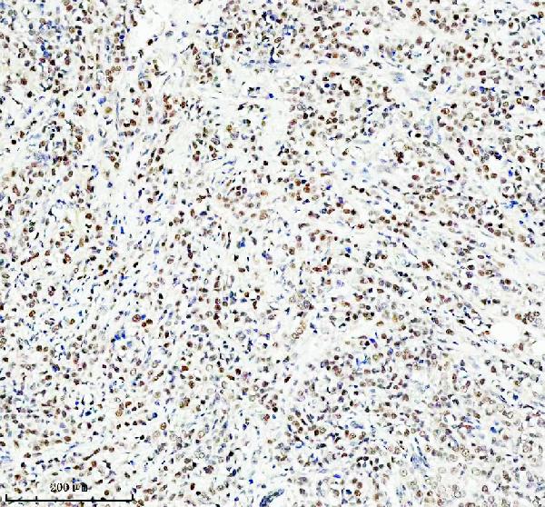
Immunohistochemistry protocol sheet for NEDD9 (IHC for NEDD9): expected localisation Cytoplasmic, membranous and nuclear tissue staining (HPA tissue IHC), antibody PB9289, validated IHC image, and IHC protocol steps
Printable NEDD9 IHC protocol sheet — expected localisation Cytoplasmic, membranous and nuclear tissue staining (HPA tissue IHC), antibody PB9289, controls and protocol steps. Open the full NEDD9 IHC guide →

NEDD9 Immunohistochemistry Experimental Design Guide

Expected localisation, validated protocols, controls and antibodies — the at-a-glance facts below, then the full design guide.

Must know before staining
Expected localisation Cytoplasmic, membranous and nuclear tissue staining (HPA tissue IHC)
Staining pattern Widespread cytoplasmic, membranous and nuclear signal (HPA tissue IHC)
Antigen retrieval EDTA pH 8.0 HIER, heat-mediated (datasheet PB9289)
Positive control ⓘ Adipose tissue+4 more · see all
Negative control ⓘ None in HPA (detected in all 45 tissues); use no-primary + isotype controls
Important caveats
Reasons your staining may differ from the expected pattern.
Fixation Keep paraffin-section fixation consistent across samples. (standard IHC practice; not target-specific)
Caveat Staining–RNA discordance and splice discrepancy (HPA tissue IHC)
Regulation SMAD3–ITCH promotes NEDD9 degradation (UniProt)
Isoform / epitope 3 isoforms and a p55 form; verify epitope coverage (UniProt)
Section 1

Recommended NEDD9 IHC & IF Protocols

The catalog antibody’s IHC-P protocol is accompanied by published NEDD9 staining protocols for lung adenocarcinoma, cervical carcinoma, and gastric tissue (PMC3493698; PMC3776827; PMC4360801).

Recommended immunohistochemistry (IHC-P) protocol parameters
SampleParaffin-embedded human breast cancer tissue; fixative not specified (datasheet PB9289)
FixationImage fixative and duration unreported (datasheet PB9289); verify before use.
Sectioning4–5 µm sections on charged slides (standard)
DeparaffinisationXylene, graded ethanol series to water (standard)
Antigen retrievalHeat retrieval: EDTA pH 8.0 (datasheet PB9289); 20 min, 95–100 °C (standard)
Peroxidase block3% H2O2, 10 min, room temperature (standard)
Blocking10% goat serum (datasheet PB9289)
Primary antibodyRabbit anti-NEDD9, 2-5μg/ml (datasheet PB9289)
Primary incubationOvernight at 4 °C (datasheet PB9289)
DetectionHRP-conjugated secondary, DAB chromogen (datasheet PB9289)
CounterstainHematoxylin, blue, dehydrate and mount (standard)
Expected resultNEDD9-positive staining in adipocytes of adipose tissue (HPA tissue IHC: High). HPA tissue profile: Widely expressed cytoplasmic, membranous and nuclear expression. No signal in the no-primary control.
💡Decision noteStart with heat-mediated EDTA at pH 8.0 for the catalog antibody (datasheet: PB9289); the published excerpts do not specify antigen retrieval (PMC3493698; PMC3776827; PMC4360801).
Section 2

What Is the Expected NEDD9 Staining Pattern?

NEDD9 can localise to cytoplasm, cell cortex, focal adhesions and nucleus; it has no transmembrane segment (UniProt Q14511). In paraffin tissue sections, expect cytoplasmic, membranous and nuclear staining across several cell types, including adipocytes, glial cells and glandular cells (HPA tissue IHC). Treat that pattern cautiously: HPA rates tissue staining Uncertain because antibody staining and RNA expression have low consistency, with a possible splice or transcript discrepancy (HPA tissue IHC).

What am I looking at on my slide?
Cytoplasmic staining, with nuclear or cell-edge staining, in adipocytes, glia or glandular cells.These compartments fit the reported tissue pattern and NEDD9 localisation (HPA tissue IHC; UniProt Q14511). Judge the labelled cell population and morphology together; compartment alone cannot establish specificity, given HPA's Uncertain tissue IHC reliability (HPA tissue IHC).
Signal is confined to extracellular material, a lumen or a sharply outlined cell surface.An exclusively external pattern is difficult to reconcile with NEDD9's reported intracellular locations and lack of a transmembrane segment (UniProt Q14511). Check tissue morphology and detection controls before calling it NEDD9; a cell-edge signal can still be plausible (UniProt Q14511).
Strong staining appears in an unexpected cell population while the expected population is unlabelled.Check cell identity and compare with the matched HPA tissue entry before inferring cross-reactivity or endogenous detection activity (HPA tissue IHC; general IHC practice). HPA reports broad expression and no negative tissue examples, so an unlisted positive population is not, by itself, proof of an artefact (HPA tissue IHC).
Colour covers the section diffusely and obscures cell boundaries.This is not an interpretable cell-specific pattern (general IHC practice). Review the no-primary control, blocking and detection steps, then reassess labelled cells against the cytoplasmic, membranous and nuclear tissue profile; diffuse colour cannot establish NEDD9 localisation (HPA tissue IHC; general IHC practice).
No staining is visible in a tissue and cell population reported as High by HPA.First verify that the relevant cells are present and the detection run worked (HPA tissue IHC; general IHC practice). Absence may reflect assay performance or biological variation; HPA's Uncertain IHC reliability prevents treating a single High entry as a guaranteed positive control (HPA tissue IHC).
💡Expected NEDD9 appearanceA plausible positive is cell-resolved cytoplasmic staining, sometimes nuclear or cell-edge, with strong signal in an HPA-listed High population such as adipocytes or glia; diffuse section-wide colour or exclusively extracellular signal warrants control checks (HPA tissue IHC; UniProt Q14511; general IHC practice).
How each factor affects the staining
Tissue and cell choiceHPA lists High staining in adipocytes, glia, cerebellar molecular-layer cells and several glandular populations, but Low staining in bronchial respiratory epithelium and smooth muscle (HPA tissue IHC). Match the comparison to the named cells; HPA lists no negative tissue (HPA tissue IHC).
Location and topologyNEDD9 has reported cytoplasmic, nuclear, cortical and focal-adhesion locations, plus no transmembrane segment (UniProt Q14511). Cell-edge signal is plausible, but a membrane outline alone does not establish a membrane-spanning antigen (UniProt Q14511).
Antibody validationThe listed tissue IHC antibodies are rated Uncertain, whereas the listed ICC antibodies are Supported for that application (HPA antibodies). Those labels apply to their respective assays; ICC support does not upgrade tissue IHC confidence (HPA antibodies).
Isoforms and phosphorylationUniProt lists 3 isoforms and multiple phosphorylation sites (UniProt Q14511). The supplied record gives no antibody epitope or evidence that these features alter paraffin-section staining, so do not predict an isoform-specific or phosphorylation-dependent IHC pattern (UniProt Q14511).
Detection backgroundEndogenous detection activity can produce chromogenic colour independent of primary-antibody binding; a no-primary control and detection-system controls help identify it (general IHC practice). The supplied HPA and UniProt records do not assign NEDD9 a tissue-specific background mechanism (HPA tissue IHC; UniProt Q14511).
Why is my staining missing, weak or wrong?
SituationLikely causeNext action
A reported High population gives no chromogenic signal.Cells may be absent from the section, or the assay may have failed; a High HPA entry is an uncertain reference, not a guaranteed result (HPA tissue IHC; general IHC practice).Confirm the cell population on the counterstain and check a working positive run and detection controls. Optimise retrieval and primary dilution empirically for the antibody, without assuming a known NEDD9 fixation effect (general IHC practice).
The entire section has diffuse brown colour.Background from blocking, washing or the detection system can obscure cell-specific staining (general IHC practice).Compare the no-primary control, review blocking and washes, and adjust detection or primary concentration using control sections; score only resolvable cells (general IHC practice).
Signal is exclusively luminal or extracellular.That distribution conflicts with the reported intracellular locations and lack of a transmembrane segment (UniProt Q14511).Inspect morphology and no-primary controls, then compare staining with a cell-resolved positive region; retain any cell-edge finding separately for review (UniProt Q14511; general IHC practice).
Unexpected cells stain strongly.Broad tissue expression, misidentified cells, cross-reactivity or endogenous detection activity are possible; HPA provides no negative tissue list (HPA tissue IHC; general IHC practice).Identify the cells on the counterstain, consult the corresponding HPA cell-level entry and review no-primary controls before assigning the signal to NEDD9 (HPA tissue IHC; general IHC practice).
Nuclear signal seems to conflict with cytoplasmic signal.Both compartments are reported for NEDD9; the supported ICC-IF locations are nucleoplasm and cytosol (UniProt Q14511; HPA subcellular).Score nuclear and cytoplasmic staining separately in IHC, and interpret each against tissue morphology and controls; ICC-IF localisation alone does not validate an IHC antibody (HPA subcellular; HPA antibodies; general IHC practice).
Can ICC-IF resolve an ambiguous IHC location?HPA reports supported nucleoplasm and cytosol localisation in ICC-IF, while tissue IHC antibody ratings remain Uncertain (HPA subcellular; HPA antibodies).Use the ICC-IF localisation as a compartment comparison only. Assess the paraffin-section result with its own cell morphology and controls; this section supplies no IF/ICC protocol option (HPA subcellular; HPA antibodies; general IHC practice).

Sample controls for NEDD9 IHC & IF

🧪Run adipose tissue first: adipocytes should show NEDD9 staining (HPA: High in adipocytes). HPA detects NEDD9 in all 45 scored tissues, so there is no supported negative tissue; use no-primary and isotype controls, and treat any unstained non-adipocyte cells on the positive slide as an internal background reference rather than a confirmed biological negative (HPA: no negative tissue rows; High in adipocytes).
Positive control tissue: Adipose tissue (Adipocytes, HPA High)
Negative control tissue: None in HPA: NEDD9 is detected in all 45 scored tissues. Use a no-primary (secondary-only) and an isotype control instead.
ICC-IF cell lines (HPA subcellular resource): HPA ICC-IF images show NEDD9 in Hep-G2, U-251MG, U2OS, EFO-21, SK-MEL-30, RPTEC/TERT1, hTERT-RPE1 (serum starved), with annotated localisation: Nucleoplasm (supported), Cytosol (supported) (HPA subcellular).
Technical controls: Include a no-primary, secondary-only slide, a host- and class-matched rabbit IgG isotype control, and NEDD9-knockout material as a biological negative where available (catalog caption: rabbit primary; standard IHC practice). Quench endogenous peroxidase before HRP/DAB detection and assess background in the adipose section (catalog caption: HRP/DAB; standard IHC practice).
⚠️Feasibility: A target-specific fixation window or fixation effect is unreported, and the selected PB9289 paraffin-section caption does not state a fixative (catalog caption: fixative unreported). Heat retrieval in EDTA at pH 8.0 is documented for that antibody, but a requirement for retrieval has not been established (catalog caption: EDTA pH 8.0). The supplied evidence does not compare frozen sections with IF for ease of use; in paraffin-processed adipose tissue, lipid extraction can leave empty-looking adipocytes that complicate interpretation (standard histology practice).

HPA tissue IHC evidence for NEDD9

Comprehensive Human Protein Atlas IHC scoring per tissue (reliability: Uncertain — Low consistency between antibody staining and RNA expression data. Caution, Splice and/or transcript discrepancy exists.). Rows are taken directly from the HPA tissue chart — click any row's HPA link to view the source.

Positive expression · recommended positive controls

TissueCell typeLevelEvidenceSource
Adipose tissue Adipocytes High Protein (IHC) HPA →
Caudate Glial cells High Protein (IHC) HPA →
Cerebellum Cells in molecular layer High Protein (IHC) HPA →
Cerebral cortex Glial cells High Protein (IHC) HPA →
Gallbladder Glandular cells High Protein (IHC) HPA →

Undetected expression · recommended negative controls

TissueCell typeLevelEvidenceSource
None in HPA: NEDD9 is detected in all 45 scored tissues. Use a no-primary (secondary-only) and an isotype control instead.
Section 3

Advanced NEDD9 IHC Tips

These NEDD9 questions address paraffin-section chromogenic IHC with PB9289; the IF question covers a separate exploratory application.

Which retrieval conditions should I start with for NEDD9 paraffin-section IHC?
Use heat-mediated antigen retrieval in EDTA at pH 8.0 for PB9289 paraffin-section IHC (datasheet PB9289). The documented image used this retrieval before overnight incubation with 2 μg/ml primary antibody at 4°C (datasheet PB9289). Keep retrieval conditions consistent across comparison sections, because changes in heating can alter staining intensity and tissue morphology (standard IHC practice). If staining is weak, check reagent performance and section integrity before testing an alternative retrieval buffer on adjacent sections (standard IHC practice). Compare any alternative against the EDTA condition using the same detection and exposure conditions (standard IHC practice).
How should I handle fixation when optimizing NEDD9 IHC?
The PB9289 image shows paraffin-embedded human breast cancer tissue, but its fixative is unreported (datasheet PB9289). Target-specific sensitivity to fixation chemistry or duration is therefore unknown for this antibody (datasheet PB9289). Record the fixative and processing history for each specimen, and compare sections processed alike when assessing staining differences (standard IHC practice). Start with heat-mediated EDTA retrieval at pH 8.0 and the documented 2 μg/ml primary concentration (datasheet PB9289). If a differently fixed specimen stains weakly, test matched controls and retrieval conditions before attributing the difference to NEDD9 abundance (standard IHC practice).
Which NEDD9 staining compartments should I expect in tissue sections?
Assess cytoplasmic and nuclear staining, while allowing for membrane-associated patterns reported in tissue IHC (HPA: widely expressed cytoplasmic, membranous and nuclear expression). NEDD9 is also annotated at the cell cortex, focal adhesions, lamellipodia and ciliary structures (UniProt Q14511 subcellular location). Resolve staining by cell type and compartment rather than treating every brown region as equivalent (standard IHC practice). Supported nucleoplasmic and cytosolic locations in cell imaging provide additional context, although they do not validate a PB9289 tissue pattern (HPA: supported nucleoplasm and cytosol; datasheet PB9289). Use a counterstain and inspect adjacent morphology to distinguish nuclear signal from overlapping cytoplasm (standard IHC practice).
Can this IHC antibody distinguish NEDD9 isoforms or phosphorylation states?
NEDD9 has 3 listed isoforms, and its record includes a p55 form (UniProt Q14511 isoforms and processing). The supplied PB9289 image does not map the recognized epitope or establish isoform selectivity (datasheet PB9289). NEDD9 also has annotated phosphotyrosine sites, including residues 92, 164 and 166 (UniProt Q14511 modified residues). Treat chromogenic intensity as antibody-reactive NEDD9 staining, without assigning an isoform or phosphorylation state from this image alone (datasheet PB9289; standard IHC interpretation). If that distinction matters, obtain epitope information and validate the intended specificity with suitable independent controls (standard IHC practice).
How should I explore NEDD9 localisation with multiplex IF?
For a separate IF experiment, pair NEDD9 with a validated marker for the expected cell type, such as adipocytes, which show high tissue staining in the HPA profile (HPA: High in adipocytes; standard IF practice). Choose a fluorophore in a channel with low measured tissue autofluorescence and use single-stain controls to assess spectral spillover (standard IF practice). NEDD9 has no transmembrane segment and has reported intracellular locations, so permeabilisation is appropriate when the antibody recognizes an intracellular epitope (UniProt Q14511 topology and subcellular location; standard IF practice). The PB9289 paraffin-section image documents chromogenic IHC, so optimize IF staining and controls separately (datasheet PB9289).
What should I check when NEDD9 IHC shows diffuse brown background?
First compare the stained section with a no-primary control to assess detection-system background (standard IHC practice). The documented PB9289 workflow used 10% goat serum blocking, 2 μg/ml primary antibody, an HRP-linked secondary and DAB chromogen (datasheet PB9289). Check blocking, wash steps and primary concentration together when diffuse staining obscures cell boundaries (standard IHC practice). Include a peroxidase-blocking step and inspect the no-primary control for endogenous enzyme signal; these are general chromogenic IHC measures, not NEDD9-specific findings (standard IHC practice). Because NEDD9 may occupy several compartments, judge background against cell morphology and the control rather than excluding all cytoplasmic staining (UniProt Q14511 subcellular location; standard IHC practice).
How should I score NEDD9 staining across tissue sections? ⚠ ANSWER MARKED FOR VERIFICATION
Choose a scoring unit before review: an H-score for intensity and proportion, percentage of positive cells, or positive-cell density per mm² (standard IHC practice). Score the same cell population and compartment in every section, recording nuclear and cytoplasmic signal separately when both are present (HPA: cytoplasmic and nuclear expression; standard IHC practice). Normalize counts to the number of eligible cells or the measured viable tissue area, as appropriate to the chosen metric (standard IHC practice). Keep retrieval, antibody concentration, DAB development and imaging conditions consistent across samples (standard IHC practice). Interpret small differences cautiously because HPA rates its tissue IHC reliability as uncertain (HPA: Uncertain).
How can I distinguish convincing NEDD9 staining from artefact?
Look for cell-associated staining in plausible compartments, including cytoplasm and nucleus, and compare its distribution with tissue morphology (HPA: cytoplasmic and nuclear expression; standard IHC practice). High staining in adipocytes or glial cells is compatible with the HPA profile, but the listed tissue IHC reliability is uncertain (HPA: High in adipocytes and glial cells; HPA: Uncertain). Treat isolated section-edge signal, necrotic areas and no-primary-control staining as possible artefacts requiring review (standard IHC practice). Check unexpected compartment or cell-type patterns against an independent control before calling them NEDD9-specific (standard IHC practice). Assess endogenous peroxidase contribution when brown signal persists without primary antibody (standard IHC practice).
Boster reagents

Best NEDD9 / Enhancer of filamentation 1 IHC Antibodies

PB9289 has IHC data from a human breast cancer paraffin section and IF data from HeLa cells; listed reactivity covers human, monkey, mouse and rat (PB9289 IHC/IF captions; catalog).

Real IHC data IHC analysis of HEF1 using anti-HEF1 antibody (PB9289). HEF1 was detected in a paraffin-embedded section of human breast cancer tissue. Heat mediated antigen retrieval was performed in EDTA buffer (pH 8.0, epitope retrieval solution). The tissue section was blocked with 10% goat serum. The tissue section was then incubated with 2 μg/ml rabbit anti-HEF1 Antibody (PB9289) overnight at 4°C. Peroxidase Conjugated Goat Anti-rabbit IgG was used as secondary antibody and incubated for 30 minutes at 37°C. The tissue section was developed using HRP Conjugated Rabbit IgG Super Vision Assay Kit (Catalog # SV0002) with DAB as the chromogen.
Anti-HEF1/NEDD9 Antibody ®
Cat # PB9289

PB9289 will render with IHC evidence from a human breast cancer paraffin section (PB9289 IHC caption). It is also listed for IF/ICC and has an IF image from HeLa cells (catalog; PB9289 IF caption).

Which to pick: For tissue IHC, choose PB9289: its caption documents EDTA retrieval at pH 8.0 and DAB detection on a paraffin section; the fixative is unreported (PB9289 IHC caption). For IF/ICC, PB9289 is listed for both applications and has an IF image from HeLa cells; its host is rabbit, while clonality is unreported (catalog; PB9289 IF caption). For cross-species work, PB9289 lists human, monkey, mouse and rat reactivity, though its supplied IHC and IF images document only human breast cancer tissue and HeLa cells, respectively (catalog; PB9289 IHC/IF captions).

Each figure is that product's own IHC / IF validation image from its datasheet.

References

  1. UniProt Consortium. UniProt entry Q14511 (CASL_HUMAN, Enhancer of filamentation 1).
  2. Human Protein Atlas. NEDD9 tissue IHC expression (reliability: Uncertain).
  3. Human Protein Atlas. NEDD9 subcellular location (ICC-IF): Mainly localized to the nucleoplasm and cytosol..
  4. Human Protein Atlas. NEDD9 antibody validation summary (5 antibodies).
  5. Role of NEDD9 in invasion and metastasis of lung adenocarcinoma. Experimental and therapeutic medicine 2012 — PMC3493698.
  6. POSITIVE EXPRESSION OF NEDD9 IN HEAD AND NECK CANCER IS RELATED TO BETTER SURVIVAL PERIOD. Acta clinica Croatica 2019 — PMC7314307.
  7. The overexpression of scaffolding protein NEDD9 promotes migration and invasion in cervical cancer via tyrosine phosphorylated FAK and SRC. PloS one 2013 — PMC3776827.
  8. Elevated expression of NEDD9 is associated with metastatic activity in gastric cancer. OncoTargets and therapy 2015 — PMC4360801.
  9. PubMed PMID:8668148 — UniProt-cited evidence.
  10. PubMed PMID:8879209 — UniProt-cited evidence.
  11. PubMed PMID:14702039 — UniProt-cited evidence.