This website uses cookies to ensure you get the best experience on our website.


- Table of Contents
Plan NEK9 paraffin-section IHC around cytoplasmic staining in a few tissues (HPA tissue IHC). This guide covers consistent fixation, the catalog antibody’s 2–5 μg/ml IHC range (datasheet A04660-1), chromogenic detection and cell-level scoring.

Expected localisation, validated protocols, controls and antibodies — the at-a-glance facts below, then the full design guide.
| Expected localisation | Tissue staining is cytoplasmic (HPA tissue IHC); nuclear localisation is reported (UniProt) | |
| Staining pattern | Cytoplasmic staining in a few tissues, including glandular cells (HPA tissue IHC) | |
| Antigen retrieval | EDTA pH 8.0 HIER, heat-mediated (datasheet A04660-1) | |
| Positive control | Adrenal gland+4 more · see all | |
| Negative control | Adipose tissue+4 more · see all |
| Fixation | Keep paraffin-section fixation consistent across samples. (standard IHC practice; not target-specific) | |
| Caveat | Antibody staining has low consistency with RNA expression (HPA tissue IHC) | |
| Regulation | Staining-intensity regulation is unreported (UniProt) | |
| Isoform / epitope | No isoforms, signal peptide or propeptide are listed (UniProt) |
The catalog antibody protocol uses EDTA pH 8.0 retrieval (datasheet: A04660-1). The published gastric cancer IHC protocols below use citrate retrieval (PMC7797683; PMC10344862).
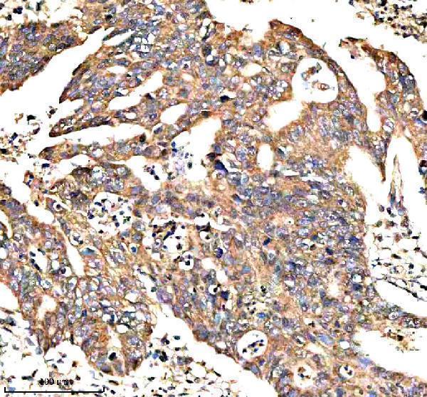
| Sample | Paraffin-embedded human colon cancer tissue; fixative not specified (datasheet A04660-1) |
| Fixation | Image fixative and duration unreported (datasheet A04660-1); verify before use. |
| Sectioning | 4–5 µm sections on charged slides (standard) |
| Deparaffinisation | Xylene, graded ethanol series to water (standard) |
| Antigen retrieval | Heat retrieval: EDTA pH 8.0 (datasheet A04660-1); 20 min, 95–100 °C (standard) |
| Peroxidase block | 3% H2O2, 10 min, room temperature (standard) |
| Blocking | 10% goat serum (datasheet A04660-1) |
| Primary antibody | Rabbit anti-NEK9, 2-5 μg/ml (datasheet A04660-1) |
| Primary incubation | Overnight at 4 °C (datasheet A04660-1) |
| Detection | HRP-conjugated secondary, DAB chromogen (datasheet A04660-1) |
| Counterstain | Hematoxylin, blue, dehydrate and mount (standard) |
| Expected result | NEK9-positive staining in glandular cells of adrenal gland (HPA tissue IHC: Medium). HPA tissue profile: Cytoplasmic expression in a few tissues. No signal in the no-primary control. |
NEK9 is cytoplasmic and nuclear and has no transmembrane segment (UniProt Q8TD19). In paraffin section IHC, expect chiefly cytoplasmic staining in selected cell populations, including glandular, follicle, decidual and lymphoid cells reported by HPA (HPA tissue IHC). HPA rates the tissue profile Approved but reports low consistency between antibody staining and RNA expression; interpret the pattern alongside controls (HPA tissue IHC).
| Cytoplasmic staining in adrenal, duodenal, fallopian tube, rectal or seminal vesicle glandular cells. | This matches HPA's medium staining in those specific cell populations (HPA tissue IHC). Score the identified cells and their cytoplasm; adjacent cells need not share the same intensity. |
| Predominantly membrane-rim staining, with little cytoplasmic signal. | A membrane-only pattern is unexpected because NEK9 has no transmembrane segment and is annotated in cytoplasm and nucleus (UniProt Q8TD19). Check morphology and staining controls before calling it NEK9. |
| Strong staining in adipocytes or bone marrow hematopoietic cells. | HPA reports these cells as not detected (HPA tissue IHC). Unexpected signal may reflect cross-reactivity or endogenous detection activity; inspect a no-primary control and confirm cell identity. |
| Color covers stroma, empty spaces or much of the section without clear cell boundaries. | Diffuse color is difficult to assign to NEK9-positive cells. Assess background with a no-primary control and compare with the cell-restricted cytoplasmic profile (HPA tissue IHC). |
| No staining in adrenal glandular cells despite an interpretable section. | HPA reports medium staining in these cells (HPA tissue IHC). A negative result warrants checking the IHC-validated antibody, detection controls and tissue preservation before interpreting biological absence. |
| Cell population and tissue choice | HPA reports medium staining in ovarian follicle cells, placental decidual cells and appendix lymphoid tissue, alongside selected glandular cells (HPA tissue IHC). Choose a documented cell population when judging a control section. |
| Low-level and negative comparators | Kidney tubule cells, lung macrophages and pancreatic endocrine cells are reported at low levels; adipocytes and bone marrow hematopoietic cells are not detected (HPA tissue IHC). Weak or absent staining must be judged in the named cells. |
| Protein location and processing | NEK9 is annotated in cytoplasm and nucleus, lacks a transmembrane segment and has no signal peptide or propeptide (UniProt Q8TD19). These annotations support intracellular interpretation; they do not establish antigen retrieval needs or fixation sensitivity. |
| Evidence and antibody status | The listed antibody, HPA001405, has IHC status Approved, while HPA describes low consistency with RNA expression (HPA antibody record; HPA tissue IHC). Approved should not be read as an Enhanced, independently reproduced tissue pattern. |
| RNA abundance versus slide staining | UniProt describes NEK9 as most abundant in heart, liver, kidney and testis, whereas HPA reports low staining in kidney tubule cells and low tissue RNA specificity (UniProt Q8TD19; HPA tissue IHC). Do not turn an abundance statement into an IHC intensity threshold. |
| IF/ICC Q: What pattern is established? | A: UniProt annotates cytoplasm and nucleus, but HPA supplies no ICC-IF images or main location for NEK9 (UniProt Q8TD19; HPA subcellular record). Treat an IF pattern as a hypothesis for its separate guide, not an IHC-derived validation. |
| Situation | Likely cause | Next action |
|---|---|---|
| A documented medium-positive cell population is blank. | The negative result conflicts with HPA's staining report for that population (HPA tissue IHC); the specific technical cause is undetermined. | Verify cell identity, antibody and detection controls, and section quality; review the antibody's IHC-P instructions before changing general workflow settings. |
| Only kidney tubules or lung macrophages show faint color. | HPA reports low staining in these cell populations (HPA tissue IHC); faint color alone cannot establish assay failure. | Compare with a documented medium-positive population and a no-primary control, then score cell-associated signal separately from background. |
| Adipocytes or marrow hematopoietic cells stain strongly. | These cells are reported as not detected (HPA tissue IHC); cross-reactivity or endogenous detection activity is possible. | Check a no-primary control for detection background and confirm the stained cell type before assigning a NEK9-positive result. |
| Brown color appears throughout the section. | Diffuse color obscures the cell-restricted cytoplasmic profile (HPA tissue IHC); nonspecific detection background is a general IHC possibility. | Inspect a no-primary control and the counterstain, then adjust blocking or detection conditions according to the assay instructions. |
| Staining appears exclusively at cell borders. | A membrane-only result conflicts with NEK9's cytoplasmic and nuclear annotation and lack of transmembrane segment (UniProt Q8TD19). | Recheck section morphology and control staining; require convincing intracellular signal before treating the border pattern as NEK9. |
| A nuclear signal appears alongside cytoplasmic staining. | Nuclear localization is annotated by UniProt, while HPA summarizes tissue IHC as cytoplasmic (UniProt Q8TD19; HPA tissue IHC). | Record nuclear and cytoplasmic staining separately, check controls, and avoid claiming that the nuclear IHC pattern is HPA-confirmed. |


Comprehensive Human Protein Atlas IHC scoring per tissue (reliability: Approved — Low consistency between antibody staining and RNA expression data.). Rows are taken directly from the HPA tissue chart — click any row's HPA link to view the source.
| Tissue | Cell type | Level | Evidence | Source |
|---|---|---|---|---|
| Adrenal gland | Glandular cells | Medium | Protein (IHC) | HPA → |
| Appendix | Lymphoid tissue | Medium | Protein (IHC) | HPA → |
| Duodenum | Glandular cells | Medium | Protein (IHC) | HPA → |
| Fallopian tube | Glandular cells | Medium | Protein (IHC) | HPA → |
| Ovary | Follicle cells | Medium | Protein (IHC) | HPA → |
| Tissue | Cell type | Level | Evidence | Source |
|---|---|---|---|---|
| Adipose tissue | Adipocytes | Not detected | Protein (IHC) | HPA → |
| Bone marrow | Hematopoietic cells | Not detected | Protein (IHC) | HPA → |
| Breast | Adipocytes | Not detected | Protein (IHC) | HPA → |
| Caudate | Glial cells | Not detected | Protein (IHC) | HPA → |
| Cerebellum | Cells in granular layer | Not detected | Protein (IHC) | HPA → |
Use the catalog antibody’s paraffin-section IHC conditions as a starting point, then assess NEK9 staining by compartment, cell type, and controls.
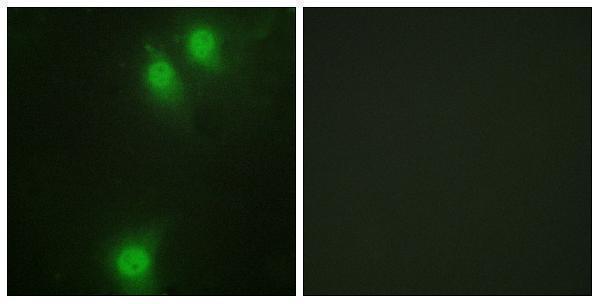
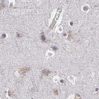
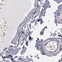
Two NEK9 antibodies have IHC images from human paraffin sections; IF images cover human paraffin sections and HeLa cells (catalog image captions).
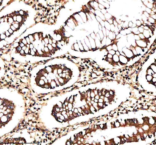
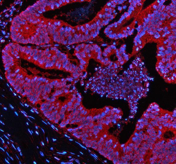
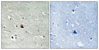
A04660-1 has IHC images from human colon, colon cancer, and breast cancer paraffin sections, plus IF images from human colon and breast cancer paraffin sections (A04660-1 image captions). P04660 targets phospho-NEK9 Thr210 and has an IHC image from human brain paraffin sections and an IF image from HeLa cells (P04660 title and image captions).
Which to pick: Choose A04660-1 for total NEK9 tissue IHC or tissue IF in human paraffin sections (A04660-1 applications, reactivity, and image captions). Choose P04660 for phospho-NEK9 Thr210 IHC or ICC/IF; it is listed as polyclonal and reactive with human, mouse, and rat, although its IHC image shows only human brain (P04660 title, dilution data, reactivity, and image captions). Both IHC captions describe paraffin sections and antigen retrieval; neither reports the fixative (A04660-1 and P04660 IHC image captions).