This website uses cookies to ensure you get the best experience on our website.


- Table of Contents
Plan chromogenic NENF IHC on paraffin sections with the catalog antibody starting at 10 μg/mL (datasheet: IHC-P). Use the cytoplasmic and membranous tissue pattern as a provisional reference because HPA rates its tissue staining reliability as uncertain (HPA tissue IHC).

Expected localisation, validated protocols, controls and antibodies — the at-a-glance facts below, then the full design guide.
| Expected localisation | Cytoplasmic and membranous tissue staining (HPA tissue IHC) | |
| Staining pattern | Glandular cells show cytoplasmic and membranous staining (HPA tissue IHC) | |
| Antigen retrieval | Citrate pH 6.0 HIER, 95–98 °C, 20 min (rule: cytoplasmic / membrane antigen) | |
| Positive control | Adrenal gland+4 more · see all | |
| Negative control | Adipose tissue+4 more · see all |
| Fixation | Keep fixation consistent across sections. (standard IHC practice; not target-specific) | |
| Caveat | Secreted NENF may stain away from its RNA source (HPA tissue IHC) | |
| Regulation | Tumor overexpression reported (UniProt) | |
| Isoform / epitope | No isoforms; the mature chain begins at residue 32 (UniProt) |
The catalog antibody has an IHC-P protocol (datasheet); the published options below cover hepatocellular carcinoma and colorectal tissue staining (PMC12964953; PMC10605590).
| Sample | Tissue sections; selected-image fixative not specified (standard IHC workflow) |
| Fixation | Image fixative and duration unreported (datasheet A11147); verify before use. |
| Sectioning | 4–5 µm sections on charged slides (standard) |
| Deparaffinisation | Xylene, graded ethanol series to water (standard) |
| Antigen retrieval | Heat-induced epitope retrieval in citrate buffer, pH 6.0, 20 min at 95–98 °C (standard rule: cytoplasmic / membrane antigen) |
| Peroxidase block | 3% H2O2, 10 min, room temperature (standard) |
| Blocking | 10% normal serum of the secondary host, 30 min, room temperature (standard) |
| Primary antibody | Rabbit anti-NENF, 10 μg/mL (datasheet A11147) |
| Primary incubation | Overnight at 4 °C (standard) |
| Detection | HRP-polymer secondary, DAB chromogen 5–10 min (standard) |
| Counterstain | Hematoxylin, blue, dehydrate and mount (standard) |
| Expected result | NENF-positive staining in glandular cells of adrenal gland (HPA tissue IHC: Medium). HPA tissue profile: General cytoplasmic and membranous expression. No signal in the no-primary control. |
NENF is secreted and is also reported in the endoplasmic reticulum and mitochondria, with no transmembrane segment (UniProt Q9UMX5 localization and topology). In paraffin sections, expect mainly cytoplasmic and membranous staining in glandular cells and selected neurons (HPA tissue IHC). Treat this as a provisional pattern: HPA rates tissue staining reliability Uncertain because antibody staining and RNA expression show low consistency, and secreted protein can appear away from its site of production (HPA tissue IHC).
| Moderate cytoplasmic or membranous staining in colon glandular cells, caudate neurons, or cerebellar Purkinje cells. | This matches reported positive cell types and the general tissue profile (HPA tissue IHC). Compare stained cells with tissue structure and a counterstain; intensity alone cannot establish specificity because the tissue IHC assessment is Uncertain (HPA tissue IHC). |
| Strong staining confined to nuclei, with little cytoplasmic or membranous signal. | An exclusively nuclear pattern is inconsistent with the reported tissue profile and listed subcellular locations (HPA tissue IHC; UniProt Q9UMX5 localization). Consider an artefact or off-target signal, and check control slides before interpreting it as NENF. |
| Prominent staining in adipocytes or lymph-node germinal center cells while expected glandular cells are weak. | Those cell types were reported as Not detected, whereas several glandular populations were Medium (HPA tissue IHC). The contrast raises concern for cross-reactivity or endogenous detection activity; an isolated positive cell does not resolve the uncertain antibody validation (HPA antibodies). |
| Diffuse color over cells, stroma, and empty spaces, obscuring cell boundaries. | A widespread deposit that does not follow the reported cytoplasmic and membranous cell pattern is difficult to score as specific staining (HPA tissue IHC). General IHC practice: inspect a negative control and assess blocking, washes, and chromogen development for background. |
| No signal in an adequately preserved colon section, including its glandular cells. | Colon glandular cells are reported at Medium intensity, so their absence is a useful warning sign, not proof that the specimen lacks NENF (HPA tissue IHC). The HPA assessment is Uncertain; assess tissue integrity and controls before calling the result negative (HPA tissue IHC). |
| Secretory processing and topology | NENF has a signal peptide at residues 1–31, a mature chain at 32–172, and no transmembrane segment (UniProt Q9UMX5 processing and topology). Interpret membranous staining alongside cytoplasmic staining and tissue architecture; topology alone does not identify a membrane-bound pool. |
| Intracellular localization | UniProt also lists endoplasmic reticulum and mitochondria, with localization to these compartments attributed to PINK1 and PARK7 (UniProt Q9UMX5 localization). This supports considering intracellular signal, but does not make a fine organelle pattern mandatory in chromogenic tissue IHC. |
| Tissue distribution and secretion | HPA reports Medium staining in several glandular populations and selected neurons, alongside Not detected calls in specific other cell types (HPA tissue IHC). Because NENF is secreted, tissue protein location need not match tissue RNA location (HPA tissue IHC; UniProt Q9UMX5 localization). |
| Strength of antibody evidence | Both listed antibodies, HPA026763 and HPA028488, have Uncertain IHC status (HPA antibodies). HPA also flags low agreement between antibody staining and RNA expression (HPA tissue IHC). Use the reported pattern to guide comparison, while keeping any single positive result provisional. |
| Situation | Likely cause | Next action |
|---|---|---|
| The expected glandular cells are unstained. | A weak assay signal or specimen issue is possible; the reported Medium glandular staining is itself uncertain (HPA tissue IHC). | General IHC practice: review section integrity, antibody and detection controls, and the antigen retrieval conditions used. Compare with a tissue containing a reported positive cell population (HPA tissue IHC); do not infer NENF-specific fixation sensitivity. |
| The whole section has diffuse brown background. | Nonspecific reagent binding, endogenous detection activity, or excess chromogen development can obscure a cell-associated pattern (general IHC practice). | General IHC practice: inspect a negative control, confirm appropriate blocking for the detection chemistry, and review washes and development time. Score only a distinguishable cellular pattern consistent with the HPA profile (HPA tissue IHC). |
| Signal is predominantly nuclear. | This differs from the reported cytoplasmic and membranous tissue profile and listed secretory or organelle locations (HPA tissue IHC; UniProt Q9UMX5 localization). | Check the counterstain and negative control, then repeat with reviewed detection conditions if needed (general IHC practice). Do not count an exclusively nuclear result as the expected NENF pattern. |
| Cells reported as Not detected stain strongly. | Off-target binding or endogenous detection activity is possible; HPA's IHC validation is Uncertain (HPA tissue IHC; HPA antibodies). | Compare the suspect cells with a reported Medium population in the same run, and examine negative controls (HPA tissue IHC; general IHC practice). Treat the unexpected signal as unresolved rather than confirming expression from color alone. |
| Two tissues show different apparent intensities. | HPA reports Medium, Low, and Not detected calls across cell types, while warning that its tissue IHC reliability is Uncertain (HPA tissue IHC). | Compare the same cell type and compartment under matched staining and scoring conditions (general IHC practice). Record differences as observed staining; avoid equating them directly with RNA abundance for a secreted protein (HPA tissue IHC). |
| Q: What pattern should IF/ICC show? | HPA summarizes NENF as Secreted but provides no main subcellular location or ICC-IF cell-line images (HPA subcellular). | A: Do not prescribe an IF/ICC organelle pattern from this IHC section. Use the separate IF/ICC guide for assay design; consider the secreted, endoplasmic reticulum, and mitochondrial annotations as context, not an image-validated IF result (UniProt Q9UMX5 localization; HPA subcellular). |


Comprehensive Human Protein Atlas IHC scoring per tissue (reliability: Uncertain — Low consistency between antibody staining and RNA expression data. Secreted protein, tissue location of RNA and protein is expected to differ. Pending external verification.). Rows are taken directly from the HPA tissue chart — click any row's HPA link to view the source.
| Tissue | Cell type | Level | Evidence | Source |
|---|---|---|---|---|
| Adrenal gland | Glandular cells | Medium | Protein (IHC) | HPA → |
| Appendix | Glandular cells | Medium | Protein (IHC) | HPA → |
| Breast | Glandular cells | Medium | Protein (IHC) | HPA → |
| Caudate | Neuronal cells | Medium | Protein (IHC) | HPA → |
| Cerebellum | Purkinje cells | Medium | Protein (IHC) | HPA → |
| Tissue | Cell type | Level | Evidence | Source |
|---|---|---|---|---|
| Adipose tissue | Adipocytes | Not detected | Protein (IHC) | HPA → |
| Bone marrow | Hematopoietic cells | Not detected | Protein (IHC) | HPA → |
| Hippocampus | Glial cells | Not detected | Protein (IHC) | HPA → |
| Lymph node | Germinal center cells | Not detected | Protein (IHC) | HPA → |
| Ovary | Follicle cells | Not detected | Protein (IHC) | HPA → |
Troubleshoot NENF chromogenic IHC by checking retrieval, signal location, controls, and scoring before interpreting staining intensity.
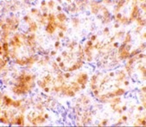
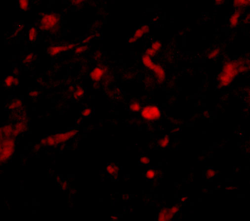
One anti-NENF antibody has IHC and IF images from rat kidney tissue (catalog image captions), with human, mouse and rat reactivity listed (catalog reactivity).
A11147 is listed for IHC-P, with an IHC image from rat kidney tissue at 10 μg/mL (catalog applications; IHC image caption). A11147 is also listed for IF, with an IF image from rat kidney tissue at 20 μg/mL (catalog applications; IF image caption).
Which to pick: Choose A11147 for paraffin-section tissue IHC because IHC-P is listed and its own IHC image shows rat kidney tissue; the fixative is unreported (catalog applications; IHC image caption). Choose A11147 for IF; ICC is not separately listed, so confirm performance for an ICC workflow (catalog applications). For work across species, A11147 lists human, mouse and rat reactivity, while the supplied images show rat kidney tissue only; clonality is unreported (catalog reactivity; catalog image captions; catalog clone field).