This website uses cookies to ensure you get the best experience on our website.


- Table of Contents
Plan paraffin-section NFAT5 IHC with the catalog antibody at 2–5 μg/mL (datasheet A01815-3). Assess nuclear and cytoplasmic staining by cell type, using the reported high goblet-cell signal as a reference (HPA tissue IHC).

Expected localisation, validated protocols, controls and antibodies — the at-a-glance facts below, then the full design guide.
| Expected localisation | Nuclear and cytoplasmic staining (HPA tissue IHC) | |
| Staining pattern | High goblet-cell signal; general nuclear and cytoplasmic pattern (HPA tissue IHC) | |
| Antigen retrieval | EDTA pH 8.0 HIER, heat-mediated (datasheet A01815-3) | |
| Positive control | Bronchus+4 more · see all | |
| Negative control | Lymph node+3 more · see all |
| Fixation | Keep fixation consistent across samples (standard IHC practice; not target-specific) | |
| Caveat | Staining and RNA show medium consistency (HPA tissue IHC) | |
| Regulation | Hypertonic nuclear shift, by similarity (UniProt) | |
| Isoform / epitope | 5 isoforms (A–E); check epitope coverage (UniProt) |
The catalog antibody’s IHC-P protocol (datasheet: A01815-3) is followed by four published NFAT5 IHC protocols (PMC7758185; PMC5355657; PMC5613209; PMC7304660).
| Sample | Paraffin-embedded human bladder cancer tissue; fixative not specified (datasheet A01815-3) |
| Fixation | Image fixative and duration unreported (datasheet A01815-3); verify before use. |
| Sectioning | 4–5 µm sections on charged slides (standard) |
| Deparaffinisation | Xylene, graded ethanol series to water (standard) |
| Antigen retrieval | Heat retrieval: EDTA pH 8.0 (datasheet A01815-3); 20 min, 95–100 °C (standard) |
| Peroxidase block | 3% H2O2, 10 min, room temperature (standard) |
| Blocking | 10% goat serum (datasheet A01815-3) |
| Primary antibody | Rabbit anti-NFAT5, 2-5μg/ml (datasheet A01815-3) |
| Primary incubation | Overnight at 4 °C (datasheet A01815-3) |
| Detection | HRP-conjugated secondary, DAB chromogen (datasheet A01815-3) |
| Counterstain | Hematoxylin, blue, dehydrate and mount (standard) |
| Expected result | NFAT5-positive staining in goblet cells of bronchus (HPA tissue IHC: High). HPA tissue profile: General nuclear and cytoplasmic expression. No signal in the no-primary control. |
NFAT5 is found in the nucleus and cytoplasm and has no transmembrane segment (UniProt O94916). In paraffin sections, expect nuclear and cytoplasmic staining, with the strongest HPA tissue observations in bronchial and nasopharyngeal goblet cells (HPA: High). Interpret intensity by cell type: HPA rates tissue IHC as Approved but reports only medium consistency between antibody staining and RNA expression (HPA: Approved; medium consistency).
| Goblet cells show clear nuclear staining, with or without cytoplasmic staining, in bronchus or nasopharynx. | This fits the reported High staining in those cell types (HPA: High in bronchial and nasopharyngeal goblet cells). Nuclear signal is consistent with NFAT5 localization; cytoplasmic signal can also be genuine (UniProt O94916; HPA: general nuclear and cytoplasmic tissue expression). Compare cells within the same section before judging intensity. |
| Signal is confined to cell borders, extracellular material, or a uniform surface rim. | A membrane-only pattern is inconsistent with the reported nuclear and cytoplasmic distribution (UniProt O94916; HPA: tissue IHC profile). NFAT5 has no transmembrane segment (UniProt O94916 topology). Check morphology and the no-primary control before treating such staining as NFAT5; cytoplasmic staining alone is not automatically erroneous. |
| Strong staining appears in lymph-node germinal-center cells while an expected positive area is weak. | HPA reports germinal-center cells as Not detected and bronchial goblet cells as High (HPA: tissue IHC). Review the cell identification and compare controls processed together. Staining in an unexpected cell type can reflect cross-reactivity or endogenous detection activity (general IHC practice), but one slide cannot establish either cause. |
| Color spreads through stroma, lumens, or most cells without a discernible nuclear or cytoplasmic pattern. | This is hard to reconcile with a cell-associated NFAT5 readout (UniProt O94916 localization; HPA: tissue IHC profile). Diffuse color can arise from nonspecific binding or detection background (general IHC practice). Inspect a no-primary control and avoid scoring the haze as positive cells. |
| Bronchial or nasopharyngeal goblet cells show no detectable signal. | These are useful positive-control cell types because HPA reports High staining there (HPA: tissue IHC); their absence prompts a run check, not an immediate biological conclusion. Confirm that goblet cells are present, then inspect the control, detection reagents, and staining conditions (general IHC practice). |
| Cell type and reference tissue | HPA reports High staining in bronchial and nasopharyngeal goblet cells, Medium in several listed cell types, and Not detected in lymph-node germinal-center cells (HPA: tissue IHC). Use those cell-level observations when selecting comparisons; do not assign one expected intensity to an entire organ. |
| Nuclear versus cytoplasmic distribution | Both compartments are reported (UniProt O94916; HPA: tissue IHC profile). UniProt notes increased nuclear distribution under hypertonic conditions by similarity (UniProt O94916); this is a biological qualifier, not a predicted intensity change for a particular paraffin section. |
| Antibody and isoform interpretation | HPA069711 has Approved IHC status, while the tissue profile has medium staining–RNA consistency (HPA: antibody validation; tissue IHC). UniProt lists five NFAT5 isoforms (UniProt O94916). Without an epitope map or isoform-specific validation, do not infer which isoforms contribute to a positive stain. |
| IF/ICC: where should signal appear? | Mainly in the nucleoplasm, with additional cytosolic signal (HPA: enhanced ICC-IF localization). This supports a compartment comparison only; the IF/ICC workflow belongs in its own guide, and these images do not establish an IHC dilution or retrieval condition. |
| Situation | Likely cause | Next action |
|---|---|---|
| No staining in the positive-control area | Goblet cells may be absent from the examined field, or the staining run may have failed (general IHC practice; HPA: High in bronchial and nasopharyngeal goblet cells). | Locate intact goblet cells, compare a concurrently processed positive control, then verify reagent delivery and detection steps (general IHC practice). |
| Weak signal in otherwise suitable goblet cells | Staining conditions may need adjustment (general IHC practice); the supplied sources do not establish NFAT5-specific fixation sensitivity. | Optimize the IHC-validated antibody's dilution and retrieval conditions against controls, following its validated IHC procedure (general IHC practice). |
| Nuclear signal is absent but cytoplasmic signal is distinct | Cytoplasmic localization is reported, so this observation alone does not prove an artefact (UniProt O94916; HPA: tissue IHC profile). | Check nuclear counterstain and cell boundaries, then compare the positive control and staining controls before assigning a localization change (general IHC practice). |
| Unexpected cells stain strongly | Possible nonspecific binding or endogenous detection activity (general IHC practice); HPA's Not detected calls apply to specified cell types, not every cell in those tissues (HPA: tissue IHC). | Confirm cell identity and inspect no-primary and detection controls; adjust blocking or detection as indicated by those controls (general IHC practice). |
| Diffuse brown background obscures cells | Nonspecific binding, residual endogenous enzyme activity, or excessive detection can obscure chromogenic IHC (general IHC practice). | Review the no-primary control; optimize blocking, antibody dilution, washing, and chromogen development one variable at a time (general IHC practice). |
| Results differ between tissues | HPA reports cell-specific levels from High to Not detected and only medium staining–RNA consistency (HPA: tissue IHC). | Score identified cell types and compartments separately, with controls from the same run; avoid calling an entire tissue positive or negative from a single field (general IHC practice). |
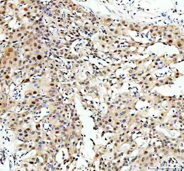
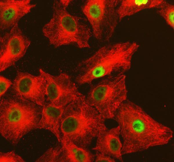
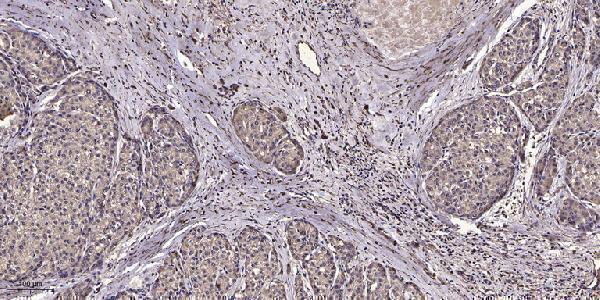
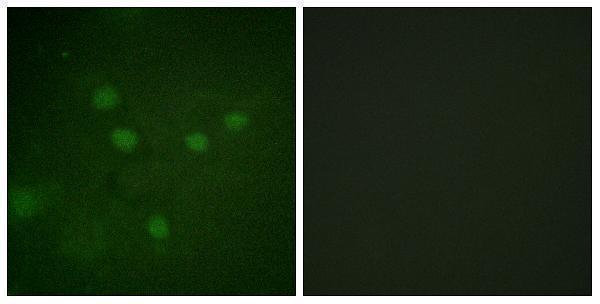


Comprehensive Human Protein Atlas IHC scoring per tissue (reliability: Approved — Medium consistency between antibody staining and RNA expression data.). Rows are taken directly from the HPA tissue chart — click any row's HPA link to view the source.
| Tissue | Cell type | Level | Evidence | Source |
|---|---|---|---|---|
| Bronchus | Goblet cells | High | Protein (IHC) | HPA → |
| Nasopharynx | Goblet cells | High | Protein (IHC) | HPA → |
| Adipose tissue | Adipocytes | Medium | Protein (IHC) | HPA → |
| Adrenal gland | Glandular cells | Medium | Protein (IHC) | HPA → |
| Appendix | Glandular cells | Medium | Protein (IHC) | HPA → |
| Tissue | Cell type | Level | Evidence | Source |
|---|---|---|---|---|
| Lymph node | Germinal center cells | Not detected | Protein (IHC) | HPA → |
| Parathyroid gland | Glandular cells | Not detected | Protein (IHC) | HPA → |
| Seminal vesicle | Glandular cells | Not detected | Protein (IHC) | HPA → |
| Soft tissue | Chondrocytes | Not detected | Protein (IHC) | HPA → |
Troubleshoot NFAT5 staining in paraffin sections by checking retrieval, background, and nuclear versus cytoplasmic signal against the documented tissue patterns.
Validated NFAT5 antibodies have IHC images from human paraffin-embedded bladder and liver cancer sections (catalog IHC captions); A01815-3 also has IF images from human bladder sections and A549 cells (catalog IF captions).
A01815-3 will render with IHC data from a human paraffin-embedded bladder cancer section; it lists human, mouse and rat reactivity and IF/ICC applications (A01815-3 caption; catalog applications/reactivity). P01815 will render with IHC data from a human paraffin-embedded liver cancer section; it lists human and mouse reactivity (P01815 caption; catalog reactivity).
Which to pick: For tissue IHC, choose A01815-3 for total NFAT5 in paraffin sections or P01815 when the target is phospho-NFAT5 Ser1197; each has its own paraffin-section IHC image (catalog titles; A01815-3 and P01815 IHC captions). For IF/ICC or rat reactivity, choose A01815-3 because those applications and rat reactivity are listed for it (catalog applications/reactivity). The IHC captions report paraffin embedding but do not report the fixative for either SKU (A01815-3 and P01815 IHC captions).