This website uses cookies to ensure you get the best experience on our website.


- Table of Contents
Plan NFIB paraffin-section IHC around the general nuclear tissue pattern (HPA tissue IHC). This guide covers fixation, tissue controls and nuclear scoring, with epitope coverage to consider across six isoforms (UniProt).

Expected localisation, validated protocols, controls and antibodies — the at-a-glance facts below, then the full design guide.
| Expected localisation | General nuclear staining (HPA tissue IHC) | |
| Staining pattern | Nuclear staining across multiple cell types (HPA tissue IHC) | |
| Antigen retrieval | Citrate pH 6 HIER, heat-mediated (datasheet A01537-1) | |
| Positive control | Heart muscle+4 more · see all | |
| Negative control | None in HPA (detected in all 45 tissues); use no-primary + isotype controls |
| Fixation | Keep fixation consistent across sections. (standard IHC practice; not target-specific) | |
| Caveat | Staining and RNA show medium consistency (HPA tissue IHC) | |
| Regulation | No expression regulator annotated (UniProt) | |
| Isoform / epitope | Six isoforms; check epitope coverage (UniProt) |
The catalog antibody’s IHC-P protocol (datasheet: A01537-1) is accompanied by published NFIB staining protocols for mouse embryonic brain and human skin specimens (PMC2802587; PMC7723069).
| Sample | Paraffin-embedded human mammary cancer tissue; fixative not specified (datasheet A01537-1) |
| Fixation | Image fixative and duration unreported (datasheet A01537-1); verify before use. |
| Sectioning | 4–5 µm sections on charged slides (standard) |
| Deparaffinisation | Xylene, graded ethanol series to water (standard) |
| Antigen retrieval | Heat retrieval: Citrate pH 6, 20 min (datasheet A01537-1) |
| Peroxidase block | 3% H2O2, 10 min, room temperature (standard) |
| Blocking | 10% goat serum (datasheet A01537-1) |
| Primary antibody | Rabbit anti-NFIB, 1μg/ml (datasheet A01537-1) |
| Primary incubation | Overnight at 4 °C (datasheet A01537-1) |
| Detection | Streptavidin-biotin complex (SABC), DAB chromogen (datasheet A01537-1) |
| Counterstain | Hematoxylin, blue, dehydrate and mount (standard) |
| Expected result | NFIB-positive staining in cardiomyocytes of heart muscle (HPA tissue IHC: High). HPA tissue profile: General nuclear expression. No signal in the no-primary control. |
NFIB is a nuclear, DNA-binding transcription factor with no transmembrane segment (UniProt O00712). In paraffin-section IHC, expect staining in nuclei across several cell types, with high staining reported in selected epithelial, alveolar and muscle cells (HPA: general nuclear expression; high in listed cells). HPA rates the tissue-IHC profile Supported, with medium consistency between antibody staining and RNA expression (HPA: tissue IHC).
| Distinct nuclear staining in pancreatic exocrine glandular cells or skin basal-layer cells, with a readable counterstain. | This fits reported high staining in those cells and the expected nuclear location (HPA: high in both cell groups; UniProt O00712: nucleus). Judge the cells individually: staining elsewhere in the section does not substitute for a nuclear signal in the intended cells (general IHC practice). |
| Strong staining chiefly in cytoplasm, at membranes, or throughout extracellular material while nuclei remain pale. | That compartment does not match NFIB's annotated nuclear location or the general nuclear tissue-IHC profile (UniProt O00712: nucleus; HPA: general nuclear expression). Treat it as suspect and check detection background and morphology before assigning it to NFIB (general IHC practice). |
| Prominent staining in a cell population outside the HPA high-staining examples, with little nuclear staining in the expected cells. | Investigate antibody cross-reactivity or endogenous detection activity (general IHC practice). HPA reports low tissue specificity and gives selected high and low examples, so an unlisted cell type alone cannot establish a false positive (HPA: low tissue specificity; tissue IHC). |
| A diffuse chromogenic haze covers nuclei and surrounding tissue, obscuring cell boundaries. | The background prevents a reliable nuclear call (general IHC practice; UniProt O00712: nucleus). Review blocking, wash and detection controls, then reassess whether discrete nuclei remain stained above the surrounding tissue (general IHC practice). |
| No nuclear staining appears in a section containing an HPA high-staining cell population. | A failed stain or sample-specific issue is possible; absence in one run does not establish absent NFIB expression (general IHC practice). Confirm the expected cell population and assay controls before interpreting the section (HPA: high in listed cell groups). |
| Compartment and topology (UniProt O00712: nucleus; no transmembrane segment). | Score nuclei in intact, identifiable cells (general IHC practice). Membrane staining has no support from the supplied NFIB topology, while the HPA tissue profile describes general nuclear expression (UniProt O00712: no transmembrane segment; HPA: tissue IHC). |
| Choice of positive reference tissue (HPA: high in listed cell groups). | HPA reports high staining in cardiomyocytes, lung alveolar type I cells, ovarian follicle cells, pancreatic exocrine cells, and several glandular or skin basal-layer cells (HPA: tissue IHC). Pick a listed population whose morphology can be identified on the section (general IHC practice). |
| Lower-staining reference populations (HPA: low in listed cell groups). | HPA lists cells in testicular seminiferous ducts, smooth muscle, splenic red pulp and bone-marrow hematopoietic cells as low, with no negative examples supplied (HPA: tissue IHC). A low category is a relative comparison, not a guaranteed blank control (HPA: tissue IHC). |
| Strength of the tissue evidence (HPA: Supported; medium RNA–staining consistency). | Use the observed IHC pattern as a guide, not an absolute rule for every specimen (HPA: tissue IHC). The listed antibody HPA003956 has Supported IHC validation; the supplied record does not assign it Enhanced IHC validation (HPA: antibody validation). |
| Isoforms and epitope uncertainty (UniProt O00712: six isoforms). | The record lists isoforms 1–6 but supplies no antibody epitope map or isoform-specific IHC result (UniProt O00712; HPA: supplied antibody record). Do not infer that one isoform, phosphorylation state or retrieval condition explains a differing stain from these data. |
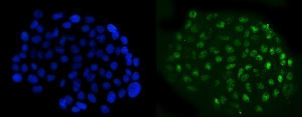
| IF/ICC Q: Where should NFIB appear? (HPA: subcellular ICC-IF). | A: HPA supports nucleoplasm and nucleoli fibrillar-center localization in ICC-IF images from A-431, U-251MG and U2OS (HPA: subcellular ICC-IF). This provides a localization cross-check; the supplied ICC-IF record does not define a paraffin-section IF protocol. |
| Situation | Likely cause | Next action |
|---|---|---|
| No stain in pancreatic exocrine cells or another identified HPA high-staining population (HPA: tissue IHC). | The assay or sample may have failed; this result alone does not identify which step is responsible (general IHC practice). | Check section integrity, primary-antibody inclusion and detection controls; then review retrieval and antibody concentration using the actual IHC protocol (general IHC practice). The supplied sources specify no NFIB dilution or retrieval setting. |
| Cytoplasmic or membrane signal dominates while nuclei are weak (UniProt O00712: nucleus). | Background or off-target binding is possible, but appearance alone cannot identify the mechanism (general IHC practice). | Inspect a control lacking primary antibody, verify nuclear counterstain and cell boundaries, and assess whether nuclear signal persists independently of diffuse color (general IHC practice). |
| Many unrelated structures share a uniform brown deposit (general IHC practice). | Endogenous peroxidase activity or nonspecific detection can contribute to chromogenic background (general IHC practice). | Review the detection-system controls, endogenous-enzyme blocking where applicable, wash steps and reagent exposure; compare with a section lacking primary antibody (general IHC practice). |
| The slide is faint despite identifiable HPA high-staining cells (HPA: tissue IHC). | Low assay sensitivity is possible, though the supplied sources do not establish NFIB-specific fixation sensitivity (general IHC practice; HPA: tissue IHC scope). | Verify counterstain, detection controls and the actual catalog antibody's IHC-P instructions before changing retrieval or dilution (general IHC practice). Do not assign a target-specific fixation cause from this record. |
| A low-staining reference population shows visible nuclear color (HPA: low in listed cell groups). | HPA's low category does not mean absent protein; staining also depends on how the local run is read (HPA: tissue IHC; general IHC practice). | Compare nuclear intensity and cell identity with a high-staining reference in the same run; avoid treating the low population as a mandatory negative control (HPA: tissue IHC; general IHC practice). |
| An unexpected cell population stains strongly while the reference cells do not (HPA: high in listed cell groups). | Possible cross-reactivity or assay background merits review; HPA's low tissue specificity also limits conclusions from cell identity alone (general IHC practice; HPA: tissue IHC). | Check the no-primary control, compartment and morphology, then repeat with appropriate assay controls before reporting the new cell pattern as NFIB (general IHC practice; UniProt O00712: nucleus). |
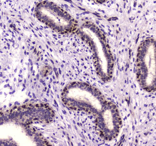
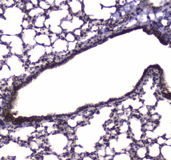
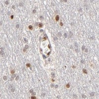


Comprehensive Human Protein Atlas IHC scoring per tissue (reliability: Supported — Medium consistency between antibody staining and RNA expression data.). Rows are taken directly from the HPA tissue chart — click any row's HPA link to view the source.
| Tissue | Cell type | Level | Evidence | Source |
|---|---|---|---|---|
| Heart muscle | Cardiomyocytes | High | Protein (IHC) | HPA → |
| Lung | Alveolar cells type I | High | Protein (IHC) | HPA → |
| Ovary | Follicle cells | High | Protein (IHC) | HPA → |
| Pancreas | Exocrine glandular cells | High | Protein (IHC) | HPA → |
| Prostate | Glandular cells | High | Protein (IHC) | HPA → |
| Tissue | Cell type | Level | Evidence | Source |
|---|---|---|---|---|
| None in HPA: NFIB is detected in all 45 scored tissues. Use a no-primary (secondary-only) and an isotype control instead. | ||||
Troubleshoot NFIB staining in paraffin sections by checking nuclear localisation, processing conditions, cell identity, and matched controls before interpreting chromogenic signal.
Anti-NFIB antibodies have IHC images from human mammary cancer, mouse lung and rat cardiac muscle paraffin sections, plus an IF image from A431 cells (catalog image captions).
A01537-1 has IHC images from human mammary cancer, mouse lung and rat cardiac muscle paraffin sections (A01537-1 image captions). M01537-1 has an IF image from A431 cells and lists ICC and IF applications (M01537-1 image caption; catalog applications).
Which to pick: Choose rabbit polyclonal A01537-1 for tissue IHC; its images document citrate retrieval and chromogenic detection in paraffin sections across human, mouse and rat samples (A01537-1 image captions; catalog host). Choose mouse monoclonal M01537-1 for IF/ICC in A431 cells (M01537-1 image caption; catalog clone 4D6E4); for cross-species tissue IHC, A01537-1 has images for all three species (A01537-1 image captions). The A01537-1 captions identify paraffin sections but do not report the fixative (A01537-1 image captions).