Click image to see more details
-

-
-
-
-
+1
Product Info Summary
| SKU: | M01537-1 |
|---|---|
| Size: | 100 μg/vial |
| Reactive Species: | Human, Mouse, Rat |
| Host: | Mouse |
| Application: | Flow Cytometry, IF, ICC, WB |
Customers Who Bought This Also Bought

Product info
Product Name
Anti-NFIB/NF1B2 Antibody Picoband® (monoclonal, 4D6E4)
SKU/Catalog Number
M01537-1
Size
100 μg/vial
Form
Lyophilized
Description
Boster Bio Anti-NFIB/NF1B2 Antibody Picoband® (monoclonal, 4D6E4) catalog # M01537-1. Tested in Flow Cytometry, IF, ICC, WB applications. This antibody reacts with Human, Mouse, Rat. The brand Picoband indicates this is a premium antibody that guarantees superior quality, high affinity, and strong signals with minimal background in Western blot applications. Only our best-performing antibodies are designated as Picoband, ensuring unmatched performance.
Storage & Handling
At -20°C for one year from date of receipt. After reconstitution, at 4°C for one month. It can also be aliquotted and stored frozen at -20°C for six months. Avoid repeated freezing and thawing.
Cite This Product
Anti-NFIB/NF1B2 Antibody Picoband® (monoclonal, 4D6E4) (Boster Biological Technology, Pleasanton CA, USA, Catalog # M01537-1)
Host
Mouse
Contents
Each vial contains 4 mg Trehalose, 0.9 mg NaCl and 0.2 mg Na2HPO4.
Clonality
Monoclonal
Clone Number
4D6E4
Isotype
Mouse IgG2b
Immunogen
A synthetic peptide corresponding to a sequence in the middle region of human NFIB/NF1B2, identical to the related mouse and rat sequences.
Cross-reactivity
No cross-reactivity with other proteins.
Reactive Species
M01537-1 is reactive to NFIB in Human, Mouse, Rat
Observed Molecular Weight
68 kDa
Calculated molecular weight
47.4 kDa
Background of NFIB
Nuclear factor 1 B-type is a protein that in humans is encoded by the NFIB gene. The NFIB gene is a part of the NFI gene complex that includes three other genes (NFIA, NFIC and NFIX). The NFIB gene is a protein coding gene that also serves as a transcription factor. This gene is essential in embryonic development and it works together with its gene complex to initiate tissue differentiation in the fetus. Through knockout experiments, researchers found that mice without the NFIB gene have severely underdeveloped lungs. This mutation does not seem to cause spontaneous abortions because in utero the fetus does not use its lungs for respiration. However, this becomes lethal once the fetus is born and has to take its first breath. It is thought that NFIB plays a role in down regulating the transcription factors TGF-β1 and Shh in normal gestation because they remained high in knockout experiments. The absence of NFIB also leads to insufficient amounts of surfactant being produced which is one reason why the mice cannot breathe once it is born. The knockout experiments demonstrated that NFIB has a significant role in fore-brain development. NFIB is typically found in pontine nuclei of the CNS, the cerebral cortex and the white matter of the brain and without NFIB these areas are dramatically affected.
Antibody Validation
Boster validates all antibodies on WB, IHC, ICC, Immunofluorescence, and ELISA with known positive control and negative samples to ensure specificity and high affinity, including thorough antibody incubations.
Application & Images
Applications
M01537-1 is guaranteed for Flow Cytometry, IF, ICC, WB Boster Guarantee
Recommend Dilution
| Application | Dilution | Species |
|---|---|---|
| Western blot | 0.25-0.5 μg/ml | Human |
| Immunocytochemistry/Immunofluorescence | 5 μg/ml | Human |
| Flow Cytometry (Fixed) | 1-3 μg/1x106 cells | Human, Mouse, Rat |
Tested application
Suggested blocking solution with 5% non-fat milk or BSA; (*)Recommended protein loading: 20-40 µg per lane
Validation Images & Assay Conditions
Click image to see more details
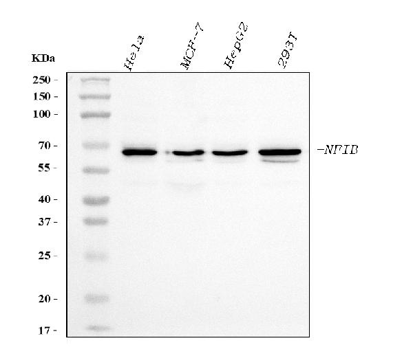
Western blot analysis of NFIB/NF1B2 using anti-NFIB/NF1B2 antibody (M01537-1).
Electrophoresis was performed on a 5-20% SDS-PAGE gel at 70V (Stacking gel) / 90V (Resolving gel) for 2-3 hours. The sample well of each lane was loaded with 30 ug of sample under reducing conditions.
Lane 1: human Hela whole cell lysates,
Lane 2: human MCF-7 whole cell lysates,
Lane 3: human HepG2 whole cell lysates,
Lane 4: human 293T whole cell lysates.
After electrophoresis, proteins were transferred to a nitrocellulose membrane at 150 mA for 50-90 minutes. Blocked the membrane with 5% non-fat milk/TBS for 1.5 hour at RT. The membrane was incubated with mouse anti-NFIB/NF1B2 antigen affinity purified monoclonal antibody (Catalog # M01537-1) at 0.5 μg/mL overnight at 4°C, then washed with TBS-0.1%Tween 3 times with 5 minutes each and probed with a goat anti-mouse IgG-HRP secondary antibody at a dilution of 1:10000 for 1.5 hour at RT. The signal is developed using an Enhanced Chemiluminescent detection (ECL) kit (Catalog # EK1001) with Tanon 5200 system. A specific band was detected for NFIB/NF1B2 at approximately 68 kDa. The expected band size for NFIB/NF1B2 is at 68 kDa.

Click image to see more details
IF analysis of NFIB/NF1B2 using anti-NFIB/NF1B2 antibody (M01537-1).
NFIB/NF1B2 was detected in an immunocytochemical section of A431 cells. Enzyme antigen retrieval was performed using IHC enzyme antigen retrieval reagent (AR0022) for 15 mins. The cells were blocked with 10% goat serum. And then incubated with 5 μg/mL mouse anti-NFIB/NF1B2 Antibody (M01537-1) overnight at 4°C. DyLight®488 Conjugated Goat Anti-Mouse IgG (BA1126) was used as secondary antibody at 1:100 dilution and incubated for 30 minutes at 37°C. The section was counterstained with DAPI. Visualize using a fluorescence microscope and filter sets appropriate for the label used.

Click image to see more details
Flow Cytometry analysis of A431 cells using anti-NFIB/NF1B2 antibody (M01537-1).
Overlay histogram showing A431 cells stained with M01537-1 (Blue line). To facilitate intracellular staining, cells were fixed with 4% paraformaldehyde and permeabilized with permeabilization buffer. The cells were blocked with 10% normal goat serum. And then incubated with mouse anti-NFIB/NF1B2 Antibody (M01537-1, 1 μg/1x106 cells) for 30 min at 20°C. DyLight®488 conjugated goat anti-mouse IgG (BA1126, 5-10 μg/1x106 cells) was used as secondary antibody for 30 minutes at 20°C. Isotype control antibody (Green line) was mouse IgG (1 μg/1x106) used under the same conditions. Unlabelled sample without incubation with primary antibody and secondary antibody (Red line) was used as a blank control.

Click image to see more details
Flow Cytometry analysis of C6 cells using anti-NFIB/NF1B2 antibody (M01537-1).
Overlay histogram showing C6 cells stained with M01537-1 (Blue line). To facilitate intracellular staining, cells were fixed with 4% paraformaldehyde and permeabilized with permeabilization buffer. The cells were blocked with 10% normal goat serum. And then incubated with mouse anti-NFIB/NF1B2 Antibody (M01537-1, 1 μg/1x106 cells) for 30 min at 20°C. DyLight®488 conjugated goat anti-mouse IgG (BA1126, 5-10 μg/1x106 cells) was used as secondary antibody for 30 minutes at 20°C. Isotype control antibody (Green line) was mouse IgG (1 μg/1x106) used under the same conditions. Unlabelled sample without incubation with primary antibody and secondary antibody (Red line) was used as a blank control.

Click image to see more details
Flow Cytometry analysis of Neuro-2a cells using anti-NFIB/NF1B2 antibody (M01537-1).
Overlay histogram showing Neuro-2a cells stained with M01537-1 (Blue line). To facilitate intracellular staining, cells were fixed with 4% paraformaldehyde and permeabilized with permeabilization buffer. The cells were blocked with 10% normal goat serum. And then incubated with mouse anti-NFIB/NF1B2 Antibody (M01537-1, 1 μg/1x106 cells) for 30 min at 20°C. DyLight®488 conjugated goat anti-mouse IgG (BA1126, 5-10 μg/1x106 cells) was used as secondary antibody for 30 minutes at 20°C. Isotype control antibody (Green line) was mouse IgG (1 μg/1x106) used under the same conditions. Unlabelled sample without incubation with primary antibody and secondary antibody (Red line) was used as a blank control.
Specific Publications For Anti-NFIB/NF1B2 Antibody Picoband® (monoclonal, 4D6E4) (M01537-1)
Loading publications
Recommended Resources
Here are featured tools and databases that you might find useful.
- Boster's Pathways Library
- Protein Databases
- Bioscience Research Protocol Resources
- Data Processing & Analysis Software
- Photo Editing Software
- Scientific Literature Resources
- Research Paper Management Tools
- Molecular Biology Software
- Primer Design Tools
- Bioinformatics Tools
- Phylogenetic Tree Analysis
Customer Reviews
Have you used Anti-NFIB/NF1B2 Antibody Picoband® (monoclonal, 4D6E4)?
Share your experimental results or join a short interview to earn up to $1,000 in product credits or other rewards.
0 Reviews For Anti-NFIB/NF1B2 Antibody Picoband® (monoclonal, 4D6E4)
Customer Q&As
Have a question?
Find answers in Q&As, reviews.
Can't find your answer?
Submit your question
