This website uses cookies to ensure you get the best experience on our website.


- Table of Contents
Plan NFKB2 paraffin-section IHC around cytoplasmic staining and high expression in lymphoid tissues (HPA tissue IHC). Use the IHC-validated antibody at 0.5–1 μg/ml (datasheet PB9150), and consider p100-to-p52 processing when interpreting staining (UniProt).

Expected localisation, validated protocols, controls and antibodies — the at-a-glance facts below, then the full design guide.
| Expected localisation | Cytoplasmic staining in most tissues (HPA tissue IHC) | |
| Staining pattern | Cytoplasmic staining; highest in lymphoid tissues (HPA tissue IHC) | |
| Antigen retrieval | EDTA pH 8.0 HIER, heat-mediated (datasheet PB9150) | |
| Positive control | Appendix+4 more · see all | |
| Negative control | Cerebellum+4 more · see all |
| Fixation | Keep fixation consistent across sections. (standard IHC practice; not target-specific) | |
| Caveat | A C-terminal epitope may miss processed p52 (UniProt) | |
| Regulation | Noncanonical signaling processes p100 (UniProt) | |
| Isoform / epitope | 3 isoforms; p52 lacks the p100 C-terminus (UniProt) |
The catalog antibody’s IHC-P protocol uses EDTA pH 8.0 heat retrieval (datasheet: PB9150). The published meningioma protocol below provides a citrate pH 6.0 alternative (PMC7445252).
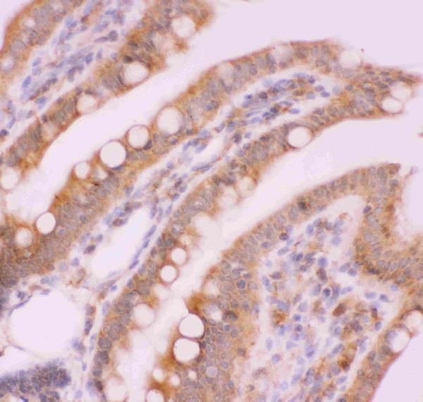
| Sample | Paraffin-embedded rat intestine tissue; fixative not specified (datasheet PB9150) |
| Fixation | Image fixative and duration unreported (datasheet PB9150); verify before use. |
| Sectioning | 4–5 µm sections on charged slides (standard) |
| Deparaffinisation | Xylene, graded ethanol series to water (standard) |
| Antigen retrieval | Heat retrieval: EDTA pH 8.0 (datasheet PB9150); 20 min, 95–100 °C (standard) |
| Peroxidase block | 3% H2O2, 10 min, room temperature (standard) |
| Blocking | 10% goat serum (datasheet PB9150) |
| Primary antibody | Rabbit anti-NFKB2, 0.5-1μg/ml (datasheet PB9150) |
| Primary incubation | Overnight at 4 °C (datasheet PB9150) |
| Detection | Streptavidin-biotin complex (SABC), DAB chromogen (datasheet PB9150) |
| Counterstain | Hematoxylin, blue, dehydrate and mount (standard) |
| Expected result | NFKB2-positive staining in lymphoid tissue of appendix (HPA tissue IHC: High). HPA tissue profile: Cytoplasmic expression in most tissues, highest expression in lymphoid tissues. No signal in the no-primary control. |
NFKB2 is expected mainly in the cytoplasm across many tissues, with the strongest tissue profile in lymphoid tissue (HPA: Enhanced tissue IHC; cytoplasmic expression in most tissues). Nuclear staining can also fit its biology: inactive complexes can remain cytoplasmic, while active NF-κB complexes can enter nuclei (UniProt Q00653: subcellular location and function). NFKB2 has no transmembrane segment, so a crisp membrane rim is unexpected (UniProt Q00653: topology).
| Cytoplasmic staining in lymphoid cells, including appendix lymphoid tissue and lymph node non-germinal center cells (HPA: High in both). | This fits the reported IHC profile; germinal center cells in tonsil are another high-staining reference (HPA: High in tonsil germinal center cells). Compare matched tissue compartments, since a whole-section score can hide differences between cell populations (standard IHC practice). |
| Nuclear staining accompanies cytoplasmic staining in some cells. | Nuclear localization is biologically plausible after pathway activation (UniProt Q00653: subcellular location and function). A single IHC image cannot establish that p100 was processed to p52 or that transcription is active (UniProt Q00653: p100/p52 processing; standard IHC interpretation). |
| A sharp membrane outline or extracellular deposit dominates the signal. | That compartment conflicts with the reported cytoplasmic and nuclear localization and the absence of a transmembrane segment (HPA: tissue IHC and subcellular profile; UniProt Q00653: topology). Treat it as suspect staining and check morphology and controls (standard IHC practice). |
| Strong signal appears in an unexpected cell population, such as cardiomyocytes or prostate glandular cells (HPA: Not detected in those cells). | The mismatch raises concern for antibody cross-reactivity or endogenous chromogen-generating activity, but a new biological context cannot be ruled out from this comparison alone (HPA: tissue IHC; standard IHC interpretation). Confirm cell identity and inspect detection controls (standard IHC practice). |
| The section shows diffuse haze, or a known high-staining compartment has no signal. | Diffuse haze limits compartment scoring and may reflect nonspecific staining or detection background (standard IHC practice). No signal in appendix lymphoid tissue or tonsil germinal center cells conflicts with their reported high staining; assess section quality and assay controls before calling the sample negative (HPA: High in both; standard IHC practice). |
| Cell population and tissue | HPA reports High staining in several lymphoid compartments, but also in placental decidual cells and testicular spermatogonia; its tissue profile has low RNA tissue specificity (HPA: tissue IHC and RNA specificity). Select and score the stated cells, not tissue names alone (standard IHC practice). |
| p100 processing and antibody epitope | NFKB2 p100 spans residues 1–900; p52 spans 1–454 (UniProt Q00653: chains). Whether an antibody detects both forms depends on its binding site, which is not supplied here; compartment staining alone cannot distinguish them (UniProt Q00653: processing; standard IHC interpretation). |
| Isoforms and validation | UniProt lists three isoforms, while HPA rates tissue IHC reliability Enhanced and lists two IHC Enhanced antibodies, HPA008422 and HPA023900 (UniProt Q00653: isoforms; HPA: antibodies and tissue IHC). Those ratings support the reported profile but do not establish which isoform a given antibody recognizes (HPA: validation; UniProt Q00653: isoforms). |
| Antigen retrieval | Retrieval conditions can affect staining in paraffin-section IHC (standard IHC practice). No NFKB2-specific retrieval requirement or fixation sensitivity is supplied by these records; evaluate conditions against positive tissue and background controls without assigning any change to NFKB2 epitope masking (standard IHC practice). |
| IF/ICC Q&A: Where should signal appear? | HPA reports enhanced nucleoplasm and cytosol localization in its ICC-IF images (HPA: subcellular profile). This supports interpreting those compartments in IF/ICC, but it does not supply an IF/ICC protocol option or convert an IF image into evidence of a particular IHC tissue pattern (HPA: subcellular and tissue profiles). |
| Situation | Likely cause | Next action |
|---|---|---|
| No signal in appendix lymphoid tissue or tonsil germinal center cells (HPA: High in both). | The expected positive compartment may be absent from the section, or an IHC step may have failed; the image alone cannot distinguish these causes (HPA: tissue IHC; standard IHC practice). | Verify the relevant cells are present, then check the positive control, detection reagents and the antibody's validated IHC conditions (standard IHC practice). |
| Broad brown haze obscures cell boundaries. | Nonspecific antibody binding, excess detection signal or residual endogenous enzyme activity can raise chromogenic background (standard IHC practice). | Inspect the no-primary and detection controls; review blocking, washes, antibody concentration and endogenous enzyme blocking as applicable (standard IHC practice). |
| Most apparent signal forms crisp cell-membrane rims. | This distribution is discordant with NFKB2's reported cytoplasmic/nuclear location and lack of a transmembrane segment (HPA: localization; UniProt Q00653: topology). | Recheck cell borders against the counterstain and review negative controls; do not score the rim alone as NFKB2 positivity (standard IHC practice). |
| Strong staining occurs in cardiomyocytes or prostate glandular cells (HPA: Not detected in those cells). | Cross-reactivity or endogenous detection activity is possible; HPA's reference pattern does not identify the cause in this section (HPA: tissue IHC; standard IHC interpretation). | Confirm cell identity and compare no-primary and positive-tissue controls before reporting an unexpected positive population (standard IHC practice). |
| Nuclear signal is present, but cytoplasmic signal is weak. | Nuclear NFKB2 is plausible, yet compartment intensity alone cannot establish activation, processing or an assay fault (UniProt Q00653: subcellular location and processing). | Record nuclear and cytoplasmic staining separately; compare cell populations and controls before making a mechanistic claim (standard IHC practice). |
| Two lymphoid samples give different intensities despite comparable staining runs. | HPA reports High staining in specified cell populations, not a uniform intensity for every cell in each tissue (HPA: appendix, lymph node and tonsil IHC). | Score the named compartments separately and check that morphology, section quality and controls permit a fair comparison (standard IHC practice). |


Comprehensive Human Protein Atlas IHC scoring per tissue (reliability: Enhanced — Medium consistency between antibody staining and RNA expression data. Paired antibodies with high similarity supports the protein profile.). Rows are taken directly from the HPA tissue chart — click any row's HPA link to view the source.
| Tissue | Cell type | Level | Evidence | Source |
|---|---|---|---|---|
| Appendix | Lymphoid tissue | High | Protein (IHC) | HPA → |
| Lymph node | Non-germinal center cells | High | Protein (IHC) | HPA → |
| Placenta | Decidual cells | High | Protein (IHC) | HPA → |
| Testis | Spermatogonia cells | High | Protein (IHC) | HPA → |
| Tonsil | Germinal center cells | High | Protein (IHC) | HPA → |
| Tissue | Cell type | Level | Evidence | Source |
|---|---|---|---|---|
| Cerebellum | Cells in granular layer | Not detected | Protein (IHC) | HPA → |
| Epididymis | Glandular cells | Not detected | Protein (IHC) | HPA → |
| Heart muscle | Cardiomyocytes | Not detected | Protein (IHC) | HPA → |
| Liver | Cholangiocytes | Not detected | Protein (IHC) | HPA → |
| Ovary | Follicle cells | Not detected | Protein (IHC) | HPA → |
Troubleshoot NFKB2 staining in paraffin sections by checking retrieval, compartment, cell identity and controls before interpreting signal intensity.
The catalog includes paraffin-section IHC images from rat and mouse intestine and human lung and mammary cancer samples (PB9150 image captions); IF and ICC appear in antibody application lists (catalog applications).
PB9150 will render with its rat intestine paraffin-section IHC figure (PB9150 figure caption). Its additional IHC captions document human lung cancer, human mammary cancer and mouse intestine paraffin sections; its catalog lists Human, Mouse and Rat reactivity (PB9150 image captions; catalog reactivity).
Which to pick: Choose PB9150 for tissue IHC: its own captions document paraffin-section staining, but do not report the fixative (PB9150 image captions). For IF/ICC, M01228-1 lists both applications, while M01228-2 lists IF only; neither has an IF image in the supplied catalog (catalog applications and image alts). All three SKUs list Human, Mouse and Rat reactivity; PB9150 has paraffin-section IHC images across those species, while M01228-1 and M01228-2 are rabbit monoclonals (catalog reactivity and clone fields; PB9150 image captions).