This website uses cookies to ensure you get the best experience on our website.


- Table of Contents
Real validated NFKB2 Western blot protocols, expected-band and isoform facts, troubleshooting for weak or shifted signal, and recommended anti-NFKB2 WB antibodies. Everything you need to plan the experiment before you commit precious samples.

Expected bands, validated protocols, controls and antibodies — the at-a-glance facts below, then the full design guide.
| Expected band | ~96.7 kDa | |
| Observed band | ~110 kDa | |
| Gel | 5–20% (catalog M01228-1) | |
| Positive control | Appendix (IHC candidate; verify WB) +4 more | |
| Negative control | Cerebellum (IHC candidate; verify WB) |
| PTM | Phosphorylated + Ubl conjugation | |
| Caveat | Phosphorylation-state controls | |
| Gene-set association | MSigDB Hallmark membership | |
| Isoform | 3 isoform(s) |
The M01228-1 protocol combines labelled catalog values with standard starting conditions. Published comparisons retain their own sample, reagent and detection scope.
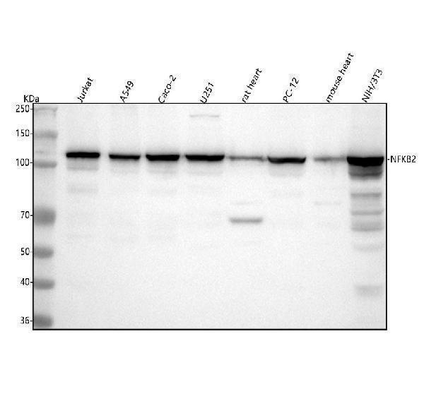
| Sample / lysate | human Jurkat, human A549, human CACO-2 (catalog M01228-1) |
| Gel % | 5–20% (catalog M01228-1) |
| Load | 30 ug; reducing conditions (catalog M01228-1) |
| Transfer | a nitrocellulose membrane at 150 mA for 50-90 minutes (catalog M01228-1) |
| Membrane | nitrocellulose membrane (catalog M01228-1) |
| Blocking | 5% non-fat milk/TBS for 1.5 hour at RT (catalog M01228-1) |
| Primary antibody | M01228-1 · 1:1000 (catalog M01228-1) |
| Primary incubation | overnight at 4°C (catalog M01228-1) |
| Secondary antibody | goat anti-rabbit IgG-HRP, 1:500 (catalog M01228-1) |
| Secondary incubation | 1.5 hour at RT (catalog M01228-1) |
| Wash | TBS-0.1%Tween 3 times with 5 minutes each (catalog M01228-1) |
| Detection | ECL (catalog M01228-1) |
NFKB2 p100 is predicted at 96.7 kDa, while an antibody QC blot shows approximately 110 kDa; the cause of this difference is not established.
| Band near 110 kDa | Empirical p100/p52 antibody band; its difference from the predicted mass is unexplained |
| Band near 96.7 kDa | Consistent with the predicted mass of full-length p100; confirm its identity |
| Band below full-length p100 | Could represent the p52 form; confirm with an antibody that recognizes the relevant region |
| Multiple bands | Could reflect p100, p52, or isoforms 1, 3, and 4; their separation is not established |
| Predicted full-length p100 mass | 96.7 kDa is the sequence-based reference; the empirical band is approximately 110 kDa |
| Phosphorylation at Ser713, Ser715, and Ser717 | May affect migration, but no visible size change is established |
| Isoforms 1, 3, and 4 | May differ in size; individual masses and resolvable band positions are not supplied |
| p52 homodimer | Could appear higher if the complex survives electrophoresis; no dimer band size is established |
| Situation | Likely cause | Next action |
|---|---|---|
| Band higher than expected | The approximately 110 kDa empirical band exceeds the 96.7 kDa predicted mass for an undetermined reason | Compare with the antibody QC band and verify identity using NFKB2 depletion or an independent antibody |
| Band lower than expected | The antibody may detect the p52 form | Check antibody epitope coverage and compare with a p100-specific antibody |
| Multiple bands | p100, p52, or isoforms 1, 3, and 4 may contribute | Use epitope-specific antibodies and NFKB2 depletion to assign bands |
| Weak or no signal | NFKB2 occurs in both nucleus and cytoplasm, so recovery may vary with extraction | Check extraction of both compartments and confirm sample loading |
| Fragments below expected size | A smaller band could be p52 or sample degradation | Compare with a p52-reactive antibody and repeat preparation with protease inhibitors |
Comprehensive Human Protein Atlas IHC scoring per tissue. Rows are taken directly from the HPA tissue chart — click any row's HPA link to view the source.
| Tissue | Cell type | Level | Evidence | Source |
|---|---|---|---|---|
| Appendix | lymphoid tissue | High | Protein (IHC) | HPA → |
| Lymph node | non-germinal center cells | High | Protein (IHC) | HPA → |
| Placenta | decidual cells | High | Protein (IHC) | HPA → |
| Skin | endothelial cells | High | Protein (IHC) | HPA → |
| Testis | spermatogonia cells | High | Protein (IHC) | HPA → |
| Tissue | Cell type | Level | Evidence | Source |
|---|---|---|---|---|
| Cerebellum | cells in granular layer | Not detected | Protein (IHC) | HPA → |
| Epididymis | glandular cells | Not detected | Protein (IHC) | HPA → |
| Heart muscle | cardiomyocytes | Not detected | Protein (IHC) | HPA → |
| Liver | cholangiocytes | Not detected | Protein (IHC) | HPA → |
| Ovary | follicle cells | Not detected | Protein (IHC) | HPA → |
Deeper troubleshooting and optimisation questions for NFKB2, answered from its protein features.
Catalog antibodies with Western blot application and product-specific WB images. Evaluate suitability with the reported sample, controls and experimental conditions.
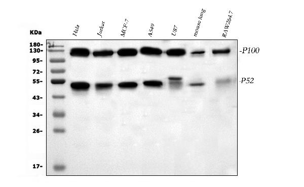
Two the supplier anti-NFKB2 antibodies have WB images. M01228-1 shows an approximately 110 kDa band in human, rat, and mouse samples; PB9150 shows approximately 52 and 120 kDa bands in human and mouse samples. These images document the listed conditions, not broader validation.
Which to pick: Choose M01228-1 for a documented rat sample or an approximately 110 kDa band. Choose PB9150 if you need an image showing both the 52 kDa active form and 120 kDa precursor. Both list human, mouse, and rat reactivity; PB9150’s image lacks rat samples.