This website uses cookies to ensure you get the best experience on our website.


- Table of Contents
Plan NIFK paraffin IHC around its nucleolar tissue pattern (HPA tissue IHC). Compare high staining in colon glandular cells with undetected staining in adipocytes (HPA tissue IHC), and start with the catalog antibody’s 1:50–1:200 IHC dilution range (datasheet: A04797).

Expected localisation, validated protocols, controls and antibodies — the at-a-glance facts below, then the full design guide.
| Expected localisation | Nucleolar staining in tissue sections (HPA tissue IHC) | |
| Staining pattern | Widespread nucleolar staining across cell types (HPA tissue IHC) | |
| Antigen retrieval | Tris-EDTA pH 9.0 HIER, 95–98 °C, 20 min (rule: nuclear antigen) | |
| Positive control | Caudate+4 more · see all | |
| Negative control | Adipose tissue+4 more · see all |
| Fixation | Keep fixation consistent across tissue samples (standard IHC practice; not target-specific) | |
| Caveat | Antibody staining has medium RNA concordance (HPA tissue IHC) | |
| Regulation | Low tissue specificity (HPA tissue RNA) | |
| Isoform / epitope | No isoforms reported; no membrane topology (UniProt) |
The catalog antibody’s IHC-P protocol is paired with one published NIFK IHC workflow using hospital samples (PMC10553176 methods).
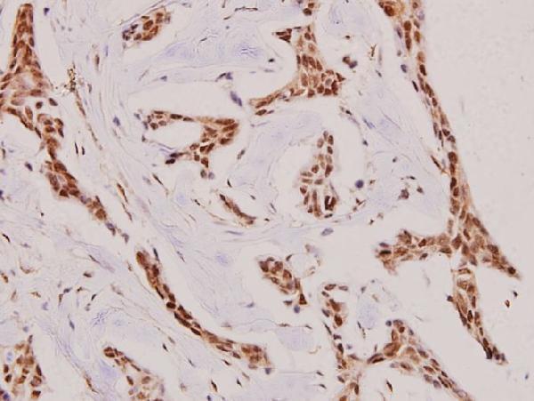
| Sample | Paraffin-embedded human breast carcinoma tissue; fixative not specified (datasheet A04797) |
| Fixation | Image fixative and duration unreported (datasheet A04797); verify before use. |
| Sectioning | 4–5 µm sections on charged slides (standard) |
| Deparaffinisation | Xylene, graded ethanol series to water (standard) |
| Antigen retrieval | Heat-induced epitope retrieval in Tris-EDTA buffer, pH 9.0, 20 min at 95–98 °C (standard rule: nuclear antigen) |
| Peroxidase block | 3% H2O2, 10 min, room temperature (standard) |
| Blocking | 10% normal serum of the secondary host, 30 min, room temperature (standard) |
| Primary antibody | Rabbit anti-NIFK, 1:50-1:200 (datasheet A04797) |
| Primary incubation | Overnight at 4 °C (standard) |
| Detection | HRP-polymer secondary, DAB chromogen 5–10 min (standard) |
| Counterstain | Hematoxylin, blue, dehydrate and mount (standard) |
| Expected result | NIFK-positive staining in neuronal cells of caudate (HPA tissue IHC: High). HPA tissue profile: Ubiquitous nucleolar expression. No signal in the no-primary control. |
NIFK is a nuclear protein concentrated in nucleoli and also localizes to mitotic chromosomes (UniProt Q9BYG3 subcellular location). In paraffin sections, expect nucleolar staining in many cell types, including strongly stained colon glandular cells and cerebral cortex neurons (HPA: ubiquitous nucleolar expression; High in both examples). Interpret intensity by cell type: HPA rates its tissue IHC profile Supported, with medium consistency between staining and RNA data (HPA: reliability). NIFK has no transmembrane segment (UniProt Q9BYG3 topology).
| Discrete nuclear dots in colon glandular cells or cerebral cortex neurons. | This fits the expected nucleolar pattern in cell types scored High by tissue IHC (HPA: tissue IHC). Judge staining within identifiable cells against the counterstained nucleus; the tissue-level label does not mean every cell must stain equally (HPA: cell-specific ratings). |
| Predominantly membrane or broad cytoplasmic staining, without convincing nucleolar signal. | Treat this as a suspect pattern: NIFK is nucleolar and lacks a transmembrane segment (UniProt Q9BYG3 subcellular location and topology). Mitotic chromosome staining is an expected exception to an exclusively nucleolar pattern (UniProt Q9BYG3 subcellular location). |
| Strong staining in adipocytes or smooth muscle cells. | These cell types were scored Not detected, so strong signal merits a specificity and detection-background check (HPA: adipocytes and smooth muscle cells). A single unexpected cell does not prove cross-reactivity; compare its compartment and the same-run controls (standard IHC practice). |
| Uniform haze across nuclei, cytoplasm, and extracellular areas. | This obscures the discrete nucleolar pattern and suggests nonspecific staining or detection background (HPA: ubiquitous nucleolar profile; standard IHC practice). Read cell-type intensity only after the background is controlled (standard IHC practice). |
| No detectable signal in colon glandular cells on the test slide. | Colon glandular cells are scored High, making this a useful positive-reference failure (HPA: colon glandular cells). First check the same-run positive section and staining workflow; absence alone cannot establish that NIFK is absent from this specimen (standard IHC practice). |
| Tissue and cell selection | Use a High-scored cell population to judge assay performance, such as colon glandular cells or cerebral cortex neurons (HPA: tissue IHC). Adipocytes and smooth muscle cells provide reported Not detected comparisons, but neither is a universal negative control (HPA: tissue IHC). |
| Interpreting a negative cell | HPA describes ubiquitous nucleolar expression but scores some specified cells Not detected and others Low, including lung macrophages (HPA: tissue IHC). Record the cell type and compartment before treating a faint or absent cell as an assay failure (standard IHC practice). |
| Mitotic cells | Chromosome-associated staining may be appropriate in mitosis because NIFK localizes to mitotic chromosomes with MKI67 (UniProt Q9BYG3 subcellular location). HPA ICC-IF also reports mitotic chromosome localization as supported (HPA: subcellular). |
| IHC evidence strength | HPA rates the overall tissue profile Supported with medium staining–RNA consistency; its listed antibodies have Supported IHC status (HPA: reliability and antibodies). Use the compartment and cell-type pattern together rather than treating a strong brown deposit alone as confirmation (standard IHC practice). |
| IF/ICC Q&A: what should fluorescence show? | Mainly nucleoli and nucleolar rims, with additional mitotic chromosome signal (HPA: subcellular ICC-IF). This is an interpretation reference for IF/ICC; evaluate the paraffin IHC result against the tissue IHC profile (HPA: tissue IHC and subcellular ICC-IF). |
| Situation | Likely cause | Next action |
|---|---|---|
| High-scored positive cells show no nucleolar signal. | The staining run may have failed, or the chosen cells may be difficult to identify (standard IHC practice). | Check a same-run section containing colon glandular cells, scored High, then review antigen retrieval, primary-antibody application, and detection steps using the validated IHC workflow (HPA: colon; standard IHC practice). |
| Membrane or diffuse cytoplasmic signal dominates. | The compartment conflicts with NIFK's nuclear and nucleolar localization (UniProt Q9BYG3 subcellular location). | Recheck focus and the nuclear counterstain; compare with a High-scored tissue and a primary-omission detection control before scoring the signal as NIFK (HPA: tissue IHC; standard IHC practice). |
| Strong deposits appear in adipocytes or smooth muscle cells. | Those populations are scored Not detected; cross-reactivity or endogenous detection activity is possible (HPA: tissue IHC; standard IHC practice). | Inspect staining without primary antibody, confirm cell identity, and compare nucleolar localization with a High-scored population on the same run (standard IHC practice; HPA: tissue IHC). |
| The whole section has brown haze. | Excess background can conceal nucleolar deposits (standard IHC practice). | Check reagent-only controls and the validated blocking, washing, primary-antibody dilution, and chromogen timing steps; reassess only after distinct nuclei can be read (standard IHC practice). |
| Only scattered dividing cells show chromosome-associated signal. | Mitotic chromosome localization is documented for NIFK (UniProt Q9BYG3 subcellular location; HPA: subcellular ICC-IF). | Confirm that the stained structures are in mitotic cells, then assess nucleolar staining separately in interphase cells from a High-scored population (UniProt Q9BYG3 subcellular location; HPA: tissue IHC). |
| A negative result is attributed to fixation sensitivity. | The supplied UniProt and HPA records do not report a NIFK-specific fixation effect (UniProt Q9BYG3 record; HPA: tissue IHC). | Document the processing conditions and compare same-run controls before assigning a cause; use antigen retrieval according to the validated IHC workflow (standard IHC practice). |


Comprehensive Human Protein Atlas IHC scoring per tissue (reliability: Supported — Medium consistency between antibody staining and RNA expression data.). Rows are taken directly from the HPA tissue chart — click any row's HPA link to view the source.
| Tissue | Cell type | Level | Evidence | Source |
|---|---|---|---|---|
| Caudate | Neuronal cells | High | Protein (IHC) | HPA → |
| Cerebral cortex | Neuronal cells | High | Protein (IHC) | HPA → |
| Colon | Glandular cells | High | Protein (IHC) | HPA → |
| Hippocampus | Neuronal cells | High | Protein (IHC) | HPA → |
| Lymph node | Non-germinal center cells | High | Protein (IHC) | HPA → |
| Tissue | Cell type | Level | Evidence | Source |
|---|---|---|---|---|
| Adipose tissue | Adipocytes | Not detected | Protein (IHC) | HPA → |
| Adrenal gland | Glandular cells | Not detected | Protein (IHC) | HPA → |
| Bronchus | Basal cells | Not detected | Protein (IHC) | HPA → |
| Nasopharynx | Basal cells | Not detected | Protein (IHC) | HPA → |
| Smooth muscle | Smooth muscle cells | Not detected | Protein (IHC) | HPA → |
Troubleshoot NIFK staining in paraffin sections by checking nucleolar localisation, tissue controls and the steps that can obscure or mimic chromogenic signal.
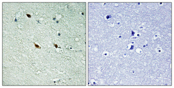
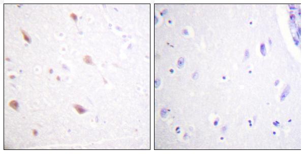
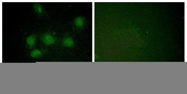
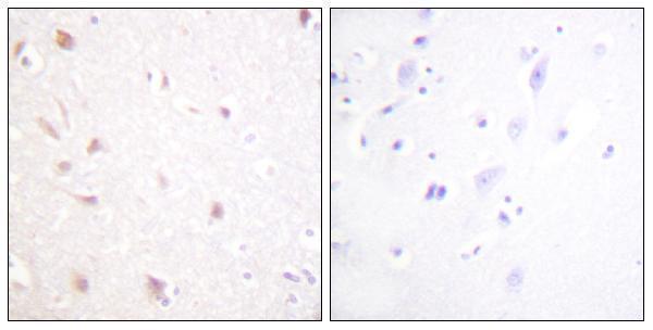
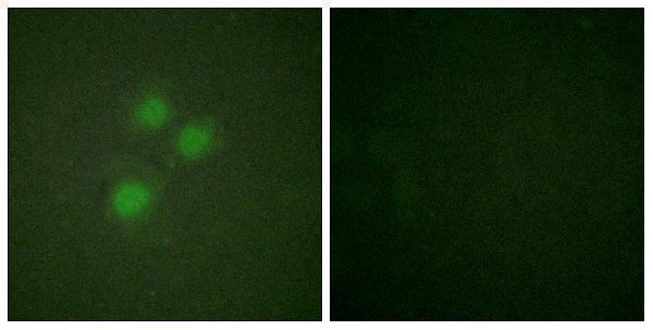
Anti-NIFK antibodies have IHC images from paraffin-embedded human breast carcinoma and brain, plus IF images from HUVEC cells; catalog reactivity includes human, mouse, and, for A04797, rat (catalog images and reactivity).
A04797 shows IHC in paraffin-embedded human breast carcinoma and lists IF, with human, mouse, and rat reactivity (A04797 IHC caption and catalog). P30452 shows phospho-Thr234 IHC in paraffin-embedded human brain and IF in HUVEC cells (P30452 image captions); A30452 shows IHC in paraffin-embedded human brain and IF in HUVEC cells (A30452 image captions).
Which to pick: For tissue IHC, choose A04797 for the demonstrated breast carcinoma sample, A30452 for NIFK in the demonstrated brain sample, or P30452 when the phospho-Thr234 target is required (each SKU’s IHC caption). For IF/ICC, A30452 and P30452 are listed for both applications, are polyclonal, and have their own HUVEC IF images (catalog applications, dilution data, and IF captions). A04797 is the cross-species choice when rat reactivity matters (catalog reactivity); the fixative is unreported in all three paraffin-section IHC captions.