This website uses cookies to ensure you get the best experience on our website.


- Table of Contents
Plan NKIRAS1 IHC-P around cytoplasmic staining, using appendix or colon glandular cells as positive tissue references (HPA tissue IHC). Start the IHC-validated antibody A11990 at 1 μg/mL (datasheet: A11990 IHC-P).

Expected localisation, validated protocols, controls and antibodies — the at-a-glance facts below, then the full design guide.
| Expected localisation | General cytoplasmic tissue staining (HPA tissue IHC) | |
| Staining pattern | High glandular-cell staining, generally cytoplasmic (HPA tissue IHC) | |
| Antigen retrieval | Citrate pH 6.0 HIER, 95–98 °C, 20 min (rule: cytoplasmic / membrane antigen) | |
| Positive control | Appendix+4 more · see all | |
| Negative control | Adipose tissue+4 more · see all |
| Fixation | Keep fixation consistent across sections. (standard IHC practice; not target-specific) | |
| Caveat | Staining has medium consistency with RNA expression (HPA tissue IHC) | |
| Regulation | Expression regulation unreported (UniProt) | |
| Isoform / epitope | One 1–192 chain; no isoforms annotated (UniProt) |
Compare the catalog antibody’s IHC-P protocol with the published NKIRAS1 staining protocol for human renal biopsies (PMC9206042).
| Sample | Tissue sections; selected-image fixative not specified (standard IHC workflow) |
| Fixation | Image fixative and duration unreported (datasheet A11990); verify before use. |
| Sectioning | 4–5 µm sections on charged slides (standard) |
| Deparaffinisation | Xylene, graded ethanol series to water (standard) |
| Antigen retrieval | Heat-induced epitope retrieval in citrate buffer, pH 6.0, 20 min at 95–98 °C (standard rule: cytoplasmic / membrane antigen) |
| Peroxidase block | 3% H2O2, 10 min, room temperature (standard) |
| Blocking | 10% normal serum of the secondary host, 30 min, room temperature (standard) |
| Primary antibody | Rabbit anti-NKIRAS1, 1 μg/mL (datasheet A11990) |
| Primary incubation | Overnight at 4 °C (standard) |
| Detection | HRP-polymer secondary, DAB chromogen 5–10 min (standard) |
| Counterstain | Hematoxylin, blue, dehydrate and mount (standard) |
| Expected result | NKIRAS1-positive staining in glandular cells of appendix (HPA tissue IHC: High). HPA tissue profile: General cytoplasmic expression. No signal in the no-primary control. |
NKIRAS1 is cytoplasmic and has no transmembrane segment (UniProt Q9NYS0: subcellular location; topology). In paraffin-section IHC, expect cytoplasmic staining in selected glandular and neuronal cells, including colon glandular cells and caudate neurons (HPA: general cytoplasmic expression; High in both). HPA rates its tissue staining Approved, with medium consistency between staining and RNA expression; interpret individual results with that limitation (HPA: reliability).
| Clear cytoplasmic stain in colon or appendix glandular cells, or caudate neurons. | This matches the reported compartment and cells (HPA: general cytoplasmic expression; High in these cells). Score the cell population and compartment, since HPA also reports low or undetected staining in other populations (HPA: tissue IHC). |
| Predominantly nuclear stain, with little cytoplasmic signal in otherwise positive cells. | A nuclear-only result conflicts with the reported cytoplasmic pattern (UniProt Q9NYS0: Cytoplasm; HPA: general cytoplasmic expression). Treat it as unresolved staining, then check controls and detection conditions before assigning NKIRAS1 localisation (general IHC practice). |
| Strong stain in cells reported as undetected, such as adipocytes or ovarian stroma cells. | That result warrants a specificity check; cross-reactivity or endogenous detection activity may contribute (general IHC practice; HPA: Not detected in these cells). These are cell-level reference observations, not proof that every cell in those tissues must be negative (HPA: tissue IHC). |
| Diffuse color across cells and empty spaces, with poor separation of cytoplasm from background. | The pattern cannot establish cytoplasmic NKIRAS1 staining (UniProt Q9NYS0: Cytoplasm; general IHC practice). Inspect the no-primary control and background before scoring intensity; diffuse color alone does not identify the stained cell population (general IHC practice). |
| No visible signal in colon or appendix glandular cells used as a positive reference. | This misses populations reported as High by HPA (HPA: colon and appendix glandular cells). Check that the relevant cells are present, then assess retrieval, primary incubation and detection with suitable controls (general IHC practice). HPA reports medium staining–RNA consistency, so one section is not definitive (HPA: reliability). |
| Which compartment should guide IHC scoring? | Use cytoplasmic staining as the main criterion (UniProt Q9NYS0: Cytoplasm; HPA: general cytoplasmic expression). HPA also describes High Purkinje-cell staining as cytoplasm/membrane; do not generalize that observation to every tissue (HPA: cerebellum). |
| How should tissue choice affect the reference comparison? | Colon and appendix glandular cells provide reported High examples; adipocytes and skeletal myocytes are reported Not detected (HPA: tissue IHC). Compare the named cells, since the HPA categories describe cell populations within tissues (HPA: tissue IHC). |
| What does the antibody-validation label support? | The listed antibody HPA077387 is Approved for IHC, while the overall tissue result has medium staining–RNA consistency (HPA: antibody validation; reliability). This supports a reference pattern, but does not establish that every unexpected signal is NKIRAS1 (general IHC practice). |
| Does the protein record suggest a membrane or shed-protein pattern? | NKIRAS1 has no transmembrane segment, signal peptide or propeptide, and its annotated chain spans residues 1–192 (UniProt Q9NYS0: topology; processing). These annotations support a primarily cytoplasmic expectation; they do not identify the antibody epitope (UniProt Q9NYS0; general IHC practice). |
| IF/ICC: what localisation is reported? | HPA reports supported cytosol localisation and uncertain endoplasmic-reticulum localisation in ICC-IF; the listed antibody is Supported for ICC (HPA: subcellular; antibody validation). Use that as an IF comparison, without treating the uncertain ER signal as a required IHC feature. |
| Situation | Likely cause | Next action |
|---|---|---|
| Colon or appendix glandular cells are unstained. | The expected High cell population may be absent from the section, or the IHC detection workflow may have failed (HPA: tissue IHC; general IHC practice). | Confirm glandular cells are present; review retrieval, primary-antibody and detection steps, and examine a suitable positive control (general IHC practice). |
| Nuclei dominate the stain in a reported positive cell population. | Nuclear-only staining conflicts with the reported cytoplasmic location (UniProt Q9NYS0: Cytoplasm; HPA: general cytoplasmic expression). | Compare with a no-primary control, inspect cytoplasmic signal separately, and avoid scoring nuclear color as a positive NKIRAS1 pattern (general IHC practice). |
| Adipocytes or skeletal myocytes stain strongly. | Those named cell populations are reported Not detected; an off-target or detection-system signal is possible (HPA: tissue IHC; general IHC practice). | Verify cell identity and examine no-primary and other appropriate specificity controls before interpreting the stain (general IHC practice). |
| Color covers tissue and empty spaces evenly. | Diffuse background obscures the cell-level cytoplasmic pattern needed for interpretation (HPA: general cytoplasmic expression; general IHC practice). | Check background controls, blocking, washes and detection development; rescore only where cell borders and cytoplasm remain interpretable (general IHC practice). |
| A section called negative contains a few stained cells. | HPA's Not detected labels apply to specified cell populations; UniProt describes NKIRAS1 as widely expressed (HPA: tissue IHC; UniProt Q9NYS0: tissue specificity). | Identify the stained cells and compare them with the matching HPA cell category before calling the tissue result discordant (HPA: tissue IHC; general IHC practice). |
| Only a membrane-like edge is visible in Purkinje cells. | HPA describes High Purkinje-cell staining as cytoplasm/membrane, while its overall tissue profile is cytoplasmic (HPA: cerebellum; tissue profile). | Look for accompanying cytoplasmic signal and compare controls; do not extend the Purkinje-cell observation to other cell types (HPA: tissue IHC; general IHC practice). |


![HPA ICC-IF BJ [Human fibroblast] — NKIRAS1 (HPA077387)](https://images.proteinatlas.org/77387/1866_A9_19_cr5b6ac765b4de7_blue_red_green.jpg)
![HPA ICC-IF BJ [Human fibroblast] — NKIRAS1 (HPA077387)](https://images.proteinatlas.org/77387/1866_A9_34_blue_red_green.jpg)
Comprehensive Human Protein Atlas IHC scoring per tissue (reliability: Approved — Medium consistency between antibody staining and RNA expression data.). Rows are taken directly from the HPA tissue chart — click any row's HPA link to view the source.
| Tissue | Cell type | Level | Evidence | Source |
|---|---|---|---|---|
| Appendix | Glandular cells | High | Protein (IHC) | HPA → |
| Caudate | Neuronal cells | High | Protein (IHC) | HPA → |
| Cerebellum | Purkinje cells - cytoplasm/membrane | High | Protein (IHC) | HPA → |
| Cerebral cortex | Neuronal cells | High | Protein (IHC) | HPA → |
| Colon | Glandular cells | High | Protein (IHC) | HPA → |
| Tissue | Cell type | Level | Evidence | Source |
|---|---|---|---|---|
| Adipose tissue | Adipocytes | Not detected | Protein (IHC) | HPA → |
| Liver | Cholangiocytes | Not detected | Protein (IHC) | HPA → |
| Ovary | Ovarian stroma cells | Not detected | Protein (IHC) | HPA → |
| Skeletal muscle | Myocytes | Not detected | Protein (IHC) | HPA → |
| Spleen | Cells in red pulp | Not detected | Protein (IHC) | HPA → |
Troubleshoot NKIRAS1 staining by checking retrieval, cellular compartment and cell type before comparing chromogenic signal across paraffin sections.
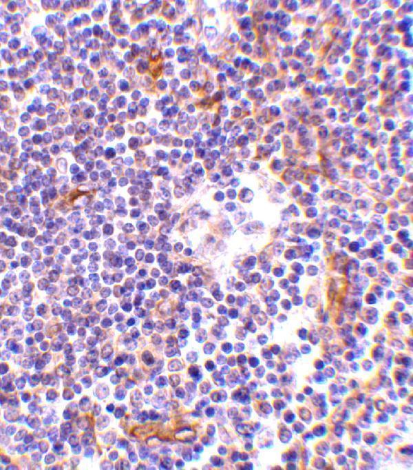
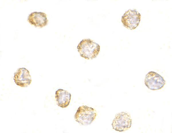
Anti-NKIRAS1 antibodies have IHC data from human lymph node tissue (A11990 image caption) and ICC data from RAW264.7 cells (A11990-1 image caption).
A11990 renders with a human lymph node IHC image at 1 μg/mL (A11990 image caption). A11990-1 renders with a RAW264.7 cell ICC image at 1 μg/mL (A11990-1 image caption).
Which to pick: Choose A11990 for paraffin-section IHC (catalog: IHC-P); its human lymph node image documents tissue IHC, but the fixative is unreported (A11990 image caption). Choose A11990-1 for ICC/IF of cells (catalog: ICC; A11990-1 image caption), and A11990 for the broader listed species reactivity of human, mouse and rat (catalog: A11990 reactivity); IHC image evidence is from human tissue only (A11990 image caption). Clonality is unreported for both antibodies (catalog: clone fields).