NKX3-1 / Homeobox protein Nkx-3.1 · IHC design guide

Design Immunohistochemistry for NKX3-1

Plan paraffin-section IHC around nuclear staining in prostate glandular cells (HPA tissue IHC). Use the catalog antibody’s IHC protocol and compare staining with tissue controls.

Evidence assembled Oct 2026 · For research use; verify linked source records and product datasheet before use
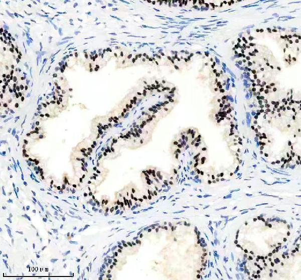
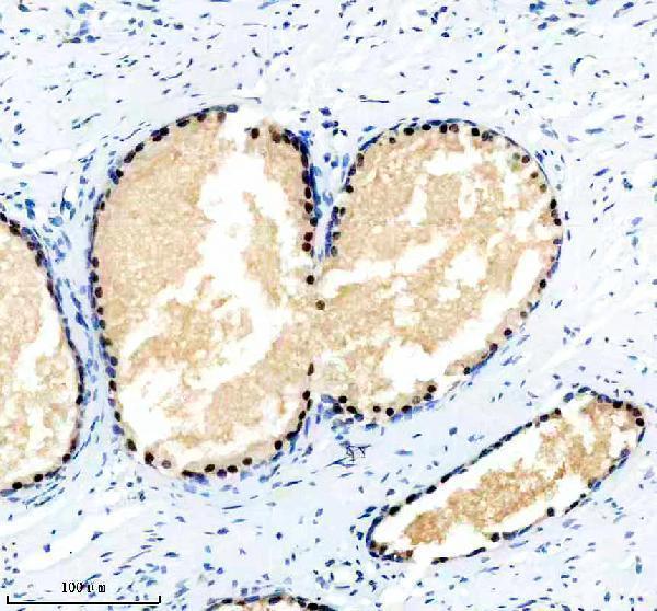
Immunohistochemistry protocol sheet for NKX3-1 (IHC for NKX3-1): expected localisation Nuclei of prostate glandular cells (HPA tissue IHC), antibody M02709, validated IHC image, and IHC protocol steps
Printable NKX3-1 IHC protocol sheet — expected localisation Nuclei of prostate glandular cells (HPA tissue IHC), antibody M02709, controls and protocol steps. Open the full NKX3-1 IHC guide →

NKX3-1 Immunohistochemistry Experimental Design Guide

Expected localisation, validated protocols, controls and antibodies — the at-a-glance facts below, then the full design guide.

Must know before staining
Expected localisation Nuclei of prostate glandular cells (HPA tissue IHC)
Staining pattern Strong nuclear staining in prostate glandular cells (HPA tissue IHC)
Antigen retrieval EDTA pH 8.0 HIER, heat-mediated (datasheet M02709)
Positive control ⓘ Prostate+2 more · see all
Negative control ⓘ Adipose tissue+4 more · see all
Important caveats
Reasons your staining may differ from the expected pattern.
Fixation Keep fixation consistent across sections (standard IHC practice; not target-specific)
Caveat Bronchus and cervix also show staining (HPA tissue IHC)
Regulation TOPORS promotes proteasomal degradation (UniProt)
Isoform / epitope Five isoforms; check antibody epitope coverage (UniProt)
Section 1

Recommended NKX3-1 IHC & IF Protocols

The catalog antibody’s IHC-P protocol (datasheet M02709) is accompanied by 3 published NKX3-1 IHC methods (PMC3072223; PMC8019685; PMC12190382).

Recommended immunohistochemistry (IHC-P) protocol parameters
SampleParaffin-embedded human prostate cancer tissue; fixative not specified (datasheet M02709)
FixationImage fixative and duration unreported (datasheet M02709); verify before use.
Sectioning4–5 µm sections on charged slides (standard)
DeparaffinisationXylene, graded ethanol series to water (standard)
Antigen retrievalHeat retrieval: EDTA pH 8.0 (datasheet M02709); 20 min, 95–100 °C (standard)
Peroxidase block3% H2O2, 10 min, room temperature (standard)
Blocking10% goat serum (datasheet M02709)
Primary antibodyRabbit monoclonal (clone 19N21) anti-NKX3-1, 1:50 (datasheet M02709)
Primary incubationOvernight at 4 °C (datasheet M02709)
DetectionHRP-conjugated secondary, DAB chromogen (datasheet M02709)
CounterstainHematoxylin, blue, dehydrate and mount (standard)
Expected resultNKX3-1-positive staining in glandular cells of prostate (HPA tissue IHC: High). HPA tissue profile: Nuclear expression mainly in the prostate gland. No signal in the no-primary control.
💡Decision noteStart with EDTA pH 8.0 heat retrieval for the catalog antibody (datasheet M02709). Use the reported retrieval conditions when reproducing a published method (PMC3072223; PMC8019685).
Section 2

What Is the Expected NKX3-1 Staining Pattern?

NKX3-1 is a nuclear protein with no transmembrane segment (UniProt Q99801). In paraffin-section IHC, expect staining mainly in prostate glandular cell nuclei (HPA: nuclear expression mainly in prostate gland; High in prostate glandular cells). HPA rates the tissue IHC profile Enhanced, while describing only medium consistency between antibody staining and RNA expression (HPA: reliability description).

What am I looking at on my slide?
Distinct nuclear staining in prostate glandular cells, with little signal outside nuclei.This matches the expected compartment and strongest reported tissue pattern (UniProt Q99801: nucleus; HPA: High in prostate glandular cells). Record the proportion and intensity of positive glandular nuclei separately; a brown counterstain or general tissue discoloration is not nuclear target staining (general IHC practice).
Staining is predominantly cytoplasmic or outlines cell membranes instead of marking nuclei.Treat this IHC pattern as a possible staining artefact and check controls before scoring it positive (UniProt Q99801: nucleus; HPA: tissue IHC profile mainly nuclear). HPA reports uncertain cytosolic localization in ICC-IF, which does not establish a cytoplasmic IHC pattern (HPA: cytosol uncertain).
Strong staining appears in an unexpected cell population or in cells of a reference negative tissue.Consider cross-reactivity or endogenous detection activity, then compare with a no-primary control and the expected cell-specific pattern (general IHC practice). For example, HPA reports NKX3-1 as Not detected in adipocytes of adipose tissue; that reference does not prove every cell in the section is negative (HPA: adipose tissue).
Diffuse color covers nuclei, cytoplasm, and tissue spaces without clear cell boundaries.This is background rather than an interpretable nuclear pattern (general IHC practice; UniProt Q99801: nucleus). Inspect the no-primary control, blocking and wash conditions, and chromogen development; background can obscure genuine glandular nuclear staining (general IHC practice).
No nuclear signal appears in a prostate glandular positive-control section.The run is inconclusive until the positive control works (HPA: High in prostate glandular cells; general IHC practice). Check section identity, antibody preparation, retrieval conditions, and detection reagents. The supplied sources do not identify an NKX3-1-specific fixation sensitivity or prescribe a retrieval condition.
💡Expected NKX3-1 appearanceCall a result positive when prostate glandular nuclei show clear, relatively strong staining (HPA: High in prostate glandular cells; UniProt Q99801: nucleus); diffuse color or isolated membrane staining is a suspect false positive (general IHC practice).
How each factor affects the staining
Tissue and cell selectionProstate glandular cells provide the strongest supplied reference; bronchial respiratory epithelial and cervical glandular cells show Medium staining (HPA: tissue IHC). A weaker signal in those tissues is therefore less decisive than a prostate nuclear control (general IHC interpretation).
Antibody evidenceHPA lists IHC validation as Enhanced for HPA078571, whereas HPA025693 has ICC Supported and no listed IHC status (HPA: antibody validation). Use an IHC-validated antibody for paraffin-section interpretation; ICC validation alone does not establish IHC performance (general assay practice).
Isoforms and epitope coverageUniProt lists five NKX3-1 isoforms (UniProt Q99801). Without an epitope map for the chosen antibody, the supplied evidence cannot say which isoforms it detects or whether isoform differences explain a weak or absent IHC result.
Processing and topologyUniProt annotates a single 1–234 protein chain, with no signal peptide, propeptide, or transmembrane segment (UniProt Q99801). Interpret a membrane-only pattern cautiously; these annotations do not establish epitope accessibility after fixation.
Retrieval and detectionAntigen retrieval, blocking, washes, and detection controls are routine paraffin-section IHC variables (general IHC practice). Optimize them against positive and no-primary controls; HPA tissue staining and UniProt annotations provide no NKX3-1-specific retrieval or fixation-effect claim.
Q: How should IF/ICC observations affect this IHC call?A: HPA supports nucleoplasmic localization and labels cytosolic localization uncertain in ICC-IF images from SiHa and U-251MG (HPA: subcellular). Use that as localization context, while scoring paraffin-section IHC against its mainly nuclear tissue pattern (HPA: tissue IHC).
Why is my staining missing, weak or wrong?
SituationLikely causeNext action
Prostate positive control is blank.A failed staining step or unsuitable assay settings are possible; the sources do not identify a target-specific fixation cause.Verify the control section and reagent sequence, then review retrieval, antibody dilution, and detector function using the IHC-validated antibody (HPA: High in prostate glandular cells; general IHC practice). Do not interpret study negatives until the control stains.
Prostate nuclei stain weakly while background remains low.The assay may have insufficient detectable signal, but weak staining alone does not identify the failing step (general IHC practice).Confirm that the prostate glandular reference is present, then optimize retrieval and antibody concentration within the chosen assay's instructions (HPA: High in prostate glandular cells; general IHC practice). Record changes against the same positive control.
Signal is mostly cytoplasmic or membranous.Nonspecific staining is possible because the expected IHC profile is mainly nuclear (HPA: tissue IHC; UniProt Q99801: nucleus).Inspect the no-primary control and compare the IHC-validated antibody's result with the nuclear reference pattern (general IHC practice; HPA: HPA078571 IHC Enhanced). Do not score cytoplasmic color alone as a positive nuclear result.
Unexpected cells stain strongly.Cross-reactivity or endogenous detection activity may account for a cell-type mismatch (general IHC practice).Check a no-primary control and evaluate staining by cell type, not whole-section color (general IHC practice). Compare with HPA's named reference populations; HPA reports Not detected in adrenal glandular cells (HPA: adrenal gland).
Brown haze obscures nuclear boundaries.Excess detection signal, incomplete washing, or inadequate blocking may contribute to diffuse background (general IHC practice).Review wash, blocking, and chromogen development steps alongside a no-primary control (general IHC practice). Score NKX3-1 only where nuclear staining can be distinguished from surrounding color (UniProt Q99801: nucleus; general IHC practice).
Medium-staining reference tissue appears negative while prostate stains.The expected signal is lower in bronchial respiratory epithelial or cervical glandular cells than in prostate glandular cells (HPA: Medium in bronchus and cervix; High in prostate).Check tissue identity and cell type, then report the discrepancy without treating that weaker reference as proof of assay failure (HPA: tissue IHC; general IHC interpretation). Preserve the prostate positive control and no-primary control for the run.

Sample controls for NKX3-1 IHC & IF

🧪Run prostate first: glandular cells should show nuclear staining (HPA: High in prostate glandular cells; UniProt Q99801: nucleus). Use adrenal glandular cells as a negative tissue comparator (HPA: Not detected in adrenal glandular cells); on the prostate slide, assess adjacent nonglandular cells for background rather than scoring them as positive glandular cells (HPA: High in prostate glandular cells).
Positive control tissue: Prostate (Glandular cells, HPA High)
Negative control tissue: Adipose tissue (HPA Not detected)
ICC-IF cell lines (HPA subcellular resource): HPA ICC-IF images show NKX3-1 in SiHa, U-251MG, with annotated localisation: Nucleoplasm (supported), Cytosol (uncertain) (HPA subcellular).
Technical controls: Include no-primary (secondary-only) and primary-matched isotype controls, matching the primary antibody’s host species and clonality, plus NKX3-1 knockout material as a biological negative if available (standard IHC practice). Block endogenous peroxidase before HRP/DAB detection in prostate sections; for IF, assess tissue autofluorescence with a no-primary control (selected-SKU IHC caption: HRP/DAB detection; standard IHC/IF practice).
⚠️Feasibility: The selected M02709 paraffin-section example used heat retrieval in EDTA at pH 8.0, but its fixative is unreported; a target-specific fixation window and retrieval dependency are also unreported (selected-SKU IHC caption). The supplied evidence does not establish whether frozen sections or IF/ICC are easier than paraffin IHC; for IF localisation, nucleoplasmic signal is supported while cytosolic signal is uncertain (HPA subcellular). No prostate-specific artefact is reported in the supplied evidence (selected-SKU IHC caption; HPA prostate row).

HPA tissue IHC evidence for NKX3-1

Comprehensive Human Protein Atlas IHC scoring per tissue (reliability: Enhanced — Medium consistency between antibody staining and RNA expression data.). Rows are taken directly from the HPA tissue chart — click any row's HPA link to view the source.

Positive expression · recommended positive controls

TissueCell typeLevelEvidenceSource
Prostate Glandular cells High Protein (IHC) HPA →
Bronchus Respiratory epithelial cells Medium Protein (IHC) HPA →
Cervix Glandular cells Medium Protein (IHC) HPA →

Undetected expression · recommended negative controls

TissueCell typeLevelEvidenceSource
Adipose tissue Adipocytes Not detected Protein (IHC) HPA →
Adrenal gland Glandular cells Not detected Protein (IHC) HPA →
Appendix Glandular cells Not detected Protein (IHC) HPA →
Bone marrow Hematopoietic cells Not detected Protein (IHC) HPA →
Breast Adipocytes Not detected Protein (IHC) HPA →
Section 3

Advanced NKX3-1 IHC Tips

Troubleshoot NKX3-1 chromogenic IHC by checking nuclear staining in the expected cells, the documented retrieval conditions, and controls run on the same batch.

What should I change when nuclear staining is weak after antigen retrieval?
Start with heat-mediated retrieval in EDTA buffer at pH 8.0 (datasheet M02709). The documented paraffin-section image used that retrieval before an overnight incubation at 4°C with the catalog antibody at 1:50 (datasheet M02709). Check that sections remain covered by buffer throughout heating and cooling, then compare a known positive prostate section processed in the same batch (standard IHC practice; HPA: high in prostate glandular cells). If nuclear signal remains weak, compare retrieval heating and cooling conditions on adjacent sections while holding detection and exposure to DAB constant (standard IHC practice). Judge improvement by cleaner glandular-cell nuclei, since NKX3-1 is nuclear (UniProt Q99801; HPA: nuclear expression mainly in prostate).
Could fixation explain weak or uneven NKX3-1 staining?
Target-specific fixation sensitivity is unknown because the selected paraffin-section caption does not report a fixative (datasheet M02709). Record the fixative, fixation duration, and processing history for each block before attributing a staining difference to NKX3-1 biology (standard IHC practice). Compare adjacent sections from the same block using the documented EDTA pH 8.0 retrieval and 1:50 primary dilution (datasheet M02709; standard IHC practice). Include a prostate glandular-cell control in every run and assess whether its nuclear staining changes alongside the specimen (HPA: high in prostate glandular cells; standard IHC practice). Uneven staining across a section warrants inspection of tissue preservation and processing records before changing antibody conditions (standard IHC practice).
How should I assess apparent cytoplasmic NKX3-1 staining in a paraffin section?
Score nuclear staining first: NKX3-1 is annotated in the nucleus, and tissue IHC shows nuclear expression mainly in prostate glandular cells (UniProt Q99801; HPA: tissue IHC). The subcellular IF record supports nucleoplasm but marks cytosol uncertain, so cytoplasmic chromogen alone is insufficient evidence of target localisation (HPA: subcellular). Check whether the brown signal follows cell borders, folds, damaged areas, or diffuse DAB deposition, and review a no-primary control (standard IHC practice). Compare intact glandular-cell nuclei in the same section and a positive control processed together (HPA: high in prostate glandular cells; standard IHC practice). Document nuclear and cytoplasmic patterns separately rather than combining them into one positive score (standard IHC practice).
Can a negative stain rule out every NKX3-1 isoform?
No: the record lists 5 NKX3-1 isoforms, but the supplied antibody caption does not define its recognized epitope or isoform coverage (UniProt Q99801; datasheet M02709). Therefore, a negative paraffin-section result cannot establish that every isoform is absent (UniProt Q99801; standard IHC interpretation). The annotated protein has no transmembrane segment, signal peptide, glycosylation sites, or modified residues, which does not resolve antibody accessibility in processed tissue (UniProt Q99801). Verify the antibody's immunogen or epitope documentation before making isoform-specific claims, and compare a known positive prostate section under identical retrieval and detection conditions (standard IHC practice; HPA: high in prostate glandular cells). Report results as staining with this antibody unless isoform recognition has been established independently (standard IHC practice).
How can IF help resolve uncertain nuclear staining in a multiplex experiment?
Use a glandular epithelial marker to identify the expected positive cells, then assess whether NKX3-1 signal overlaps their nuclei (HPA: high in prostate glandular cells; UniProt Q99801: nucleus; standard IF practice). Choose spectrally separated fluorophores and inspect unstained tissue for autofluorescence before interpreting a weak channel (standard IF practice). Because NKX3-1 lacks a transmembrane segment and has a supported nucleoplasmic location, use permeabilisation compatible with access to intracellular nuclear epitopes (UniProt Q99801; HPA: subcellular; standard IF practice). Include single-marker and secondary-only controls to check bleed-through and nonspecific fluorescence (standard IF practice). The HPA IF record marks cytosol uncertain, so require convincing nuclear localisation before treating cytoplasmic fluorescence as corroboration (HPA: subcellular).
What causes diffuse brown staining despite a nuclear positive control?
Check a no-primary control for detection-system background and a secondary-only control when the workflow permits (standard IHC practice). The documented image used a peroxidase-conjugated secondary and DAB, so verify the peroxidase block and monitor chromogen development for diffuse deposition (datasheet M02709; standard IHC practice). Recheck the documented 10% goat-serum block and 1:50 primary dilution before increasing antibody concentration (datasheet M02709). Inspect folds, edges, and damaged areas separately from intact glandular-cell nuclei because these regions can trap reagents or accentuate staining (standard IHC practice). Accept a run only when the positive control shows interpretable nuclear signal and its negative controls remain suitably clean (UniProt Q99801: nucleus; standard IHC practice).
How should I score heterogeneous NKX3-1 staining across prostate glands? ⚠ ANSWER MARKED FOR VERIFICATION
Define the glandular-cell compartment before scoring because HPA reports high expression in prostate glandular cells and predominantly nuclear tissue staining (HPA: tissue IHC). Record the percentage of positive nuclei and nuclear intensity separately; an H-score can combine intensity categories with their percentages when the scoring scheme is stated (standard IHC practice). Normalise counts to the number of evaluable glandular-cell nuclei, or express positive-cell density per mm² of annotated viable tissue (standard IHC practice). Exclude folds, necrosis, and poorly preserved regions using the same criteria across specimens (standard IHC practice). Keep retrieval and DAB development consistent across batches, and use a shared control section to track staining drift (standard IHC practice).
When is NKX3-1 staining convincing rather than artefactual?
Convincing staining is nuclear in intact glandular cells and agrees with a positive prostate control processed in the same run (UniProt Q99801: nucleus; HPA: high in prostate glandular cells; standard IHC practice). Treat isolated cytoplasmic signal cautiously because the tissue profile is mainly nuclear and the IF cytosolic location is uncertain (HPA: tissue IHC; HPA: subcellular). Review edge staining, folds, and necrotic areas for processing artefacts, and check a no-primary control for endogenous-enzyme or detection background (standard IHC practice). Medium bronchial or cervical staining and low testis staining are reported, so cell context matters when interpreting a positive outside prostate (HPA: tissue IHC). Report compartment, cell type, control results, and staining extent together rather than treating any brown deposit as NKX3-1 positivity (standard IHC practice).
Boster reagents

Best NKX3-1 / Homeobox protein Nkx-3.1 IHC Antibodies

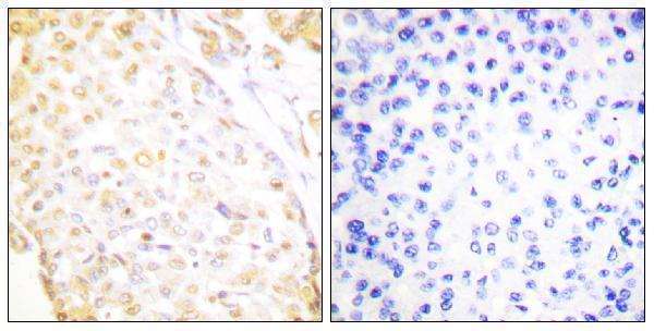
Catalog images show NKX3-1 IHC in paraffin-embedded human prostate cancer and breast carcinoma (M02709 and A30460 captions); A30460 also lists IF/ICC and human, mouse, and rat reactivity (A30460 applications/reactivity).

Real IHC data IHC analysis of NKX3-1 using anti-NKX3-1 antibody (M02709). NKX3-1 was detected in a paraffin-embedded section of human prostate cancer tissue. Heat mediated antigen retrieval was performed in EDTA buffer (pH 8.0, epitope retrieval solution). The tissue section was blocked with 10% goat serum. The tissue section was then incubated with 1: 50 rabbit anti-NKX3-1 Antibody (M02709) overnight at 4°C. Peroxidase Conjugated Goat Anti-rabbit IgG was used as secondary antibody and incubated for 30 minutes at 37°C. The tissue section was developed using HRP Conjugated Rabbit IgG Super Vision Assay Kit (Catalog # SV0002) with DAB as the chromogen.
Anti-NKX3.1 Rabbit Monoclonal Antibody
Cat # M02709
Real IHC data Immunohistochemistry analysis of paraffin-embedded human breast carcinoma, using NKX3.1 Antibody. The picture on the right is blocked with the synthesized peptide.
Anti-NKX3.1 Antibody
Cat # A30460

M02709 will render with an IHC image of paraffin-embedded human prostate cancer tissue (M02709 image caption). A30460 will render with an IHC image of paraffin-embedded human breast carcinoma; its catalog also lists IF/ICC, but supplies no IF image (A30460 image caption; A30460 applications and IF image alts).

Which to pick: For human prostate tissue IHC, M02709 provides a rabbit monoclonal antibody with a documented 1:50 paraffin-section staining example; the fixative is unreported (M02709 catalog entry and image caption). For IF/ICC, choose A30460 because those applications are listed for this rabbit polyclonal antibody; its supplied figure documents IHC rather than IF (A30460 catalog entry and image caption). A30460 also lists mouse and rat reactivity alongside human, with paraffin-section IHC shown only in human breast carcinoma; that caption does not report the fixative (A30460 reactivity and image caption).

Each figure is that product's own IHC / IF validation image from its datasheet.

References

  1. UniProt Consortium. UniProt entry Q99801 (NKX31_HUMAN, Homeobox protein Nkx-3.1).
  2. Human Protein Atlas. NKX3-1 tissue IHC expression (reliability: Enhanced).
  3. Human Protein Atlas. NKX3-1 subcellular location (ICC-IF): Localized to the nucleoplasm and cytosol..
  4. Human Protein Atlas. NKX3-1 antibody validation summary (2 antibodies).
  5. Diagnostic and Clinicopathological Utility of High-Molecular-Weight Cytokeratin (HMWCK) and NKX3.1 Immunohistochemistry in Prostatic Adenocarcinoma. Cureus 2026 — PMC13381367.
  6. NKX3.1 as a marker of prostatic origin in metastatic tumors. The American journal of surgical pathology 2010 — PMC3072223.
  7. Skene's Gland Derivatives in the Female Genital Tract and Cervical Adenoid Basal Carcinoma are Consistently Positive With Prostatic Marker NKX3.1. International journal of gynecological pathology : official journal of the International Society of Gynecological Pathologists 2021 — PMC8019685.
  8. Deletions of LPL and NKX3.1 in Prostate Cancer Progression: Game Changers or By-Standers in Tumor Evolution. Biomolecules 2025 — PMC12190382.
  9. PubMed PMID:9226374 — UniProt-cited evidence.
  10. PubMed PMID:9537602 — UniProt-cited evidence.
  11. PubMed PMID:11137288 — UniProt-cited evidence.