This website uses cookies to ensure you get the best experience on our website.


- Table of Contents
Plan NKX3-2 IHC-P around nuclear tissue staining (HPA tissue IHC), starting the catalog antibody at 5 μg/mL (datasheet). Keep fixation consistent and interpret staining with the reported low agreement with RNA expression in mind (HPA tissue IHC).

Expected localisation, validated protocols, controls and antibodies — the at-a-glance facts below, then the full design guide.
| Expected localisation | Nuclear staining in tissues (HPA tissue IHC) | |
| Staining pattern | Nuclear staining across all tissues (HPA tissue IHC) | |
| Antigen retrieval | Tris-EDTA pH 9.0 HIER, 95–98 °C, 20 min (rule: nuclear antigen) | |
| Positive control | Adipose tissue+4 more · see all | |
| Negative control | None in HPA (detected in all 45 tissues); use no-primary + isotype controls |
| Fixation | Keep fixation consistent across sections. (standard IHC practice; not target-specific) | |
| Caveat | Low consistency between staining and RNA expression (HPA tissue IHC) | |
| Regulation | No stimulus-linked regulation annotated (UniProt) | |
| Isoform / epitope | No isoforms or processing annotated; epitope effects unknown (UniProt) |
The catalog antibody’s IHC-P protocol is accompanied by one published NKX3-2 IHC protocol using human hepatocellular carcinoma sections (PMC11104445).
| Sample | Tissue sections; selected-image fixative not specified (standard IHC workflow) |
| Fixation | Image fixative and duration unreported (datasheet A07429); verify before use. |
| Sectioning | 4–5 µm sections on charged slides (standard) |
| Deparaffinisation | Xylene, graded ethanol series to water (standard) |
| Antigen retrieval | Heat-induced epitope retrieval in Tris-EDTA buffer, pH 9.0, 20 min at 95–98 °C (standard rule: nuclear antigen) |
| Peroxidase block | 3% H2O2, 10 min, room temperature (standard) |
| Blocking | 10% normal serum of the secondary host, 30 min, room temperature (standard) |
| Primary antibody | Rabbit anti-NKX3-2, 5 μg/mL (datasheet A07429) |
| Primary incubation | Overnight at 4 °C (standard) |
| Detection | HRP-polymer secondary, DAB chromogen 5–10 min (standard) |
| Counterstain | Hematoxylin, blue, dehydrate and mount (standard) |
| Expected result | NKX3-2-positive staining in adipocytes of adipose tissue (HPA tissue IHC: Medium). HPA tissue profile: Nuclear expression in all tissues. No signal in the no-primary control. |
NKX3-2 is a nuclear transcriptional repressor with no transmembrane segment (UniProt P78367). In paraffin IHC, expect nuclear staining in tissue cells, including the glandular cells, adipocytes, respiratory epithelial cells and hematopoietic cells reported at medium intensity by HPA (HPA: tissue IHC). HPA describes nuclear expression across tissues, but rates its IHC evidence Approved with low consistency between antibody staining and RNA expression; interpret a positive result with that limitation (HPA: tissue IHC reliability).
| Discrete nuclear staining in appendix glandular cells, with limited cytoplasmic signal. | This fits the expected compartment and an HPA-reported medium-staining cell population (UniProt P78367: nucleus; HPA: appendix glandular cells, Medium). Assess the nuclei alongside the tissue structure and counterstain; intensity alone does not confirm specificity (general IHC practice; HPA: low IHC–RNA consistency). |
| Predominantly cytoplasmic or membrane-associated staining, with little nuclear signal. | This conflicts with the reported nuclear location and absence of a transmembrane segment (UniProt P78367: nucleus, topology). Treat it as possible nonspecific staining or detection artefact; inspect the negative control and repeat with adjusted detection conditions before interpreting it as NKX3-2 (general IHC practice). |
| Strong signal in a cell population outside the HPA-reported pattern. | Check cell identity and the nuclear compartment first (general IHC practice; HPA: nuclear expression). Unexpected staining can reflect cross-reactivity or endogenous detection activity, but HPA reports nuclear expression across tissues, so an unlisted cell population is not, by itself, proof of a false positive (general IHC practice; HPA: tissue IHC profile). |
| Broad haze or widespread signal that obscures nuclear boundaries. | The pattern cannot be scored reliably as nuclear staining (general IHC practice; UniProt P78367: nucleus). Excess primary antibody, incomplete blocking or residual detection reagent are general IHC possibilities; compare the negative control and optimize one staining step at a time (general IHC practice). |
| No nuclear signal in appendix glandular cells or another HPA medium-staining population. | Check tissue preservation, retrieval, antibody application and detection with appropriate controls (general IHC practice). HPA reports medium staining in these populations, but its low IHC–RNA consistency limits how confidently one section can serve as a positive control (HPA: tissue IHC, reliability). |
| Subcellular location and topology | NKX3-2 is nuclear and has no transmembrane segment (UniProt P78367). Score staining within nuclei; membrane-dominant signal does not match the supplied target annotation (general IHC practice; UniProt P78367: topology). |
| Tissue pattern and evidence strength | HPA reports nuclear expression across tissues, medium staining in its listed cell populations and low staining in spleen red pulp cells (HPA: tissue IHC). Its Approved rating includes low consistency with RNA expression, so tissue intensity is a guide, not a specificity test (HPA: reliability). |
| Antibody validation | The supplied antibody record lists HPA027564 as Approved for IHC and ICC, without an Enhanced IHC designation (HPA: antibodies). Interpret its staining with slide controls and morphology; the supplied record does not establish independent reproduction of the IHC pattern (HPA: antibodies; general IHC practice). |
| Processing and epitope limits | UniProt lists no signal peptide, propeptide, annotated glycosylation sites or modified residues, and one chain spanning residues 1–333 (UniProt P78367). The supplied sources give no antibody epitope or target-specific retrieval sensitivity, so they do not justify a target-specific retrieval setting (UniProt P78367; HPA: antibodies). |
| IF/ICC cross-check? | HPA reports approved nucleoplasmic localization by ICC-IF, with images from HAP1, THP-1, U2OS and NIH 3T3 (HPA: subcellular, antibodies). Use that compartment as a comparison when reviewing IF/ICC results; those cell images do not establish an IHC intensity threshold (HPA: subcellular; general IHC practice). |
| Situation | Likely cause | Next action |
|---|---|---|
| Expected nuclear staining is absent in appendix glandular cells. | The section or general IHC workflow may have failed; HPA's medium appendix result is qualified by low IHC–RNA consistency (general IHC practice; HPA: appendix, reliability). | Check tissue morphology, positive and negative controls, retrieval, primary antibody application and detection in that order (general IHC practice). Avoid declaring biological absence from one failed section (general IHC practice). |
| Signal is cytoplasmic or outlines cell membranes. | The compartment conflicts with UniProt's nuclear location and lack of a transmembrane segment (UniProt P78367). Nonspecific antibody or detection staining is possible (general IHC practice). | Compare a negative control, verify nuclear boundaries with the counterstain and adjust antibody or detection conditions if control staining persists (general IHC practice). |
| Nuclei and surrounding tissue share a diffuse brown haze. | Background can obscure the reported nuclear pattern (UniProt P78367: nucleus). Incomplete blocking, excess antibody or residual reagent are general IHC possibilities (general IHC practice). | Inspect the negative control, improve blocking or washes as indicated, and titrate the primary antibody within its validated IHC use (general IHC practice; HPA: antibody IHC Approved). |
| Unlisted cells stain strongly while expected cells are weak. | Cell identification, cross-reactivity or endogenous detection activity may explain the difference (general IHC practice). HPA also reports nuclear expression across tissues, so an unlisted cell is not automatically negative (HPA: tissue IHC profile). | Confirm morphology and nuclear location, then compare negative controls and HPA-listed cell populations on the same run (general IHC practice; HPA: tissue IHC). |
| Spleen red pulp appears weak or absent. | HPA reports low staining in red pulp cells, rather than a negative designation (HPA: spleen red pulp, Low; HPA: negative list empty). | Use a listed medium-staining population to assess whether the run produced detectable nuclear signal; do not use weak spleen staining alone to diagnose assay failure (HPA: tissue IHC; general IHC practice). |
| The slide stains, but the result remains hard to interpret. | An Approved IHC rating coexists with low consistency between staining and RNA expression; the supplied antibody record does not show Enhanced IHC validation (HPA: reliability, antibodies). | Record compartment, cell identity and control behavior, then report the staining as an observed pattern with the HPA evidence limitation (general IHC practice; HPA: tissue IHC reliability). |


Comprehensive Human Protein Atlas IHC scoring per tissue (reliability: Approved — Low consistency between antibody staining and RNA expression data.). Rows are taken directly from the HPA tissue chart — click any row's HPA link to view the source.
| Tissue | Cell type | Level | Evidence | Source |
|---|---|---|---|---|
| Adipose tissue | Adipocytes | Medium | Protein (IHC) | HPA → |
| Adrenal gland | Glandular cells | Medium | Protein (IHC) | HPA → |
| Appendix | Glandular cells | Medium | Protein (IHC) | HPA → |
| Bone marrow | Hematopoietic cells | Medium | Protein (IHC) | HPA → |
| Breast | Adipocytes | Medium | Protein (IHC) | HPA → |
| Tissue | Cell type | Level | Evidence | Source |
|---|---|---|---|---|
| None in HPA: NKX3-2 is detected in all 45 scored tissues. Use a no-primary (secondary-only) and an isotype control instead. | ||||
Troubleshoot chromogenic NKX3-2 IHC in paraffin sections by checking nuclear staining, tissue controls and workflow variables (UniProt P78367; HPA tissue IHC).
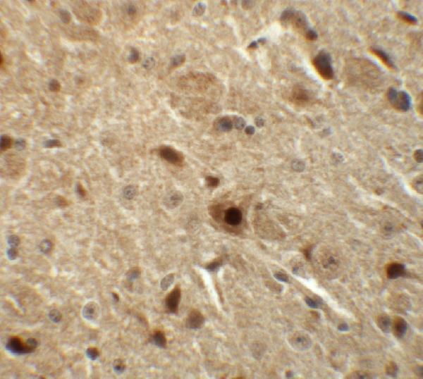
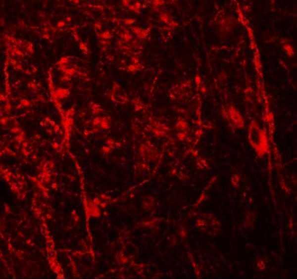
A07429 has mouse brain tissue IHC and IF images (catalog image captions); its listed reactivity covers human, mouse and rat (catalog reactivity).
A07429 has an IHC image from mouse brain tissue at 5 μg/mL (catalog IHC image caption). Its IF image is also from mouse brain tissue, at 20 μg/mL (catalog IF image caption).
Which to pick: Choose A07429 for paraffin-section IHC: IHC-P is listed, and its own IHC image shows mouse brain tissue (catalog applications; catalog IHC image caption); the fixative is unreported (catalog IHC image caption). Choose A07429 for tissue IF based on its mouse brain IF image; ICC validation is unreported (catalog IF image caption; catalog applications). For work across species, A07429 lists human, mouse and rat reactivity, though the supplied IHC and IF images show mouse tissue only (catalog reactivity; catalog image captions).