NOS1 / Nitric oxide synthase 1 · IHC design guide

Design Immunohistochemistry for NOS1

Plan chromogenic NOS1 IHC in paraffin sections using cytoplasmic staining in neurons and renal distal tubules as the expected readout (HPA tissue IHC). The guide covers fixation consistency, controls, and the catalog antibody’s 0.5–1 μg/ml IHC dilution (datasheet).

Evidence assembled Oct 2026 · For research use; verify linked source records and product datasheet before use
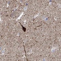
Immunohistochemistry protocol sheet for NOS1 (IHC for NOS1): expected localisation Neuronal and distal tubular cytoplasm (HPA tissue IHC), antibody PA1329, validated IHC image, and IHC protocol steps
Printable NOS1 IHC protocol sheet — expected localisation Neuronal and distal tubular cytoplasm (HPA tissue IHC), antibody PA1329, controls and protocol steps. Open the full NOS1 IHC guide →

NOS1 Immunohistochemistry Experimental Design Guide

Expected localisation, validated protocols, controls and antibodies — the at-a-glance facts below, then the full design guide.

Must know before staining
Expected localisation Neuronal and distal tubular cytoplasm (HPA tissue IHC)
Staining pattern Cytoplasmic staining in some neurons and renal distal tubules (HPA tissue IHC)
Antigen retrieval Citrate pH 6.0 HIER, 95–98 °C, 20 min (rule: cytoplasmic / membrane antigen)
Positive control ⓘ Caudate+4 more · see all
Negative control ⓘ Adipose tissue+4 more · see all
Important caveats
Reasons your staining may differ from the expected pattern.
Fixation Keep fixation consistent across paraffin sections (standard IHC practice; not target-specific)
Caveat Skeletal muscle RNA enrichment contrasts with low myocyte staining (HPA tissue IHC)
Regulation No expression regulator specified (UniProt)
Isoform / epitope 5 isoforms; no transmembrane segment; check epitope coverage (UniProt)
Section 1

Recommended NOS1 IHC & IF Protocols

The catalog antibody has a paraffin-section protocol (page protocol). Published NOS1 IHC methods below cover renal tumours, lung cancer specimens, and rat spinal cord (PMC2409527; PMC5882435; PMC8956269).

Recommended immunohistochemistry (IHC-P) protocol parameters
SampleTissue sections; selected-image fixative not specified (standard IHC workflow)
FixationImage fixative and duration unreported (datasheet PA1329); verify before use.
Sectioning4–5 µm sections on charged slides (standard)
DeparaffinisationXylene, graded ethanol series to water (standard)
Antigen retrievalHeat-induced epitope retrieval in citrate buffer, pH 6.0, 20 min at 95–98 °C (standard rule: cytoplasmic / membrane antigen)
Peroxidase block3% H2O2, 10 min, room temperature (standard)
Blocking10% normal serum of the secondary host, 30 min, room temperature (standard)
Primary antibodyRabbit anti-NOS1, 0.5-1μg/ml (datasheet PA1329)
Primary incubationOvernight at 4 °C (standard)
DetectionHRP-polymer secondary, DAB chromogen 5–10 min (standard)
CounterstainHematoxylin, blue, dehydrate and mount (standard)
Expected resultNOS1-positive staining in neuronal cells of caudate (HPA tissue IHC: High). HPA tissue profile: Cytoplasmic expression in a subset of neurons and renal distal tubules. No signal in the no-primary control.
💡Decision noteFor paraffin sections, start with citrate pH 6.0 HIER at 95–98 °C for 20 min (page retrieval). Use a paper’s stated conditions when reproducing its protocol.
Section 2

What Is the Expected NOS1 Staining Pattern?

NOS1 stains neuronal and distal-tubule cytoplasm (HPA: tissue IHC, Enhanced); it lacks a transmembrane segment (UniProt P29475).

What am I looking at on my slide?
Caudate neurons stain strongly (HPA: High).This fits the neuronal cytoplasmic pattern (HPA: tissue IHC).
Kidney distal tubules stain (HPA: Medium).This fits the reported renal pattern (HPA: tissue IHC).
IHC signal is nuclear only.Discordant with tissue cytoplasmic staining; check for artefact (HPA: tissue IHC).
Adipocytes stain in adipose tissue.Unexpected (HPA: Not detected); check cross-reactivity or endogenous detection activity.
Staining is diffuse across the section.Background may obscure cell-specific signal; review blocking and washes (general IHC practice).
💡Expected NOS1 appearanceCorrect: High caudate neuronal cytoplasmic staining; adipocyte staining is suspect (HPA: tissue IHC).
How each factor affects the staining
Tissue selectionCaudate neurons are High; kidney distal tubules Medium; muscle myocytes Low (HPA: tissue IHC).
Cellular locationSarcolemma and dendritic spine enrichment are noted by similarity (UniProt P29475).
Isoforms5 isoforms are listed; isoform 3 is testis restricted (UniProt P29475).
Antibody validationHPA069509 and CAB002167 have Enhanced IHC validation (HPA: antibodies).
Why is my staining missing, weak or wrong?
SituationLikely causeNext action
No caudate neuronal signal (HPA: High).The run may have failed (general IHC practice).Check the positive control, retrieval and antibody dilution (general IHC practice).
Nuclear-only IHC staining.Discordant with the tissue cytoplasmic profile (HPA: tissue IHC).Compare with a positive tissue control and secondary-only control (general IHC practice).
Adipocytes stain in adipose tissue.Unexpected (HPA: Not detected); cross-reactivity or detection activity is possible.Check a negative control and endogenous enzyme blocking (general IHC practice).
Diffuse chromogenic background.Incomplete blocking or washing is possible (general IHC practice).Review blocking, washes and detection controls (general IHC practice).
Weak skeletal-muscle myocyte staining.Low staining is reported in myocytes (HPA: tissue IHC).Use caudate neurons as a stronger positive control (HPA: High).
What localisation should IF/ICC show?Plasma membrane is main; nucleoplasm is additional (HPA: ICC-IF).Interpret IF/ICC with its separate application guide (HPA: ICC-IF).

Sample controls for NOS1 IHC & IF

🧪Run caudate first: neuronal cells should stain strongly (HPA: High in caudate neuronal cells). Use adipose tissue as the negative tissue (HPA: adipocytes Not detected); on the caudate slide, cells without neuronal morphology should remain at background relative to the no-primary control (standard IHC control practice).
Positive control tissue: Caudate (Neuronal cells, HPA High)
Negative control tissue: Adipose tissue (HPA Not detected)
ICC-IF cell lines (HPA subcellular resource): HPA ICC-IF images show NOS1 in HaCaT, U2OS, with annotated localisation: Plasma membrane (supported) (HPA subcellular).
Technical controls: Include no-primary (secondary-only) and host-species- and clonality-matched isotype controls, plus NOS1-knockout tissue or a peptide-block control if available (standard IHC control practice). For caudate chromogenic IHC, quench endogenous peroxidase and check the no-primary slide for pigment or residual background (standard brain IHC practice).
⚠️Feasibility: A NOS1-specific fixation window and antigen-retrieval dependency are unreported in the supplied evidence; the selected rat-brain IHC(P) caption also leaves the fixative unreported (PA1329 caption). The evidence does not establish whether frozen sections or IF are easier than paraffin IHC (supplied application evidence). Neuronal lipofuscin can cause autofluorescence if an IF follow-up is performed (standard brain IF practice).

HPA tissue IHC evidence for NOS1

Comprehensive Human Protein Atlas IHC scoring per tissue (reliability: Enhanced — Medium consistency between antibody staining and RNA expression data.). Rows are taken directly from the HPA tissue chart — click any row's HPA link to view the source.

Positive expression · recommended positive controls

TissueCell typeLevelEvidenceSource
Caudate Neuronal cells High Protein (IHC) HPA →
Cerebellum Molecular layer cells - cytoplasm/membrane High Protein (IHC) HPA →
Cerebral cortex Neuronal cells Medium Protein (IHC) HPA →
Hippocampus Neuronal cells Medium Protein (IHC) HPA →
Kidney Distal tubules Medium Protein (IHC) HPA →

Undetected expression · recommended negative controls

TissueCell typeLevelEvidenceSource
Adipose tissue Adipocytes Not detected Protein (IHC) HPA →
Adrenal gland Glandular cells Not detected Protein (IHC) HPA →
Appendix Glandular cells Not detected Protein (IHC) HPA →
Bone marrow Hematopoietic cells Not detected Protein (IHC) HPA →
Breast Adipocytes Not detected Protein (IHC) HPA →
Section 3

Advanced NOS1 IHC Tips

Troubleshoot NOS1 staining in paraffin section IHC using the stated retrieval setting, the catalog antibody’s rat brain image, and cell and compartment patterns (page setting; PA1329 caption; HPA tissue IHC).

What retrieval should I start with for weak NOS1 staining in paraffin sections?
Start with citrate buffer at pH 6.0, heated to 95–98 °C for 20 min (page retrieval setting). If staining remains weak, compare a modestly adjusted heating time on adjacent sections while keeping the buffer and detection conditions fixed (standard IHC practice). Include a rat brain section as a process control because the selected antibody has a rat brain IHC(P) image (PA1329 caption). Score neuronal cytoplasmic staining separately from diffuse section background, since HPA reports NOS1 in a subset of neurons (HPA tissue IHC). Record retrieval conditions for every slide so any gain in signal can be weighed against tissue damage and background (standard IHC practice).
How should I troubleshoot fixation when NOS1 staining varies between paraffin blocks?
The supplied evidence does not establish NOS1-specific sensitivity to a fixative or fixation duration, so treat that effect as unknown (supplied evidence). The rat brain IHC(P) caption does not state a fixative, and its paraffin designation cannot establish an FFPE fixation condition (PA1329 caption). Compare blocks with documented processing histories, using the same section thickness, citrate pH 6.0 retrieval and detection run (page retrieval setting; standard IHC practice). Check morphology and staining together, because damaged or detached tissue can make a weak chromogenic result difficult to interpret (standard IHC practice). Do not infer fixation sensitivity from NOS1 localisation or phosphorylation sites (UniProt P29475).
Which staining compartments support a plausible NOS1 result?
Look for staining associated with neuronal cytoplasm and processes, while assessing whether membrane-associated signal fits the structure being examined (HPA tissue IHC; UniProt P29475 subcellular). NOS1 has no transmembrane segment, yet UniProt places it at the sarcolemma and dendritic spines through cellular associations (UniProt P29475 topology and subcellular). HPA reports cytoplasmic staining in a subset of neurons and renal distal tubules, so a uniform stain across every cell deserves scrutiny (HPA tissue IHC). HPA subcellular data also support plasma membrane localisation and approve an additional nucleoplasmic location (HPA subcellular). Compare cellular distribution with morphology and controls before calling isolated nuclear colour target-specific (standard IHC practice).
Can a missing NOS1 stain reflect isoform or epitope coverage?
NOS1 has 5 listed isoforms, and their tissue distributions differ, so expression expectations depend on which isoforms an antibody can detect (UniProt P29475 isoforms and tissue specificity). Isoform 3 is reported only in testis, whereas isoform 4 is reported in testis, skeletal muscle, lung and kidney, with low brain expression (UniProt P29475 tissue specificity). The supplied antibody caption identifies rat brain IHC(P) but gives no epitope or isoform coverage (PA1329 caption). Ask for the antibody’s immunogen or mapped epitope before interpreting a negative section as absence of every NOS1 isoform (standard IHC practice). Keep retrieval and detection identical when comparing sections (standard IHC practice).
How can I check NOS1 by IF alongside this chromogenic IHC result?
Use IF as a separate assay and multiplex NOS1 with a marker for the expected neuronal cell type when assessing brain sections (HPA tissue IHC; standard IF practice). Choose a fluorophore channel after inspecting tissue autofluorescence, and include single-label and no-primary controls to identify bleed-through or intrinsic signal (standard IF practice). Permeabilise sufficiently to reach a cytoplasmic or intracellular membrane-facing epitope, while optimising against loss of membrane-associated pattern; NOS1 has no transmembrane segment (UniProt P29475 topology; standard IF practice). HPA provides ICC/IF images in HaCaT and U2OS cells, but the supplied PA1329 evidence is a rat brain IHC(P) caption (HPA subcellular; PA1329 caption).
What should I change when NOS1 DAB staining is widespread?
First compare the stained section with a no-primary control and examine tissue edges, folds and damaged areas for colour unrelated to cell structure (standard IHC practice). Apply a peroxidase block before DAB detection and use an appropriate protein block, because endogenous enzyme activity and nonspecific binding can produce chromogenic background (standard IHC practice). Then titrate the primary antibody and adjust wash conditions one variable at a time; no working dilution is supplied for PA1329 here (PA1329 caption; standard IHC practice). Judge improvement against neuronal staining in the rat brain IHC(P) example and the subset pattern reported in tissue (PA1329 caption; HPA tissue IHC).
How should I score NOS1 staining across neuronal regions? ⚠ ANSWER MARKED FOR VERIFICATION
Define the anatomical region and eligible cell type before scoring, because HPA describes NOS1 in a subset of neurons rather than all cells (HPA tissue IHC). For chromogenic sections, report the percentage of positive eligible cells and an intensity-based H-score, or count positive cells per mm² when cell boundaries permit reliable counting (standard IHC practice). Normalise counts to the sampled tissue area and report the number of eligible cells assessed; keep illumination, thresholding and background correction consistent (standard IHC practice). Score renal distal tubules separately from neuronal regions because both are reported positive compartments but have different tissue architecture (HPA tissue IHC; standard IHC practice).
How do I distinguish credible NOS1 staining from artefact?
A credible brain result should follow identifiable neuronal cells and a plausible cytoplasmic or process-associated distribution, consistent with the rat brain IHC(P) image and HPA neuronal profile (PA1329 caption; HPA tissue IHC). In kidney, assess distal tubules rather than treating staining in every structure as expected NOS1 expression (HPA tissue IHC). Distrust colour concentrated at section edges, in necrotic areas or across unrelated cells, and compare it with a no-primary control (standard IHC practice). If the control is coloured, revisit peroxidase blocking and detection before interpreting DAB as NOS1 (standard IHC practice). HPA rates its tissue evidence Enhanced while noting medium staining–RNA consistency, so interpret discordant regions cautiously (HPA tissue IHC).
Boster reagents

Best NOS1 / Nitric oxide synthase 1 IHC Antibodies

PA1329 has real rat brain paraffin-section IHC data (PA1329 image caption) and listed human, mouse, and rat reactivity (catalog: reactivity).

Real IHC data Anti-nNOS(neuronal) antibody, PA1329, IHC(P) IHC(P): Rat Brain Tissue
Anti-nNOS (neuronal)/NOS1 Antibody ®
Cat # PA1329

PA1329 is listed for IHC and reacts with human, mouse, and rat (catalog: applications/reactivity). Its own image shows IHC(P) on rat brain tissue; no IF application or image is listed (PA1329 image caption; catalog: applications/images).

Which to pick: Choose PA1329 for tissue IHC in paraffin sections based on its rat brain IHC(P) image; the fixative is unreported (PA1329 image caption). No listed SKU is validated for IF/ICC (catalog: applications/images). PA1329 is the cross-species option on the listed reactivity of human, mouse, and rat; its rabbit host is reported, but clonality is unreported (catalog: host/clone/reactivity).

Each figure is that product's own IHC / IF validation image from its datasheet.

References

  1. UniProt Consortium. UniProt entry P29475 (NOS1_HUMAN, Nitric oxide synthase 1).
  2. Human Protein Atlas. NOS1 tissue IHC expression (reliability: Enhanced).
  3. Human Protein Atlas. NOS1 subcellular location (ICC-IF): Mainly localized to the plasma membrane. In addition localized to the nucleoplasm..
  4. Human Protein Atlas. NOS1 antibody validation summary (3 antibodies).
  5. Loss of NOS1 expression in high-grade renal cell carcinoma associated with a shift of NO signalling. British journal of cancer 2004 — PMC2409527.
  6. CXCL14 and NOS1 expression in specimens from patients with stage I-IIIA nonsmall cell lung cancer after curative resection. Medicine 2018 — PMC5882435.
  7. Novel oxygen sensing mechanism in the spinal cord involved in cardiorespiratory responses to hypoxia. Science advances 2022 — PMC8956269.
  8. NOS1 upregulates ABCG2 expression contributing to DDP chemoresistance in ovarian cancer cells. Oncology letters 2019 — PMC6341833.
  9. PubMed PMID:7528745 — UniProt-cited evidence.
  10. PubMed PMID:7515942 — UniProt-cited evidence.
  11. PubMed PMID:7678401 — UniProt-cited evidence.