NOSTRIN / Nostrin · IHC design guide

Design Immunohistochemistry for NOSTRIN

Plan chromogenic IHC for NOSTRIN using tissue staining patterns and control choices supported by HPA tissue IHC. The guide also notes four isoforms and elevated placental protein in pre-eclampsia (UniProt).

Evidence assembled Oct 2026 · For research use; verify linked source records and product datasheet before use
Immunohistochemistry protocol sheet for NOSTRIN (IHC for NOSTRIN): expected localisation Cytoplasmic and membranous tissue staining (HPA tissue IHC), antibody A06014, validated IHC image, and IHC protocol steps
Printable NOSTRIN IHC protocol sheet — expected localisation Cytoplasmic and membranous tissue staining (HPA tissue IHC), antibody A06014, controls and protocol steps. Open the full NOSTRIN IHC guide →

NOSTRIN Immunohistochemistry Experimental Design Guide

Expected localisation, validated protocols, controls and antibodies — the at-a-glance facts below, then the full design guide.

Must know before staining
Expected localisation Cytoplasmic and membranous tissue staining (HPA tissue IHC)
Staining pattern Cytoplasmic and membranous staining across tissues (HPA tissue IHC)
Antigen retrieval Tris-EDTA pH 9.0 HIER, 95–98 °C, 20 min (rule: nuclear antigen)
Positive control ⓘ Colon+4 more · see all
Negative control ⓘ Nasopharynx+1 more · see all
Important caveats
Reasons your staining may differ from the expected pattern.
Fixation Keep fixation consistent across sections. (standard IHC practice; not target-specific)
Caveat Staining has medium concordance with RNA expression (HPA tissue IHC)
Regulation Higher in pre-eclamptic placenta (UniProt)
Isoform / epitope Four isoforms; epitope coverage is unknown (UniProt)
Section 1

Recommended NOSTRIN IHC & IF Protocols

Compare the catalog antibody’s IHC-P protocol with the published mouse prostate IHC protocol (PMC11491235).

Recommended immunohistochemistry (IHC-P) protocol parameters
SampleTissue sections; selected-image fixative not specified (standard IHC workflow)
FixationImage fixative and duration unreported (datasheet A06014); verify before use.
Sectioning4–5 µm sections on charged slides (standard)
DeparaffinisationXylene, graded ethanol series to water (standard)
Antigen retrievalHeat-induced epitope retrieval in Tris-EDTA buffer, pH 9.0, 20 min at 95–98 °C (standard rule: nuclear antigen)
Peroxidase block3% H2O2, 10 min, room temperature (standard)
Blocking10% normal serum of the secondary host, 30 min, room temperature (standard)
Primary antibodyRabbit anti-NOSTRIN, 5 μg/mL (datasheet A06014)
Primary incubationOvernight at 4 °C (standard)
DetectionHRP-polymer secondary, DAB chromogen 5–10 min (standard)
CounterstainHematoxylin, blue, dehydrate and mount (standard)
Expected resultNOSTRIN-positive staining in endothelial cells of colon (HPA tissue IHC: High). HPA tissue profile: Ubiquitous cytoplasmic and membranous expression. No signal in the no-primary control.
💡Decision noteStart with Tris-EDTA pH 9.0 HIER at 95–98 °C for 20 min (page retrieval); compare the published citrate retrieval for mouse prostate (PMC11491235).
Section 2

What Is the Expected NOSTRIN Staining Pattern?

NOSTRIN is expected mainly in cytoplasmic and membranous regions on tissue IHC, including colon endothelial cells, lung macrophages, kidney glomerular cells and duodenal glandular cells (HPA tissue IHC). It has no transmembrane segment; UniProt also lists cytoplasmic vesicles, cytoskeleton and nucleus (UniProt Q8IVI9 topology/localisation). HPA rates tissue IHC Approved, with medium consistency between staining and RNA data (HPA tissue IHC).

What am I looking at on my slide?
Cytoplasmic or membranous staining in colon endothelial cells, with a clear counterstain (HPA: High in colon endothelial cells).This fits the reported tissue pattern (HPA tissue IHC). Judge localisation within the endothelial cells, since tissue-wide signal alone does not establish a cell-specific positive result (standard IHC interpretation).
Strong staining confined to nuclei, with little cytoplasmic signal in otherwise positive cells.Recheck the pattern: HPA describes ubiquitous cytoplasmic and membranous tissue staining (HPA tissue IHC). UniProt also lists the nucleus, so nuclear signal alone cannot be declared false without controls (UniProt Q8IVI9 localisation; standard IHC interpretation).
Strong signal in nasopharyngeal basal cells or ovarian stroma cells.Those cells are reported as Not detected (HPA tissue IHC). Check for nonspecific binding or endogenous chromogenic activity before assigning the signal to NOSTRIN (standard IHC troubleshooting).
Diffuse colour across cells, stroma and blank spaces, obscuring cell boundaries.The distribution cannot be scored confidently against HPA’s cell-level observations (HPA tissue IHC; standard IHC interpretation). Review blocking, washes and a no-primary control (standard IHC practice).
No staining in colon endothelial cells or lung macrophages despite preserved tissue morphology.Both are reported High (HPA tissue IHC). A negative result in one section is inconclusive; check a positive-control section and the IHC workflow before interpreting absence in the specimen (standard IHC practice).
💡Expected NOSTRIN appearanceCall positive when cytoplasmic or membranous colour is resolved in a reported positive cell type, such as High-staining colon endothelial cells (HPA tissue IHC); widespread colour without cell boundaries is suspect (standard IHC interpretation).
How each factor affects the staining
Tissue and cell choiceHigh examples include duodenal glandular cells, kidney glomerular cells and lung macrophages (HPA tissue IHC). Low-staining glial cells in hippocampus are a less decisive positive control (HPA tissue IHC; standard IHC interpretation).
Compartment and topologyHPA describes cytoplasmic and membranous tissue staining (HPA tissue IHC). UniProt lists membrane, vesicle, cytoskeleton and nuclear locations, with no transmembrane segment (UniProt Q8IVI9 topology/localisation).
Evidence strengthTissue IHC is Approved, with medium staining-to-RNA consistency (HPA tissue IHC). HPA044384 is IHC Approved; the supplied antibody list does not establish IHC validation for HPA076885 (HPA antibodies).
IF/ICC Q&A: What pattern is reported?Mainly cytosol, with additional vesicles, both Approved (HPA subcellular ICC-IF). This is ICC-IF localisation evidence; assess paraffin-section results against tissue IHC observations (HPA subcellular ICC-IF; HPA tissue IHC).
Why is my staining missing, weak or wrong?
SituationLikely causeNext action
A reported High tissue has no interpretable signal.The section may have failed at retrieval, antibody incubation or chromogenic detection (standard IHC troubleshooting).Run a positive-control section, verify the detection reagents, and check the catalog antibody’s IHC-P instructions for its validated conditions (standard IHC practice).
Colour spreads through the section without a cellular pattern.Residual detection activity, inadequate blocking or incomplete washing can raise background (standard IHC troubleshooting).Compare with a no-primary control; review endogenous enzyme blocking, wash steps and detection exposure (standard chromogenic IHC practice).
Only nuclei stain strongly.The result diverges from HPA’s dominant tissue pattern, although UniProt lists nuclear localisation (HPA tissue IHC; UniProt Q8IVI9 localisation).Inspect reported positive cell types and controls; avoid calling nuclear-only staining specific from compartment agreement alone (standard IHC interpretation).
Cells reported Not detected appear strongly positive.Nonspecific antibody binding or endogenous detection activity may explain the signal (HPA tissue IHC; standard IHC troubleshooting).Compare nasopharyngeal basal cells or ovarian stroma with a no-primary control and a reported positive cell type in the run (HPA tissue IHC; standard IHC practice).
A low-staining tissue gives an unclear negative result.HPA reports Low staining in hippocampal glial cells and several other cell groups (HPA tissue IHC).Use a reported High cell type to check assay performance before treating a Low example as evidence that staining failed (HPA tissue IHC; standard IHC interpretation).
Staining appears in expected cells but is difficult to score.Crowded chromogen or counterstain can obscure boundaries and compartment assignment (standard IHC interpretation).Review morphology and detection intensity on a positive-control section; score only cells whose staining and boundaries can be resolved (standard IHC practice).

Sample controls for NOSTRIN IHC & IF

🧪Run kidney first and score cells in glomeruli, where NOSTRIN staining is High (HPA: kidney, cells in glomeruli, High); use ovarian stroma as the negative tissue (HPA: ovary, ovarian stroma cells, Not detected). On the kidney slide, adjacent nonglomerular cells can serve as candidate internal negatives if independently verified to lack NOSTRIN; they should show no specific chromogen above background.
Positive control tissue: Colon (Endothelial cells, HPA High)
Negative control tissue: Nasopharynx (HPA Not detected)
ICC-IF cell lines (HPA subcellular resource): HPA ICC-IF images show NOSTRIN in CACO-2, HEL, U2OS, with annotated localisation: Cytosol (approved) (HPA subcellular).
Technical controls: Include a no-primary (secondary-only) control, an isotype control matched to the primary antibody’s host species, class and clonality, and a biological negative such as NOSTRIN knockout tissue or a validated peptide block. In kidney, check for endogenous peroxidase signal and, if using avidin–biotin detection, endogenous biotin signal in the negative controls (standard IHC practice).
⚠️Feasibility: A target-specific fixation window and retrieval dependency are unreported in the supplied evidence; test antigen retrieval conditions on paraffin sections (standard IHC practice). The selected A06014 mouse-kidney IHC caption reports 5 μg/ml but does not report a fixative (selected-SKU tissue-IHC caption: fixative not stated). HPA reports cytosolic and vesicular ICC-IF localization (HPA: subcellular), but the supplied evidence does not establish that frozen sections or IF are easier than paraffin IHC; kidney endogenous peroxidase or biotin may complicate chromogenic interpretation (standard IHC practice).

HPA tissue IHC evidence for NOSTRIN

Comprehensive Human Protein Atlas IHC scoring per tissue (reliability: Approved — Medium consistency between antibody staining and RNA expression data.). Rows are taken directly from the HPA tissue chart — click any row's HPA link to view the source.

Positive expression · recommended positive controls

TissueCell typeLevelEvidenceSource
Colon Endothelial cells High Protein (IHC) HPA →
Duodenum Glandular cells High Protein (IHC) HPA →
Endometrium Glandular cells Medium Protein (IHC) HPA →
Fallopian tube Ciliated cells (tip of cilia) High Protein (IHC) HPA →
Kidney Cells in glomeruli High Protein (IHC) HPA →

Undetected expression · recommended negative controls

TissueCell typeLevelEvidenceSource
Nasopharynx Basal cells Not detected Protein (IHC) HPA →
Ovary Ovarian stroma cells Not detected Protein (IHC) HPA →
Section 3

Advanced NOSTRIN IHC Tips

Troubleshoot NOSTRIN staining by checking retrieval, cell type and compartment together, then score signal against matched controls.

What retrieval conditions should I start with for NOSTRIN paraffin sections?
Start with Tris-EDTA pH 9.0 heat-induced epitope retrieval at 95–98 °C for 20 min (page retrieval setting). Cool the sections consistently, then compare the same tissue across retrieval runs using one antibody dilution and detection time (standard IHC practice). If staining is weak, test a shorter or longer heating interval on adjacent sections while checking whether tissue morphology deteriorates (standard IHC practice). Score signal in glomerular cells alongside nearby tissue, since HPA reports high staining in kidney glomeruli but only medium consistency between staining and RNA data (HPA tissue IHC: kidney; HPA reliability).
Could fixation explain weak or uneven NOSTRIN staining?
Target-specific fixation sensitivity is unknown from the supplied evidence, so record the fixative and fixation duration for every specimen rather than assigning weak signal to a NOSTRIN-specific fixation effect (supplied NOSTRIN evidence; standard IHC practice). The mouse kidney IHC caption reports 5 µg/ml antibody but does not state its fixative (A06014 tissue-IHC caption). Compare adjacent sections from similarly processed blocks before changing retrieval, primary concentration or detection time (standard IHC practice). If staining varies across a section, inspect tissue integrity and section attachment, then repeat staining with a matched positive-control section in the same run (standard IHC practice).
Which staining compartments fit NOSTRIN in tissue sections?
Assess cytoplasmic and membranous signal first, because HPA describes a ubiquitous cytoplasmic and membranous tissue profile (HPA tissue IHC). Fine cytoplasmic puncta may also fit the reported vesicular location, while selected actin structures and the nucleus appear in the UniProt localisation record (HPA subcellular; UniProt Q8IVI9 localisation). NOSTRIN has no transmembrane segment, so a membrane-associated pattern need not form a continuous membrane outline (UniProt Q8IVI9 topology; standard IHC interpretation). Compare cellular staining with no-primary and matched tissue controls before accepting isolated nuclear or uniformly dark membrane staining as specific (standard IHC practice).
How should isoforms and epitope location affect my antibody choice?
Check the antibody’s stated immunogen or mapped epitope against all 4 listed NOSTRIN isoforms before interpreting a negative section (UniProt Q8IVI9 isoforms; standard IHC practice). The protein contains an F-BAR region at residues 1–260, REM-1 at 292–372 and SH3 at 438–497 (UniProt Q8IVI9 domains). Phosphoserines at residues 114 and 479 are listed, but their effects on this antibody’s staining are unreported (UniProt Q8IVI9 modified residues; supplied antibody evidence). If epitope coverage is unknown, document that limitation and compare independently validated antibodies or orthogonal expression evidence before calling an isoform absent (standard IHC practice).
How can I use IF to check an ambiguous NOSTRIN IHC pattern?
For a separate IF experiment, multiplex NOSTRIN with a validated marker for the cell population being examined; HPA reports high tissue staining in colon endothelial cells and kidney glomerular cells (HPA tissue IHC: colon; HPA tissue IHC: kidney). Choose fluorophores after examining unstained tissue autofluorescence, and include single-color controls to assess channel spillover (standard IF practice). Because NOSTRIN has no transmembrane segment and is reported in the cytosol and vesicles, optimize permeabilisation for access to intracellular epitopes when the antibody’s epitope permits it (UniProt Q8IVI9 topology; HPA subcellular; standard IF practice). Compare the cellular pattern with chromogenic sections without assuming that IF staining validates the IHC antibody (standard IHC/IF practice).
What should I check when the whole section looks brown?
Run a no-primary control through the same chromogenic workflow to expose detection-system and tissue background (standard IHC practice). Include a peroxidase-blocking step before horseradish peroxidase detection, and keep DAB development time constant across comparison sections (standard IHC practice). Review blocking, washes and primary-antibody concentration if diffuse brown signal obscures cells; the selected mouse kidney caption reports 5 µg/ml, without establishing that concentration for other tissues or protocols (A06014 tissue-IHC caption; standard IHC practice). Compare the suspected signal with morphology and expected cellular compartments, since HPA reports cytoplasmic and membranous tissue staining for NOSTRIN (HPA tissue IHC).
How should I quantify NOSTRIN staining across samples? ⚠ ANSWER MARKED FOR VERIFICATION
Define the cell population and compartment before scoring, then use an H-score based on the percentage of cells at each intensity or report the percentage of positive cells (standard IHC practice). For spatially restricted populations, report positive-cell density per mm² of viable tissue or defined region (standard IHC practice). Normalize each measure to the corresponding eligible cell count or analyzed viable area, and apply one threshold and DAB development time across the comparison set (standard IHC practice). Keep endothelial, glandular and glomerular populations separate because HPA reports staining in these distinct cell types (HPA tissue IHC: colon; HPA tissue IHC: duodenum; HPA tissue IHC: kidney).
How do I distinguish convincing NOSTRIN staining from artefact?
Look for staining in intact cells with a plausible cytoplasmic or membranous pattern, supported by the reported vesicular localisation where puncta are present (HPA tissue IHC; HPA subcellular). Check the identified cell type: HPA reports high staining in kidney glomerular cells, whereas ovarian stroma cells are listed as not detected (HPA tissue IHC: kidney; HPA tissue IHC: ovary). Treat section-edge concentration, necrotic areas and diffuse signal in the no-primary control as possible artefacts, including endogenous enzyme activity in chromogenic detection (standard IHC practice). Interpret discordant cases cautiously because the HPA tissue staining assessment has medium consistency with RNA expression data (HPA reliability).
Boster reagents

Best NOSTRIN / Nostrin IHC Antibodies

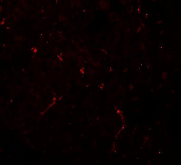
A06014 has IHC and IF images from mouse kidney tissue (catalog image captions); Human, Mouse, and Rat are listed as reactive species (catalog reactivity).

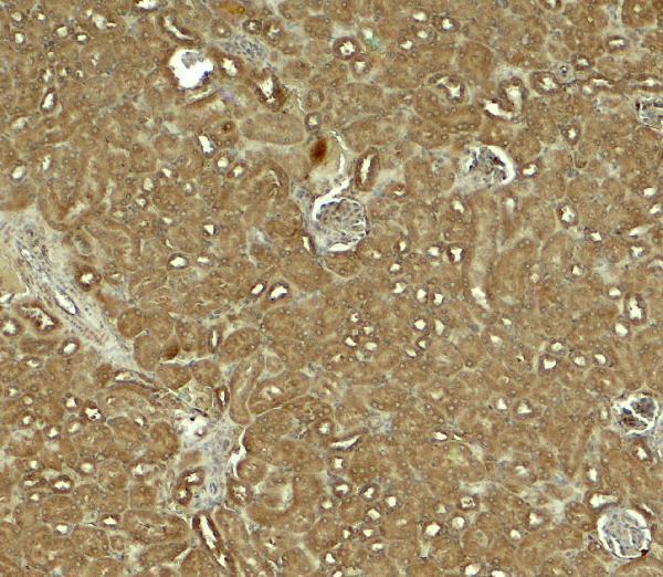
Real IHC data Immunohistochemistry of NOSTRIN in mouse kidney tissue with NOSTRIN antibody at 5 μg/ml.
Anti-NOSTRIN Antibody
Cat # A06014

A06014 is listed for IHC-P, with an IHC image from mouse kidney tissue at 5 μg/mL (catalog applications; IHC image caption). Its IF image is also from mouse kidney tissue, at 20 μg/mL (IF image caption).

Which to pick: Choose A06014 for paraffin-section IHC (catalog: IHC-P); its own IHC caption documents mouse kidney tissue at 5 μg/mL, but does not report the fixative (IHC image caption). Choose A06014 for IF (catalog applications; IF image caption); ICC validation and clonality are unreported (catalog payload). For work across species, A06014 lists Human, Mouse, and Rat reactivity (catalog reactivity), while the supplied IHC and IF images document mouse kidney only (catalog image captions).

Each figure is that product's own IHC / IF validation image from its datasheet.

References

  1. UniProt Consortium. UniProt entry Q8IVI9 (NOSTN_HUMAN, Nostrin).
  2. Human Protein Atlas. NOSTRIN tissue IHC expression (reliability: Approved).
  3. Human Protein Atlas. NOSTRIN subcellular location (ICC-IF): Mainly localized to the cytosol. In addition localized to vesicles..
  4. Human Protein Atlas. NOSTRIN antibody validation summary (2 antibodies).
  5. NOSTRIN is involved in benign prostatic hyperplasia via inhibition of proliferation, oxidative stress, and inflammation in prostate epithelial cells. Translational andrology and urology 2024 — PMC11491235.
  6. CFD overexpression inhibits CESC progression and enhances TIL therapy efficacy via NOSTRIN and eNOS signaling pathway. Scientific reports 2026 — PMC13469590.
  7. PubMed PMID:12446846 — UniProt-cited evidence.
  8. PubMed PMID:14702039 — UniProt-cited evidence.
  9. PubMed PMID:15815621 — UniProt-cited evidence.