NOTCH1 / Neurogenic locus notch homolog protein 1 · IHC design guide

Design Immunohistochemistry for NOTCH1

Plan chromogenic NOTCH1 IHC in paraffin sections using the cytoplasmic and nuclear tissue pattern reported by HPA (HPA tissue IHC). This guide covers fixation, staining interpretation and how NOTCH1 processing affects the choice of epitope (UniProt).

Evidence assembled Oct 2026 · For research use; verify linked source records and product datasheet before use
Immunohistochemistry protocol sheet for NOTCH1 (IHC for NOTCH1): expected localisation Cytoplasmic and nuclear staining in most tissues (HPA tissue IHC), antibody M00033, validated IHC image, and IHC protocol steps
Printable NOTCH1 IHC protocol sheet — expected localisation Cytoplasmic and nuclear staining in most tissues (HPA tissue IHC), antibody M00033, controls and protocol steps. Open the full NOTCH1 IHC guide →

NOTCH1 Immunohistochemistry Experimental Design Guide

Expected localisation, validated protocols, controls and antibodies — the at-a-glance facts below, then the full design guide.

Must know before staining
Expected localisation Cytoplasmic and nuclear staining in most tissues (HPA tissue IHC)
Staining pattern High in adrenal glandular cells; cytoplasmic/nuclear overall (HPA tissue IHC)
Antigen retrieval Tris-EDTA pH 9.0 HIER, 95–98 °C, 20 min (rule: nuclear antigen)
Positive control ⓘ Adrenal gland+4 more · see all
Negative control ⓘ Ovary
Important caveats
Reasons your staining may differ from the expected pattern.
Fixation Matched tissue-IHC evidence does not establish the fixation claim. Validate the specimen-specific method before use. (selected-SKU IHC image M00033)
Caveat NICD release can shift signal; epitope location matters (UniProt)
Regulation Fetal spleen, brain stem and lung enriched (UniProt)
Isoform / epitope No isoforms listed; extracellular vs cytoplasmic epitope matters (UniProt)
Section 1

Recommended NOTCH1 IHC & IF Protocols

Compare the catalog antibody’s IHC-P protocol with four published NOTCH1 chromogenic IHC workflows (page protocol; PMC12940838; PMC10363259; PMC7988833; PMC5354663).

Recommended immunohistochemistry (IHC-P) protocol parameters
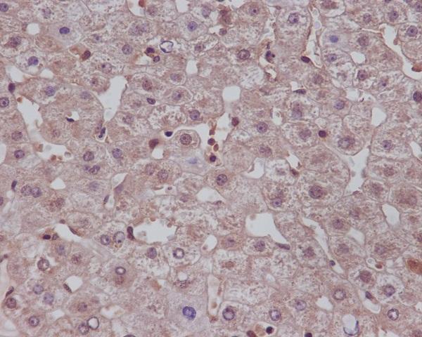
SampleParaffin-embedded human liver tissue; fixative not specified (datasheet M00033)
FixationImage fixative and duration unreported (datasheet M00033); verify before use.
Sectioning4–5 µm sections on charged slides (standard)
DeparaffinisationXylene, graded ethanol series to water (standard)
Antigen retrievalHeat-induced epitope retrieval in Tris-EDTA buffer, pH 9.0, 20 min at 95–98 °C (standard rule: nuclear antigen)
Peroxidase block3% H2O2, 10 min, room temperature (standard)
Blocking10% normal serum of the secondary host, 30 min, room temperature (standard)
Primary antibodyRabbit monoclonal (clone BGA-14) anti-NOTCH1, 1:50 (datasheet M00033)
Primary incubationOvernight at 4 °C (standard)
DetectionHRP-polymer secondary, DAB chromogen 5–10 min (standard)
CounterstainHematoxylin, blue, dehydrate and mount (standard)
Expected resultNOTCH1-positive staining in glandular cells of adrenal gland (HPA tissue IHC: High). HPA tissue profile: Cytoplasmic and nuclear expression in most tissues. No signal in the no-primary control.
💡Decision noteStart with Tris-EDTA pH 9.0 retrieval for nuclear staining (page protocol); citrate pH 6.0 is a published alternative (PMC7988833; PMC5354663).
Section 2

What Is the Expected NOTCH1 Staining Pattern?

NOTCH1 can appear at the cell membrane, in late endosomes, or in nuclei (UniProt P46531: subcellular location; transmembrane segment 1736–1756). In paraffin sections, HPA reports cytoplasmic and nuclear staining in most tissues, including high staining in selected glandular cells, rectal enterocytes, spermatogonia, and Bergmann glia (HPA: tissue IHC). Treat this as a supported reference pattern with medium consistency between staining and RNA data (HPA: reliability).

What am I looking at on my slide?
Distinct cytoplasmic or nuclear staining in the expected cells, with membrane-associated staining in Bergmann glia (HPA: tissue IHC).This fits the reported IHC pattern; interpret each compartment in the identified cell population, rather than treating nuclear staining alone as proof of pathway activation (HPA: tissue IHC; UniProt P46531: function).
A compartment outside the reported pattern dominates, such as staining confined to extracellular spaces (HPA: tissue IHC; UniProt P46531: subcellular location).Suspect nonspecific signal or a localisation artefact. Compare a known-positive section and a detection control before assigning that pattern to NOTCH1 (general IHC practice).
Strong staining appears in ovarian stromal cells, which HPA lists as not detected (HPA: ovary IHC).Investigate cross-reactivity or endogenous detection activity; the HPA observation is a reference for that cell population, not proof that every ovarian specimen must be negative (HPA: tissue IHC; general IHC practice).
Chromogen is spread across stroma, lumina, or much of the section without clear cell boundaries (general IHC practice).Diffuse background prevents a reliable compartment call. Check the detection control and review blocking, washes, and antibody concentration before scoring cells (general IHC practice).
No staining appears in a known-positive cell population, such as adrenal glandular cells reported as high (HPA: adrenal gland IHC).An absent signal does not establish absent NOTCH1. Verify the positive-control section, antibody and detection steps, then review retrieval against the antibody’s IHC instructions (HPA: adrenal gland IHC; general IHC practice).
💡Expected NOTCH1 appearanceCall a section positive when identifiable cells show cytoplasmic or nuclear staining in an HPA-positive population, potentially high in its listed cells; diffuse staining without cell boundaries is suspect (HPA: tissue IHC; general IHC practice).
How each factor affects the staining
Epitope locationNOTCH1 has an extracellular region at residues 19–1735 and a cytoplasmic region at 1757–2555; the antibody’s epitope determines which forms its staining can represent (UniProt P46531: topology; general IHC practice).
Receptor processingUniProt lists an intracellular-domain chain at residues 1754–2555 and nuclear localisation. Nuclear IHC is therefore biologically plausible, but its presence alone cannot identify the detected chain (UniProt P46531: processing, subcellular location).
Tissue and cell selectionHPA reports high staining in several specific cell populations and not detected ovarian stroma. Score the named cells within each tissue, since a tissue-wide average can obscure them (HPA: tissue IHC; general IHC practice).
Antibody evidenceTwo listed antibodies have Supported IHC validation; the antibody listed with approved ICC has no IHC status in this payload. Match validation to the antibody used (HPA: antibodies).
Antigen retrievalRetrieval is a routine paraffin-IHC variable to check when a positive control fails. No NOTCH1-specific retrieval condition or fixation sensitivity is supplied here (general IHC practice; supplied UniProt/HPA scope).
IF/ICC Q: should fluorescence be nuclear?Yes: HPA reports approved nucleoplasmic ICC-IF localisation. Use that as IF context; it does not replace the cytoplasmic and nuclear tissue-IHC reference pattern (HPA: subcellular ICC-IF; HPA: tissue IHC).
Why is my staining missing, weak or wrong?
SituationLikely causeNext action
Positive-control cells have no visible chromogen (general IHC practice).The staining run may have failed; HPA reports high adrenal glandular-cell staining as a reference (HPA: adrenal gland IHC).Check the control section, antibody application, retrieval, and detection steps before interpreting the test section as negative (general IHC practice).
Signal is weak in a cell population HPA rates high (HPA: tissue IHC).The run or specimen may differ from the HPA reference; its tissue-IHC reliability has medium staining-to-RNA consistency (HPA: reliability).Compare cell identity and a known-positive section, then review the antibody’s IHC instructions and the run controls (general IHC practice).
Ovarian stroma stains strongly despite HPA’s not detected observation (HPA: ovary IHC).Possible cross-reactivity or endogenous detection activity requires checking; the discrepant stain alone cannot distinguish them (HPA: ovary IHC; general IHC practice).Examine detection and negative controls, then assess whether staining follows identifiable cells or background structures (general IHC practice).
Nuclei stain but the researcher expected membrane staining only (HPA: tissue IHC).Nuclear localisation is reported, and receptor activation releases an intracellular domain involved in transcriptional regulation (UniProt P46531: location, function).Score nuclear staining in the appropriate cells; confirm the antibody epitope before attributing signal to a particular NOTCH1 fragment (UniProt P46531: topology, processing; general IHC practice).
Most structures carry diffuse chromogen (general IHC practice).Nonspecific background or endogenous detection activity may obscure the reported cell-level pattern (HPA: tissue IHC; general IHC practice).Review blocking, washes, antibody concentration, and detection controls; score only interpretable cells after background is resolved (general IHC practice).
A candidate IHC antibody was chosen from the ICC-IF result (HPA: antibodies).The listed ICC-approved antibody has no IHC validation status in the supplied HPA antibody record (HPA: antibodies).Check validation for the actual IHC antibody; use the HPA tissue pattern and an appropriate positive control to judge its staining (HPA: antibodies, tissue IHC; general IHC practice).

Sample controls for NOTCH1 IHC & IF

🧪Run adrenal gland first: glandular cells should stain (HPA: High in adrenal gland glandular cells). Use ovary as the negative tissue, focusing on ovarian stroma cells (HPA: Not detected in ovarian stroma cells); on the adrenal slide, assess adjacent non-glandular cells as potential internal negatives only if they lack specific staining (HPA: the supplied adrenal row identifies glandular cells, not an internal negative population).
Positive control tissue: Adrenal gland (Glandular cells, HPA High)
Negative control tissue: Ovary (HPA Not detected)
ICC-IF cell lines (HPA subcellular resource): HPA ICC-IF images show NOTCH1 in HaCaT, U2OS, with annotated localisation: Nucleoplasm (approved) (HPA subcellular).
Technical controls: Include no-primary (secondary-only) and primary host species- and clonality-matched isotype controls, plus a NOTCH1 knockout specimen or a validated immunizing-peptide block as a biological specificity control (standard IHC practice). Quench endogenous peroxidase and check for pigment that could be mistaken for chromogen in adrenal tissue (standard chromogenic IHC practice).
⚠️Feasibility: A target-specific fixation window and retrieval effect are unreported in the supplied evidence; optimize antigen retrieval for the catalog antibody on paraffin sections (standard IHC practice). The selected M00033 paraffin-section liver caption does not state a fixative, so its fixative is unreported (M00033 tissue-IHC caption). The supplied evidence does not establish that frozen sections or IF are easier; adrenal pigment can complicate chromogenic scoring, so compare staining with the no-primary slide (standard IHC practice).

HPA tissue IHC evidence for NOTCH1

Comprehensive Human Protein Atlas IHC scoring per tissue (reliability: Supported — Medium consistency between antibody staining and RNA expression data.). Rows are taken directly from the HPA tissue chart — click any row's HPA link to view the source.

Positive expression · recommended positive controls

TissueCell typeLevelEvidenceSource
Adrenal gland Glandular cells High Protein (IHC) HPA →
Appendix Endocrine cells High Protein (IHC) HPA →
Cerebellum Bergmann glia - cytoplasm/membrane High Protein (IHC) HPA →
Gallbladder Glandular cells High Protein (IHC) HPA →
Rectum Enterocytes High Protein (IHC) HPA →

Undetected expression · recommended negative controls

TissueCell typeLevelEvidenceSource
Ovary Ovarian stroma cells Not detected Protein (IHC) HPA →
Section 3

Advanced NOTCH1 IHC Tips

Troubleshoot NOTCH1 staining in paraffin sections by checking retrieval, epitope location, compartment patterns and controls before interpreting signal.

What retrieval should I try when NOTCH1 staining is weak in paraffin sections?
Start with Tris-EDTA at pH 9.0, heated to 95–98 °C for 20 min (page retrieval setting). Cool sections consistently before washing, then compare staining with a matched section processed in the same run (standard IHC practice). If signal remains weak, vary heating time in small increments while keeping the buffer fixed, because retrieval conditions can affect epitope accessibility and tissue integrity (standard IHC practice). Score membrane, cytoplasmic and nuclear staining separately: NOTCH1 spans the membrane, while its intracellular domain can enter the nucleus (UniProt P46531 topology and function). Record tissue preservation alongside signal strength so damaged edges do not drive the choice of retrieval condition (standard IHC practice).
How should I troubleshoot variable NOTCH1 staining between paraffin blocks?
The selected antibody caption documents paraffin-embedded human liver but does not report its fixative, so target-specific fixation sensitivity is unknown (M00033 tissue-IHC caption). Record each block’s fixative, fixation duration and processing history before comparing staining intensity (standard IHC practice). Stain sections from the blocks together using identical retrieval, antibody incubation and chromogenic detection conditions to limit run-to-run variation (standard IHC practice). Check morphology and staining in a reference section alongside the affected blocks, and repeat with matched controls if preservation differs (standard IHC practice). Do not assign a fixation-dependent change to NOTCH1 without a controlled comparison using known fixation conditions (M00033 tissue-IHC caption; standard IHC practice).
How should membrane, cytoplasmic and nuclear NOTCH1 staining be assessed?
Assess compartments separately because intact NOTCH1 is a membrane receptor, endosomal localisation is reported, and released intracellular domain participates in nuclear signalling (UniProt P46531 localisation and function). The tissue atlas also reports cytoplasmic and nuclear expression across most tissues (HPA tissue IHC). Examine the cell boundary at high magnification before calling diffuse cytoplasmic colour membranous staining (standard IHC practice). A nuclear signal deserves comparison with the antibody’s documented epitope, because an extracellular epitope cannot identify released intracellular domain by itself (UniProt P46531 topology; standard IHC interpretation). Include compartment-specific scores and representative images at the same exposure and counterstain conditions when comparing sections (standard IHC practice).
Can this antibody distinguish full-length NOTCH1 from its intracellular domain?
First confirm the catalog antibody’s immunogen or mapped epitope; the supplied tissue-IHC caption gives no epitope position (M00033 tissue-IHC caption). NOTCH1 has an extracellular region at residues 19–1735, a membrane span at 1736–1756, and a cytoplasmic region at 1757–2555 (UniProt P46531 topology). Its listed intracellular-domain chain covers 1754–2555, so an extracellular-epitope antibody should not be used to score that released domain (UniProt P46531 processing and topology). No alternative isoforms are listed in the supplied record; that does not establish which processed forms this antibody detects (UniProt P46531 isoform and processing records). If the epitope remains unknown, report compartment-specific NOTCH1 immunoreactivity without assigning a molecular form (standard IHC interpretation).
How can I check NOTCH1 localisation by multiplex immunofluorescence?
Choose a marker for the expected cell population and verify it in a separate channel before attributing NOTCH1 signal to that population (standard IF practice). Bergmann glia provide one tissue-specific example with high cytoplasmic or membrane staining in the tissue atlas (HPA tissue IHC). Select fluorophores after examining an unstained section for autofluorescence, and place the weaker signal in a channel with lower background (standard IF practice). If the antibody recognises the cytoplasmic side of NOTCH1, optimise permeabilisation; an extracellular epitope may require a different approach to membrane access (UniProt P46531 topology; standard IF practice). The paraffin liver caption establishes tissue IHC use, while IF conditions require their own validation (M00033 tissue-IHC caption; standard IF practice).
How can I separate nonspecific brown staining from NOTCH1 signal?
Compare the stained section with a no-primary control processed through the same detection steps (standard IHC practice). Block endogenous peroxidase before chromogenic detection, because residual enzyme activity can produce brown signal independent of antibody binding (standard IHC practice). Titrate the primary antibody and detection reagent within their validated ranges, and assess whether background increases across all compartments rather than within cells showing a plausible NOTCH1 pattern (standard IHC practice; UniProt P46531 localisation). Inspect folds, cut edges and necrotic areas separately, where pooled reagent or damaged tissue can distort interpretation (standard IHC practice). Use the same counterstain intensity when comparing sections so faint nuclear signal remains interpretable (standard IHC practice).
What is a defensible way to quantify NOTCH1 chromogenic staining? ⚠ ANSWER MARKED FOR VERIFICATION
Define the cell population and compartment before scoring, because NOTCH1 may appear at the membrane, in cytoplasm or in nuclei (UniProt P46531 localisation; HPA tissue IHC). For each compartment, record the percentage of positive cells and intensity categories, then calculate an H-score only if those categories are reproducible (standard IHC practice). Alternatively, report positive-cell density per mm² of viable tissue when cell counts or area vary across sections (standard IHC practice). Normalise comparisons to the same eligible cell population or viable tissue area, and use identical thresholds and imaging settings across groups (standard IHC practice). Document excluded folds, edges and necrotic regions before scoring (standard IHC practice).
When does a NOTCH1-positive section support a biological conclusion?
Require staining in the expected cells and a plausible compartment: NOTCH1 is membrane-associated, can enter the endosomal pathway, and signals through a released intracellular domain (UniProt P46531 localisation and function). High staining in Bergmann glia or adrenal glandular cells is consistent with reported tissue patterns, but neither pattern validates an individual slide (HPA tissue IHC; standard IHC interpretation). Treat isolated edge staining, necrotic colour and no-primary-control signal as potential artefacts, including residual endogenous peroxidase activity (standard IHC practice). Nuclear staining alone does not prove pathway activation unless the antibody’s intracellular epitope and assay controls support that inference (UniProt P46531 topology and function; standard IHC interpretation). Report localisation and control results alongside any biological interpretation (standard IHC practice).
Boster reagents

Best NOTCH1 / Neurogenic locus notch homolog protein 1 IHC Antibodies

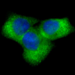
M00033 has IHC data from paraffin-embedded human liver and IF data from HeLa cells (M00033 image captions); Human, Mouse and Rat reactivity is listed for both catalog antibodies (catalog reactivity).

Real IHC data Immunohistochemical analysis of paraffin-embedded human liver, using Notch1 Antibody .
Anti-Notch1 Monoclonal Antibody
Cat # M00033

M00033 is the only SKU with an IHC figure here: its caption identifies paraffin-embedded human liver (M00033 IHC image caption). M00033 also has IF images of HeLa cells and an antibody dilution of 1:50 (M00033 IF image captions); M00033-4 lists IHC and IF applications but has no image caption in the payload (M00033-4 catalog applications and image alts).

Which to pick: For tissue IHC, pick M00033 because its own image shows staining of paraffin-embedded human liver; the caption does not report the fixative (M00033 IHC image caption). For IF/ICC, pick M00033 because its application list includes both and its IF caption identifies HeLa cells (M00033 applications and IF image caption). For Human, Mouse or Rat samples, both M00033 and M00033-4 list reactivity and are monoclonal antibodies; M00033 has the supplied IHC and IF images, while M00033-4 has no supplied images (catalog reactivity, descriptions and image alts).

Each figure is that product's own IHC / IF validation image from its datasheet.

References

  1. UniProt Consortium. UniProt entry P46531 (NOTC1_HUMAN, Neurogenic locus notch homolog protein 1).
  2. Human Protein Atlas. NOTCH1 tissue IHC expression (reliability: Supported).
  3. Human Protein Atlas. NOTCH1 subcellular location (ICC-IF): Localized to the nucleoplasm..
  4. Human Protein Atlas. NOTCH1 antibody validation summary (3 antibodies).
  5. Expression of NOTCH1 Is Correlated with Expression of Cancer Stem Cell Markers and miR-150 in Oral Epithelial Dysplasia. International journal of molecular sciences 2026 — PMC12940838.
  6. Immunohistochemical Evaluation of Notch1 in Ovarian Tumours and Its Prognostic Implications. Cureus 2023 — PMC10363259.
  7. The clinical significance of Notch1 immunoexpression in Caucasian patients with colorectal adenocarcinoma. Przeglad gastroenterologiczny 2020 — PMC7988833.
  8. Expression of Notch1 and Numb in small cell lung cancer. Oncotarget 2017 — PMC5354663.
  9. PubMed PMID:15164053 — UniProt-cited evidence.
  10. PubMed PMID:1831692 — UniProt-cited evidence.
  11. PubMed PMID:17573339 — UniProt-cited evidence.