NPC2 / NPC intracellular cholesterol transporter 2 · IHC design guide

Design Immunohistochemistry for NPC2

Plan NPC2 chromogenic IHC on paraffin sections using the catalog antibody at 0.5–1 μg/ml (datasheet A01582-3). Use epididymal glandular cells as a high-expression reference and account for secreted variants when interpreting staining (HPA tissue IHC).

Evidence assembled Oct 2026 · For research use; verify linked source records and product datasheet before use
Immunohistochemistry protocol sheet for NPC2 (IHC for NPC2): expected localisation Cytoplasmic in glandular cells and hepatocytes (HPA tissue IHC), antibody A01582-3, validated IHC image, and IHC protocol steps
Printable NPC2 IHC protocol sheet — expected localisation Cytoplasmic in glandular cells and hepatocytes (HPA tissue IHC), antibody A01582-3, controls and protocol steps. Open the full NPC2 IHC guide →

NPC2 Immunohistochemistry Experimental Design Guide

Expected localisation, validated protocols, controls and antibodies — the at-a-glance facts below, then the full design guide.

Must know before staining
Expected localisation Cytoplasmic in glandular cells and hepatocytes (HPA tissue IHC)
Staining pattern Cytoplasmic staining; strongest in epididymis (HPA tissue IHC)
Antigen retrieval EDTA pH 8.0 HIER, heat-mediated (datasheet A01582-3)
Positive control ⓘ Epididymis+4 more · see all
Negative control ⓘ Adipose tissue+4 more · see all
Important caveats
Reasons your staining may differ from the expected pattern.
Fixation Keep fixation consistent across sections (standard IHC practice; not target-specific)
Caveat Secreted variants may weaken RNA–staining correlation (HPA tissue IHC)
Regulation Highest expression in epididymis (HPA tissue IHC)
Isoform / epitope 2 isoforms; signal peptide 1–19 is removed; epitope unknown (UniProt)
Section 1

Recommended NPC2 IHC & IF Protocols

The catalog antibody’s IHC-P protocol (datasheet: A01582-3) is followed by four published NPC2 IHC methods (PMC3017059; PMC3798307; PMC11333723; PMC3406925).

Recommended immunohistochemistry (IHC-P) protocol parameters
SampleParaffin-embedded human tonsil tissue; fixative not specified (datasheet A01582-3)
FixationImage fixative and duration unreported (datasheet A01582-3); verify before use.
Sectioning4–5 µm sections on charged slides (standard)
DeparaffinisationXylene, graded ethanol series to water (standard)
Antigen retrievalHeat retrieval: EDTA pH 8.0 (datasheet A01582-3); 20 min, 95–100 °C (standard)
Peroxidase block3% H2O2, 10 min, room temperature (standard)
Blocking10% goat serum (datasheet A01582-3)
Primary antibodyRabbit anti-NPC2, 0.5-1μg/ml (datasheet A01582-3)
Primary incubationOvernight at 4 °C (datasheet A01582-3)
DetectionStreptavidin-biotin complex (SABC), DAB chromogen (datasheet A01582-3)
CounterstainHematoxylin, blue, dehydrate and mount (standard)
Expected resultNPC2-positive staining in glandular cells of epididymis (HPA tissue IHC: High). HPA tissue profile: Cytoplasmic expression in most glandular cells and in hepatocytes. Highest expression in the epididymis. No signal in the no-primary control.
💡Decision noteStart with heat-mediated EDTA pH 8.0 retrieval for the catalog antibody (datasheet: A01582-3). Optimize staining with positive and negative controls.
Section 2

What Is the Expected NPC2 Staining Pattern?

NPC2 is a soluble protein found in lysosomes, the endoplasmic reticulum and secreted material; it has no transmembrane segment (UniProt P61916: location and topology). In paraffin-section IHC, expect cytoplasmic staining in glandular cells and hepatocytes, with the strongest reported expression in epididymis (HPA: tissue IHC). HPA rates the tissue staining profile Enhanced, while noting medium consistency between staining and RNA data because secreted protein can appear away from its site of production (HPA: reliability).

What am I looking at on my slide?
Epididymal glandular cells stain strongly; kidney distal tubules, lung macrophages and testicular Leydig cells also stain strongly (HPA: High in each cell type).These provide cell-specific positive references for comparing runs. Judge the labelled cells and their cytoplasm, rather than treating a uniformly stained section as the expected result (HPA: tissue IHC; general IHC interpretation).
Staining is predominantly nuclear or sharply outlines the plasma membrane, with little cytoplasmic signal.Review this as a compartment mismatch: HPA reports cytoplasmic tissue staining, and UniProt describes soluble, lysosomal and secreted NPC2 without a transmembrane segment (HPA: tissue IHC; UniProt P61916: location and topology). Check morphology and controls before calling it an artefact (general IHC practice).
Cells reported as unstained, such as adipocytes in adipose tissue or gallbladder glandular cells, show strong chromogen (HPA: Not detected in those cells).The cell-type mismatch raises possible cross-reactivity or endogenous detection activity; it does not by itself identify the cause. Compare a no-primary control and a known-positive cell population from the same run (HPA: tissue IHC; general IHC practice).
Chromogen spreads across tissue and empty spaces, obscuring cell boundaries.Diffuse background cannot establish NPC2 localisation. Review blocking, washing and detection controls, then reassess whether positive cells retain a distinct cytoplasmic pattern (general IHC practice; HPA: cytoplasmic tissue profile).
Epididymal glandular cells show no convincing signal (HPA: High in glandular cells).A blank known-positive reference makes a negative result elsewhere hard to interpret. Check antibody use for IHC, detection reagents and run controls before scoring the test tissue (HPA: tissue IHC; general IHC practice).
💡Expected NPC2 appearanceCall a positive result when the relevant cells show distinct cytoplasmic staining—especially strong epididymal glandular-cell staining—while widespread background, nuclear-only signal or strong staining in HPA-negative cell types prompts review (HPA: tissue IHC; UniProt P61916: location and topology; general IHC interpretation).
How each factor affects the staining
Cell population and tissueHPA reports High staining in epididymal glandular cells, kidney distal tubules, lung macrophages and Leydig cells; hepatocytes are Medium. Score the named cells rather than assigning one intensity to an entire organ (HPA: tissue IHC; general IHC interpretation).
Secretion and intracellular targetingNPC2 is secreted and also localises to lysosomes and the endoplasmic reticulum; cell-surface M6PR interaction supports uptake and lysosomal targeting (UniProt P61916: location). Extracellular signal alone does not establish the producing cell (HPA: reliability note; UniProt P61916: location).
Protein processing and antibody epitopeUniProt places the signal peptide at residues 1–19 and the mature chain at 20–151 (UniProt P61916: processing). The supplied antibody records do not locate either epitope, so these positions cannot predict which processed form either antibody detects (HPA: antibody records; UniProt P61916: processing).
Molecular variantsUniProt lists 2 isoforms and glycosylation sites at residues 58 and 135 (UniProt P61916: isoforms and glycosylation). Those annotations alone do not establish distinct IHC patterns, epitope masking or fixation sensitivity (UniProt P61916: annotations).
Validation and IF/ICC questionFor IHC, HPA labels CAB032888 Enhanced and HPA000835 Supported (HPA: antibody validation). What IF/ICC pattern is established? HPA lists NPC2 as secreted but provides no main subcellular location or ICC-IF images, so its record cannot establish an IF/ICC staining pattern (HPA: subcellular record).
Why is my staining missing, weak or wrong?
SituationLikely causeNext action
The known-positive epididymal reference is blank (HPA: High in glandular cells).The run may have failed, or the antibody and detection conditions may be unsuitable; absence alone does not identify which step failed (general IHC practice).Confirm the primary antibody's IHC validation and the positive control, then inspect retrieval, primary incubation and detection steps against the run record (HPA: antibody validation; general IHC practice).
Only diffuse, weak staining remains in a tissue expected to contain positive cells (HPA: tissue IHC).Background can conceal cell-restricted signal; weak staining alone is insufficient to reassign the expected NPC2 compartment (general IHC interpretation; HPA: cytoplasmic profile).Review the no-primary control, blocking, washes and chromogen development; then compare named positive cells with adjacent tissue (general IHC practice; HPA: tissue IHC).
A HPA-negative cell type stains strongly (HPA: Not detected in adipocytes or gallbladder glandular cells).Cross-reactivity or endogenous detection activity is possible; the observation alone cannot distinguish them (general IHC interpretation).Check a no-primary control for detection background and compare with a HPA-positive cell type under the same run conditions (general IHC practice; HPA: tissue IHC).
Signal appears nuclear-only or forms a continuous membrane rim.This conflicts with HPA's cytoplasmic tissue profile and UniProt's soluble, non-transmembrane topology; the image alone cannot prove artefact (HPA: tissue IHC; UniProt P61916: topology).Recheck cell boundaries and counterstain, then compare a known-positive section and controls before scoring the compartment (general IHC practice; HPA: tissue IHC).
A test section is negative while the positive control works.NPC2 staining varies by cell type: HPA reports High, Medium, Low and Not detected populations (HPA: tissue IHC). A valid run therefore does not make every cell positive.Identify and score the specific cell population against its HPA entry; avoid calling an organ negative from an unstained population alone (HPA: tissue IHC; general IHC interpretation).
IF/ICC yields puncta or diffuse fluorescence that is difficult to assign.HPA supplies no ICC-IF images or main subcellular location for NPC2; lysosomal targeting in UniProt does not validate a particular IF image (HPA: subcellular record; UniProt P61916: location).Treat the IF/ICC appearance as unconfirmed by these sources and assess it with appropriate staining controls and the separate IF/ICC guide (HPA: subcellular record; general IF practice).

Sample controls for NPC2 IHC & IF

🧪Run epididymis first; glandular cells should stain (HPA: High in epididymal glandular cells). Use adipose tissue as a negative comparator, where adipocytes are not detected (HPA: Not detected in adipocytes); on the epididymis slide, use non-glandular areas to assess background without assuming those cells are NPC2-negative.
Positive control tissue: Epididymis (Glandular cells, HPA High)
Negative control tissue: Adipose tissue (HPA Not detected)
ICC-IF cell lines (HPA subcellular resource): HPA carries no ICC-IF cell line for NPC2; derive a cell-line control from the positive tissue's cell type (Glandular cells) and confirm it by RNA or western blot first.
Technical controls: Include no-primary (secondary-only), a rabbit isotype control matched to the primary antibody’s class and concentration, and NPC2 knockout material as a biological negative (datasheet: rabbit primary; standard IHC control practice). For the illustrated biotin-based DAB detection, block endogenous peroxidase and assess endogenous biotin background (datasheet: biotinylated secondary, streptavidin-biotin complex and DAB).
⚠️Feasibility: A target-specific fixation window and fixation effect are unreported, and the selected A01582-3 paraffin-section caption does not state the fixative (datasheet: fixative unreported). The illustrated IHC uses heat retrieval in EDTA at pH 8.0; a retrieval requirement across specimens has not been established (datasheet: EDTA retrieval). Frozen sections or IF cannot be judged easier from the supplied evidence (HPA: no ICC-IF cell-line images); because NPC2 is secreted as well as lysosomal, assess luminal staining separately from cellular staining in epididymis (UniProt P61916: secreted and lysosomal; HPA: High in epididymal glandular cells).

HPA tissue IHC evidence for NPC2

Comprehensive Human Protein Atlas IHC scoring per tissue (reliability: Enhanced — Medium consistency between antibody staining and RNA expression data. At least one protein variant secreted, tissue location of RNA and protein might differ and correlation is complex.). Rows are taken directly from the HPA tissue chart — click any row's HPA link to view the source.

Positive expression · recommended positive controls

TissueCell typeLevelEvidenceSource
Epididymis Glandular cells High Protein (IHC) HPA →
Kidney Distal tubules High Protein (IHC) HPA →
Lung Macrophages High Protein (IHC) HPA →
Testis Leydig cells High Protein (IHC) HPA →
Adrenal gland Glandular cells Medium Protein (IHC) HPA →

Undetected expression · recommended negative controls

TissueCell typeLevelEvidenceSource
Adipose tissue Adipocytes Not detected Protein (IHC) HPA →
Breast Adipocytes Not detected Protein (IHC) HPA →
Cerebellum Cells in granular layer Not detected Protein (IHC) HPA →
Cerebral cortex Endothelial cells Not detected Protein (IHC) HPA →
Cervix Glandular cells Not detected Protein (IHC) HPA →
Section 3

Advanced NPC2 IHC Tips

Troubleshoot NPC2 staining in paraffin sections by checking retrieval, cell type, compartment and controls before comparing staining intensity.

What should I change when NPC2 staining is weak after antigen retrieval?
Start with heat-mediated retrieval in EDTA at pH 8.0 (datasheet A01582-3). The catalog antibody detected NPC2 in a paraffin section of human tonsil after that retrieval, followed by 1 µg/mL primary antibody overnight at 4°C (caption A01582-3). If staining remains weak, check that sections were fully deparaffinised and that the retrieval bath reached its intended temperature throughout the run (standard IHC practice). Compare any retrieval adjustment on adjacent sections with the same positive control, because altered heating can also increase background or damage tissue morphology (standard IHC practice).
Could fixation explain weak or uneven NPC2 staining in paraffin sections?
NPC2-specific sensitivity to fixation is unknown from the supplied evidence: the paraffin-section caption does not state a fixative (caption A01582-3). Record the fixative, fixation duration and section age for each specimen, then compare adjacent sections processed with the same EDTA retrieval at pH 8.0 (datasheet A01582-3; standard IHC practice). If staining varies within a block, inspect tissue preservation and section edges before changing antibody concentration (standard IHC practice). Use a consistently processed positive control alongside each run so a processing difference is distinguishable from variation in the test tissue (standard IHC practice).
Should NPC2 appear diffuse, punctate or extracellular in chromogenic IHC?
Evaluate NPC2 primarily as cytoplasmic staining in the appropriate cells, since tissue IHC reports cytoplasmic expression in many glandular cells and hepatocytes (HPA tissue IHC). A punctate cytoplasmic pattern is biologically plausible because NPC2 is associated with lysosomes; secretion and endoplasmic-reticulum localisation are also annotated (UniProt P61916 subcellular location). Its 151-aa precursor has no transmembrane segment, so a sharp continuous plasma-membrane outline deserves scrutiny (UniProt P61916 topology and length). Compare the suspect pattern with adjacent positive tissue, morphology and a no-primary control before calling extracellular deposits or diffuse DAB precipitate specific staining (standard IHC practice).
Can processing or isoforms change what the NPC2 antibody detects?
Check the antibody’s documented immunogen or epitope before interpreting discordant staining; the supplied caption does not identify it (caption A01582-3). NPC2 has 2 annotated isoforms, a signal peptide at residues 1–19, and a mature chain spanning 20–151 (UniProt P61916 processing and isoforms). Glycosylation is annotated at residues 58 and 135, but the supplied evidence does not establish whether either site affects this antibody’s tissue staining (UniProt P61916 glycosylation; caption A01582-3). Keep retrieval, antibody concentration and detection constant while comparing sections, and avoid assigning an isoform from chromogenic staining alone (standard IHC practice).
How can I investigate an unexpected NPC2 pattern by IF?
Use IF as a separate validation experiment and multiplex NPC2 with a marker for the cell population being assessed, such as macrophages in lung sections where NPC2 staining is reported as high (HPA: High in lung macrophages). Choose spectrally separated fluorophores and inspect single-stain controls, especially when tissue autofluorescence overlaps a detection channel (standard IF practice). NPC2 has no transmembrane segment and is associated with lysosomes, so assess whether the chosen permeabilisation permits access to its intracellular epitope without disrupting morphology (UniProt P61916 topology and subcellular location; standard IF practice). The supplied evidence does not establish an IF epitope or an IF-specific fixation condition for this antibody (caption A01582-3).
How do I reduce widespread brown staining without losing NPC2 signal?
First compare the section with a no-primary control and inspect whether brown signal follows tissue edges, folds or damaged areas (standard IHC practice). The documented tonsil workflow used 10% goat serum for blocking, 1 µg/mL primary antibody overnight at 4°C, and biotinylated secondary detection with DAB (caption A01582-3). For a peroxidase-based workflow, check the peroxidase block and shorten chromogen development if the control also darkens (standard IHC practice). Because that workflow uses biotin-based detection, assess reagent-only controls before attributing diffuse brown deposits to NPC2 (caption A01582-3; standard IHC practice).
How should I quantify NPC2 staining across paraffin sections? ⚠ ANSWER MARKED FOR VERIFICATION
Define the cell population and compartment before scoring: NPC2 is reported in glandular-cell and hepatocyte cytoplasm, with high staining in epididymal glandular cells (HPA tissue IHC). For intensity differences, use an H-score based on the percentage of cells at each intensity; for prevalence, report the percentage of positive cells (standard IHC practice). If counting positive cells per mm², normalise to viable tissue area and report the number of evaluable cells or fields (standard IHC practice). Hold retrieval at EDTA pH 8.0 and apply one staining threshold across matched runs, with a shared control section to monitor drift (datasheet A01582-3; standard IHC practice).
How can I distinguish true NPC2 staining from an IHC artefact?
A credible result places signal in expected cells and compartments: high staining is reported in lung macrophages and kidney distal tubules, while NPC2 is associated with lysosomes and secretion (HPA tissue IHC; UniProt P61916 subcellular location). Treat an isolated nuclear pattern or a continuous plasma-membrane rim cautiously because NPC2 has no transmembrane segment and the reported tissue pattern is cytoplasmic (UniProt P61916 topology; HPA tissue IHC). Exclude edge staining, necrotic areas and DAB deposits that also appear in no-primary or detection-only controls (standard IHC practice). Interpret discordant cells against section morphology and matched positive controls before calling a biological difference (standard IHC practice).
Boster reagents

Best NPC2 / NPC intracellular cholesterol transporter 2 IHC Antibodies

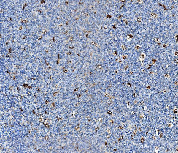
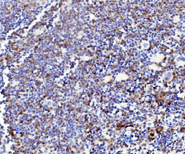
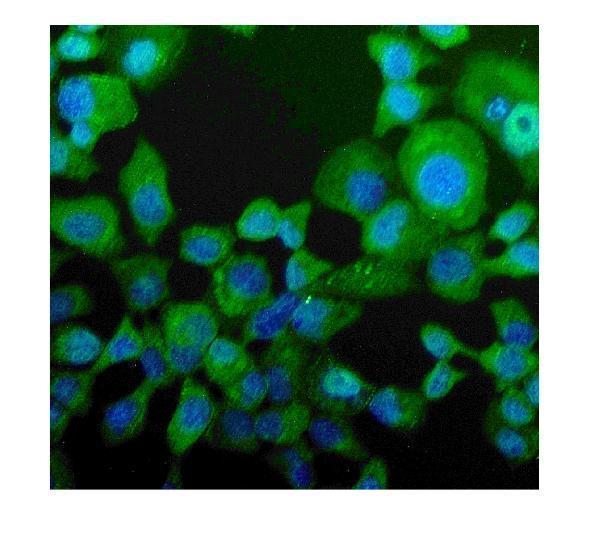
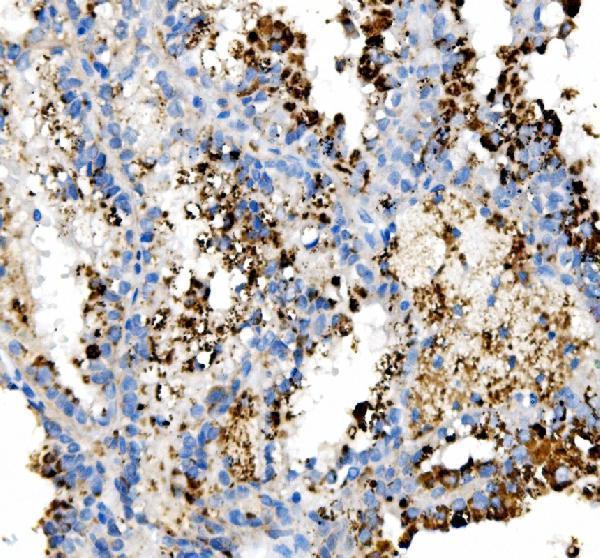
Anti-NPC2 antibodies have IHC images from human tonsil, human renal cancer, and mouse spleen paraffin sections, plus an IF image from A431 cells (catalog image captions).

Real IHC data IHC analysis of NPC2 using anti-NPC2 antibody (A01582-3). NPC2 was detected in paraffin-embedded section of human tonsil tissue. Heat mediated antigen retrieval was performed in EDTA buffer (pH8.0, epitope retrieval solution). The tissue section was blocked with 10% goat serum. The tissue section was then incubated with 1μg/ml rabbit anti-NPC2 Antibody (A01582-3) overnight at 4°C. Biotinylated goat anti-rabbit IgG was used as secondary antibody and incubated for 30 minutes at 37°C. The tissue section was developed using Strepavidin-Biotin-Complex (SABC) (Catalog # SA1022) with DAB as the chromogen.
Anti-Niemann Pick C2/NPC2 ® Antibody
Cat # A01582-3
Real IHC data IHC analysis of NPC2 using anti-NPC2 antibody (A01582-2). NPC2 was detected in a paraffin-embedded section of human renal cancer tissue. Heat mediated antigen retrieval was performed in EDTA buffer (pH 8.0, epitope retrieval solution). The tissue section was blocked with 10% goat serum. The tissue section was then incubated with 2 μg/ml rabbit anti-NPC2 Antibody (A01582-2) overnight at 4°C. Peroxidase Conjugated Goat Anti-rabbit IgG was used as secondary antibody and incubated for 30 minutes at 37°C. The tissue section was developed using HRP Conjugated Rabbit IgG Super Vision Assay Kit (Catalog # SV0002) with DAB as the chromogen.
Anti-Niemann Pick C2/NPC2 Antibody ®
Cat # A01582-2

A01582-3 has IHC images from human tonsil and mouse spleen paraffin sections and an IF image from A431 cells (A01582-3 image captions). A01582-2 has an IHC image from a human renal cancer paraffin section (A01582-2 image caption).

Which to pick: For human paraffin-section IHC, either SKU has its own IHC image; A01582-3 was used at 1 μg/ml and A01582-2 at 2 μg/ml, with fixative unreported in both captions (IHC image captions). Choose A01582-3 for IF/ICC or mouse tissue: IF and ICC are listed applications, human and mouse are listed as reactive, and its mouse spleen IHC image supports paraffin-section use (A01582-3 catalog applications, reactivity, and IHC image caption). A01582-2 is listed as polyclonal and human reactive, with IHC but no IF/ICC listed; clonality is unreported for A01582-3 (catalog dilution_raw, applications, reactivity, and clone fields).

Each figure is that product's own IHC / IF validation image from its datasheet.

References

  1. UniProt Consortium. UniProt entry P61916 (NPC2_HUMAN, NPC intracellular cholesterol transporter 2).
  2. Human Protein Atlas. NPC2 tissue IHC expression (reliability: Enhanced).
  3. Human Protein Atlas. NPC2 subcellular location (ICC-IF): Secreted.
  4. Human Protein Atlas. NPC2 antibody validation summary (2 antibodies).
  5. A novel role for Niemann-Pick disease type 2C protein in papillae formation. PloS one 2011 — PMC3017059.
  6. Characterization of Niemann-Pick Type C2 protein expression in multiple cancers using a novel NPC2 monoclonal antibody. PloS one 2013 — PMC3798307.
  7. The role of NPC2 gene in glioma was investigated based on bioinformatics analysis. Scientific reports 2024 — PMC11333723.
  8. Lung cancer signatures in plasma based on proteome profiling of mouse tumor models. Cancer cell 2011 — PMC3406925.
  9. PubMed PMID:8418812 — UniProt-cited evidence.
  10. PubMed PMID:14702039 — UniProt-cited evidence.
  11. PubMed PMID:12508121 — UniProt-cited evidence.