This website uses cookies to ensure you get the best experience on our website.


- Table of Contents
Plan NR1H3 chromogenic IHC around the reported general nuclear tissue pattern (HPA tissue IHC). This guide covers fixation consistency, nuclear scoring, and interpretation given the low consistency between antibody staining and RNA expression (HPA tissue IHC).

Expected localisation, validated protocols, controls and antibodies — the at-a-glance facts below, then the full design guide.
| Expected localisation | Nuclear tissue staining (HPA tissue IHC) | |
| Staining pattern | Nuclear staining in glandular and hematopoietic cells (HPA tissue IHC) | |
| Antigen retrieval | Tris-EDTA pH 9.0 HIER, 95–98 °C, 20 min (rule: nuclear antigen) | |
| Positive control | Adrenal gland+4 more · see all | |
| Negative control | None in HPA (detected in all 45 tissues); use no-primary + isotype controls |
| Fixation | Matched tissue-IHC evidence does not establish the fixation claim. Validate the specimen-specific method before use. (selected-SKU IHC image A03331) | |
| Caveat | Antibody staining and RNA expression show low consistency (HPA tissue IHC) | |
| Regulation | Liver-enhanced RNA expression (HPA tissue RNA) | |
| Isoform / epitope | 3 isoforms; epitope coverage is unreported (UniProt) |
The catalog antibody's IHC-P protocol is accompanied by two published NR1H3 protocols for paraffin sections (PMC13400882; PMC6343513).
| Sample | Tissue sections; selected-image fixative not specified (standard IHC workflow) |
| Fixation | Image fixative and duration unreported (datasheet A03331); verify before use. |
| Sectioning | 4–5 µm sections on charged slides (standard) |
| Deparaffinisation | Xylene, graded ethanol series to water (standard) |
| Antigen retrieval | Heat-induced epitope retrieval in Tris-EDTA buffer, pH 9.0, 20 min at 95–98 °C (standard rule: nuclear antigen) |
| Peroxidase block | 3% H2O2, 10 min, room temperature (standard) |
| Blocking | 10% normal serum of the secondary host, 30 min, room temperature (standard) |
| Primary antibody | Rabbit anti-NR1H3, 5 μg/mL (datasheet A03331) |
| Primary incubation | Overnight at 4 °C (standard) |
| Detection | HRP-polymer secondary, DAB chromogen 5–10 min (standard) |
| Counterstain | Hematoxylin, blue, dehydrate and mount (standard) |
| Expected result | NR1H3-positive staining in glandular cells of adrenal gland (HPA tissue IHC: Medium). HPA tissue profile: General nuclear expression. No signal in the no-primary control. |
NR1H3 should appear mainly in nuclei, with possible cytoplasmic staining (UniProt Q13133: nucleus and cytoplasm; HPA: general nuclear expression). HPA reports medium IHC staining in adrenal glandular cells and bone marrow hematopoietic cells, among other listed cells (HPA tissue IHC). It has no transmembrane segment, so a membrane-only pattern is unexpected (UniProt Q13133 topology). Interpret tissue staining cautiously: HPA rates its IHC Approved but reports low consistency with RNA expression (HPA tissue IHC).
| Nuclear staining in adrenal glandular cells or bone marrow hematopoietic cells. | This matches cells with medium staining in the HPA tissue images and the reported general nuclear pattern (HPA tissue IHC). Compare stained nuclei with neighboring cells and the counterstain; intensity alone does not establish specificity, given the reported low agreement between IHC staining and RNA expression (HPA tissue IHC; standard IHC practice). |
| Strong membrane-only or extracellular staining, with little nuclear signal. | That distribution conflicts with the reported nuclear and cytoplasmic localization and lack of a transmembrane segment (UniProt Q13133; HPA tissue IHC). Check the matched negative control and tissue morphology before treating the signal as NR1H3; an unexpected compartment raises concern for background or off-target binding but does not identify its cause (standard IHC practice). |
| Prominent staining in a cell population outside the pattern being assessed. | First confirm cell identity on the counterstained section; neighboring cells can be mistaken for the intended population (standard IHC practice). If the signal remains inconsistent with the HPA cell-specific observations, compare antibody-omission and detection-only controls for nonspecific binding or endogenous detection activity (HPA tissue IHC; standard IHC practice). HPA lists low staining in liver cholangiocytes, not a universal negative result (HPA tissue IHC). |
| Diffuse brown signal across cells, stroma, or slide background. | A diffuse pattern is difficult to reconcile with predominantly nuclear NR1H3 staining (HPA tissue IHC). Review background in the negative controls and assess blocking, washes, and endogenous chromogen-generating activity as general chromogenic IHC checks (standard IHC practice). Background alone cannot establish whether target protein is present (standard IHC practice). |
| No convincing nuclear signal in an HPA medium-staining cell population. | A missing signal in, for example, adrenal glandular cells warrants checking section quality, retrieval, antibody and detection steps, and an appropriate positive control (HPA tissue IHC; standard IHC practice). It does not by itself prove absence of NR1H3: HPA reports low agreement between antibody staining and RNA expression, and its listed levels describe observations rather than guaranteed results (HPA tissue IHC). |
| Choice of tissue and cell population | HPA reports medium staining in adrenal glandular, appendix glandular, bone marrow hematopoietic, bronchial respiratory epithelial, and selected glial cells, but low staining in liver cholangiocytes (HPA tissue IHC). UniProt reports strong expression in liver, kidney, and intestine at the tissue level; that statement does not specify which cells in a section will stain (UniProt Q13133). |
| IHC evidence strength | HPA calls the tissue IHC profile Approved while explicitly noting low consistency between staining and RNA expression; both statements should accompany interpretation (HPA tissue IHC). The listed antibodies HPA036443 and CAB037109 are IHC Approved, not IHC Enhanced in the supplied record (HPA antibodies). |
| Compartment and topology | Nucleus and cytoplasm are reported locations; HPA describes general nuclear IHC expression (UniProt Q13133; HPA tissue IHC). No transmembrane segment, signal peptide, propeptide, or reported glycosylation site supports an expected membrane or secreted pattern (UniProt Q13133). These annotations do not predict antigen-retrieval conditions or fixation sensitivity (UniProt Q13133). |
| Isoforms and epitope coverage | UniProt lists three NR1H3 isoforms and a ligand-binding domain at residues 209–447 (UniProt Q13133). Interpret any isoform claim only if the chosen antibody's documented epitope supports it; the supplied HPA antibody list gives no epitope positions (HPA antibodies). |
| IF/ICC: what should the signal look like? | In the separate IF/ICC assessment, HPA reports approved nucleoplasmic localization with additional cytosolic localization and images from HeLa, U-251MG, and U2OS cells (HPA subcellular). These observations support a compartment cross-check; they do not supply an IHC-P staining protocol or establish tissue-specific intensity (HPA subcellular). |
| Situation | Likely cause | Next action |
|---|---|---|
| The positive-control section is blank. | The run may have lost usable signal during retrieval, primary-antibody incubation, or chromogenic detection (standard IHC practice). | Check the control section and each run step, then compare with a documented HPA medium-staining cell population; HPA's observations are guides, not guaranteed positivity (HPA tissue IHC; standard IHC practice). |
| The section stains brown even when primary antibody is omitted. | Detection-system background or endogenous chromogen-generating activity may be contributing (standard IHC practice). | Compare detection-only and reagent controls, review endogenous-activity blocking and washes, and interpret nuclear staining only after the control background is understood (standard IHC practice). |
| Most structures show weak, diffuse color. | Nonspecific reagent binding, inadequate washing, or detection background can obscure cell boundaries (standard IHC practice). | Review blocking, washes, antibody concentration, and detection exposure with matched controls; seek a nuclear pattern in the intended cells (HPA tissue IHC; standard IHC practice). |
| Signal appears mainly at cell borders. | A border-dominant pattern conflicts with reported localization and topology; its specific cause remains unresolved (UniProt Q13133; HPA tissue IHC). | Check morphology and negative controls, then reassess antibody specificity before scoring border staining as NR1H3 (standard IHC practice). |
| Liver cholangiocytes stain weakly despite strong liver expression in UniProt. | The sources describe different resolutions: UniProt reports tissue-level expression, while HPA lists low IHC staining specifically in cholangiocytes (UniProt Q13133; HPA tissue IHC). | Record the scored cell population and its controls; avoid assigning the tissue-level statement to cholangiocytes or calling the section a failed run on that basis alone (UniProt Q13133; HPA tissue IHC). |
| Two candidate antibodies give different cell patterns. | HPA marks both listed antibodies IHC Approved and reports low staining–RNA agreement; the supplied record does not explain a discrepancy between reagents (HPA antibodies; HPA tissue IHC). | Compare each reagent's matched controls and nuclear distribution in the same cell populations; report unresolved discordance instead of assigning specificity from intensity alone (HPA tissue IHC; standard IHC practice). |


Comprehensive Human Protein Atlas IHC scoring per tissue (reliability: Approved — Low consistency between antibody staining and RNA expression data.). Rows are taken directly from the HPA tissue chart — click any row's HPA link to view the source.
| Tissue | Cell type | Level | Evidence | Source |
|---|---|---|---|---|
| Adrenal gland | Glandular cells | Medium | Protein (IHC) | HPA → |
| Appendix | Glandular cells | Medium | Protein (IHC) | HPA → |
| Bone marrow | Hematopoietic cells | Medium | Protein (IHC) | HPA → |
| Breast | Glandular cells | Medium | Protein (IHC) | HPA → |
| Bronchus | Respiratory epithelial cells | Medium | Protein (IHC) | HPA → |
| Tissue | Cell type | Level | Evidence | Source |
|---|---|---|---|---|
| None in HPA: NR1H3 is detected in all 45 scored tissues. Use a no-primary (secondary-only) and an isotype control instead. | ||||
Troubleshoot NR1H3 staining in paraffin sections by checking retrieval, nuclear localisation, antibody specificity and cell level scoring.
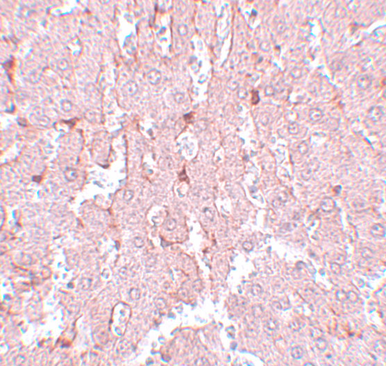
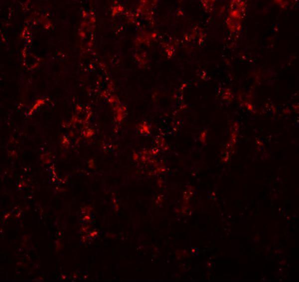
A03331 has rat liver IHC and IF images (catalog image captions); its listed reactivity covers human, mouse and rat (catalog: reactivity).
A03331 is the SKU with an IHC card, showing rat liver tissue stained at 5 μg/mL (A03331 IHC image caption). The catalog also shows rat liver IF at 20 μg/mL for A03331 (A03331 IF image caption).
Which to pick: Choose A03331 for paraffin-section IHC (catalog: IHC-P); its own IHC image shows rat liver at 5 μg/mL, and the fixative is unreported (A03331 IHC image caption). For IF/ICC, M03331 is a rabbit monoclonal with both applications listed, while A03331 has a rat liver IF image (catalog: M03331 host, clone and applications; A03331 IF image caption). Both list human, mouse and rat reactivity for cross-species planning (catalog: reactivity); the supplied images demonstrate rat liver only (A03331 IHC and IF image captions).