NUCB2 / Nucleobindin-2 · IHC design guide

Design Immunohistochemistry for NUCB2

Plan chromogenic NUCB2 IHC in paraffin sections around its predominantly cytoplasmic tissue pattern (HPA tissue IHC). Select a high-staining glandular cell population as a positive control and consider secreted variants when interpreting distribution (HPA tissue IHC).

Evidence assembled Oct 2026 · For research use; verify linked source records and product datasheet before use
Immunohistochemistry protocol sheet for NUCB2 (IHC for NUCB2): expected localisation Cytoplasmic staining in most tissues (HPA tissue IHC), antibody A02400, validated IHC image, and IHC protocol steps
Printable NUCB2 IHC protocol sheet — expected localisation Cytoplasmic staining in most tissues (HPA tissue IHC), antibody A02400, controls and protocol steps. Open the full NUCB2 IHC guide →

NUCB2 Immunohistochemistry Experimental Design Guide

Expected localisation, validated protocols, controls and antibodies — the at-a-glance facts below, then the full design guide.

Must know before staining
Expected localisation Cytoplasmic staining in most tissues (HPA tissue IHC)
Staining pattern Cytoplasmic staining across most tissues (HPA tissue IHC)
Antigen retrieval Tris-EDTA pH 9.0 HIER, 95–98 °C, 20 min (rule: nuclear antigen)
Positive control ⓘ Bone marrow+4 more · see all
Negative control ⓘ Kidney+4 more · see all
Important caveats
Reasons your staining may differ from the expected pattern.
Fixation Matched tissue-IHC evidence does not establish the fixation claim. Validate the specimen-specific method before use. (selected-SKU IHC image A02400)
Caveat Secreted variants can shift protein from its source cells (HPA tissue IHC)
Regulation Staining regulation not specified (UniProt)
Isoform / epitope 2 isoforms; cleavage yields nesfatin-1 (aa 25–106), affecting epitope choice (UniProt)
Section 1

Recommended NUCB2 IHC & IF Protocols

Compare the catalog antibody’s IHC-P protocol (guide payload) with four published NUCB2 tissue protocols (PMC3852884; PMC6169241; PMC6388962; PMC5471050).

Recommended immunohistochemistry (IHC-P) protocol parameters
SampleTissue sections; selected-image fixative not specified (standard IHC workflow)
FixationImage fixative and duration unreported (datasheet A02400); verify before use.
Sectioning4–5 µm sections on charged slides (standard)
DeparaffinisationXylene, graded ethanol series to water (standard)
Antigen retrievalHeat-induced epitope retrieval in Tris-EDTA buffer, pH 9.0, 20 min at 95–98 °C (standard rule: nuclear antigen)
Peroxidase block3% H2O2, 10 min, room temperature (standard)
Blocking10% normal serum of the secondary host, 30 min, room temperature (standard)
Primary antibodyRabbit anti-NUCB2, 1:100–1:500 starting range (standard)
Primary incubationOvernight at 4 °C (standard)
DetectionHRP-polymer secondary, DAB chromogen 5–10 min (standard)
CounterstainHematoxylin, blue, dehydrate and mount (standard)
Expected resultNUCB2-positive staining in hematopoietic cells of bone marrow (HPA tissue IHC: High). HPA tissue profile: Cytoplasmic expression in most tissues. No signal in the no-primary control.
💡Decision noteStart with Tris-EDTA pH 9.0 HIER at 95–98 °C for 20 min (page retrieval rule). The thyroid protocol specifies EDTA pH 8.0 (PMC6388962).
Section 2

What Is the Expected NUCB2 Staining Pattern?

NUCB2 staining is cytoplasmic in most tissues, with high staining in colon glandular cells (HPA tissue IHC). HPA rates tissue staining reliability Enhanced, with medium consistency between antibody staining and RNA expression (HPA). NUCB2 has no transmembrane segment (UniProt P80303 topology).

What am I looking at on my slide?
Strong cytoplasmic staining in colon glandular cells (HPA: High).Expected IHC pattern (HPA: cytoplasmic expression in most tissues).
Isolated nuclear staining.Discordant with the expected cytoplasmic IHC pattern; assess artefact (HPA; general IHC practice).
Staining in lung alveolar cells (HPA: Not detected).Unexpected; assess cross-reactivity or endogenous detection activity (HPA; general IHC practice).
Diffuse haze across the section.Possible background; compare a no-primary control (general IHC practice).
No staining in colon glandular cells (HPA: High).Unexpected; check the positive control and detection workflow (HPA; general IHC practice).
💡Expected NUCB2 appearancePositive: high colon glandular cytoplasmic staining; isolated nuclear signal is suspect (HPA).
How each factor affects the staining
Tissue and cell choiceColon glands are High; lung alveolar cells are Not detected (HPA tissue IHC).
Processing and antibody epitopeChains 25–420 and 25–106 exist; detected forms depend on the epitope (UniProt P80303).
Antibody validationCAB015931: IHC Enhanced; HPA008395 and CAB079781: IHC Supported (HPA).
IF/ICC: where should signal appear?Golgi apparatus (HPA ICC-IF: enhanced); images include A-431, U-251MG and U2OS (HPA).
Why is my staining missing, weak or wrong?
SituationLikely causeNext action
Colon glands lack signal (HPA: High).Possible retrieval or detection failure (general IHC practice).Check a known-positive section and detection controls (general IHC practice).
Signal is mainly nuclear.Discordant with cytoplasmic IHC staining (HPA).Compare a no-primary control and review localisation (general IHC practice).
Alveolar cells stain (HPA: Not detected).Possible cross-reactivity or endogenous detection activity (general IHC practice).Check no-primary and endogenous-activity controls (general IHC practice).
Diffuse section-wide haze.Possible nonspecific background (general IHC practice).Review blocking, antibody dilution and washes (general IHC practice).
Results vary by tissue.A secreted variant can complicate RNA–protein agreement (HPA).Score each cell type against its HPA tissue IHC pattern (HPA).

Sample controls for NUCB2 IHC & IF

🧪Run colon first: glandular cells should stain (HPA: High in colon glandular cells). Use skeletal muscle myocytes as the negative comparator (HPA: Not detected in skeletal muscle myocytes); on the colon slide, unlabeled neighboring cells should show only background chromogen, without assuming that every nonglandular cell is NUCB2-negative (HPA: High reported for glandular cells).
Positive control tissue: Bone marrow (Hematopoietic cells, HPA High)
Negative control tissue: Kidney (HPA Not detected)
ICC-IF cell lines (HPA subcellular resource): HPA ICC-IF images show NUCB2 in A-431, U-251MG, U2OS, with annotated localisation: Golgi apparatus (enhanced) (HPA subcellular).
Technical controls: Include a no-primary, secondary-only control; a primary-host-species- and isotype-matched control IgG; and, where available, NUCB2 knockout tissue or an immunogen-peptide block as a biological specificity control (standard IHC practice). Quench endogenous peroxidase in colon sections before chromogenic detection, and block endogenous biotin if using avidin–biotin detection (standard IHC practice).
⚠️Feasibility: A target-specific fixation window and retrieval effect are unreported in the supplied evidence; optimize antigen retrieval on paraffin sections empirically (standard IHC practice). The selected rat-brain IHC caption does not state a fixative (A02400 caption: fixative unreported). Frozen sections or IF cannot be judged easier from these data; the colon control needs attention to endogenous peroxidase and, with avidin–biotin detection, endogenous biotin (standard IHC practice).

HPA tissue IHC evidence for NUCB2

Comprehensive Human Protein Atlas IHC scoring per tissue (reliability: Enhanced — Medium consistency between antibody staining and RNA expression data. At least one protein variant secreted, tissue location of RNA and protein might differ and correlation is complex.). Rows are taken directly from the HPA tissue chart — click any row's HPA link to view the source.

Positive expression · recommended positive controls

TissueCell typeLevelEvidenceSource
Bone marrow Hematopoietic cells High Protein (IHC) HPA →
Bronchus Ciliated cells (cell body) High Protein (IHC) HPA →
Colon Glandular cells High Protein (IHC) HPA →
Duodenum Glandular cells High Protein (IHC) HPA →
Epididymis Glandular cells High Protein (IHC) HPA →

Undetected expression · recommended negative controls

TissueCell typeLevelEvidenceSource
Kidney Cells in glomeruli Not detected Protein (IHC) HPA →
Lung Alveolar cells Not detected Protein (IHC) HPA →
Ovary Follicle cells Not detected Protein (IHC) HPA →
Skeletal muscle Myocytes Not detected Protein (IHC) HPA →
Urinary bladder Urothelial cells Not detected Protein (IHC) HPA →
Section 3

Advanced NUCB2 IHC Tips

Troubleshoot NUCB2 chromogenic IHC by checking retrieval, compartment, cell type and controls before interpreting staining intensity.

How should I retrieve NUCB2 in paraffin sections when staining is weak?
Start with Tris-EDTA at pH 9.0, heated to 95–98 °C for 20 min (page retrieval setting). Let sections cool consistently, then compare the same tissue, antibody concentration and detection run across slides; uneven heating or drying can make retrieval appear unreliable (standard IHC practice). If staining remains weak, test an alternative retrieval condition on adjacent sections as a fallback, documenting both signal and tissue preservation rather than assuming stronger retrieval is better (standard IHC practice). Score the expected cytoplasmic pattern separately from diffuse background, since NUCB2 has Golgi and other intracellular annotations (HPA subcellular; UniProt P80303 localisation).
Could fixation explain weak or inconsistent NUCB2 staining?
NUCB2-specific fixation sensitivity is unknown from the supplied IHC evidence; the selected antibody’s rat brain caption reports 2.5 µg/ml but does not state a fixative (A02400 tissue-IHC caption). Record fixation duration, tissue thickness and processing history for each block before comparing staining across specimens (standard IHC practice). When material permits, compare similarly processed sections in one staining run and keep retrieval at Tris-EDTA pH 9.0, 95–98 °C for 20 min (page retrieval setting). Interpret differences alongside morphology and section integrity, and do not assign a NUCB2-specific fixation effect without a controlled comparison (standard IHC practice).
Which staining compartments fit NUCB2 biology in tissue sections?
Assess cytoplasmic staining first, because the tissue IHC profile reports cytoplasmic expression in most tissues (HPA tissue IHC). A concentrated perinuclear pattern is also plausible given the enhanced Golgi localisation in cell images and N-terminal Golgi retention annotation (HPA subcellular; UniProt P80303 localisation). NUCB2 is additionally annotated in the endoplasmic reticulum, nuclear envelope and secreted compartment, with no transmembrane segment (UniProt P80303 localisation and topology). Compare these patterns with cell morphology and a negative reagent control before calling isolated nuclear or extracellular chromogen specific; section folds and deposits can imitate compartmental staining (standard IHC practice).
How does epitope location affect interpretation of NUCB2 staining?
Check the catalog antibody’s immunogen or mapped epitope before comparing staining with other NUCB2 assays (standard IHC practice). NUCB2 has 2 annotated isoforms, a signal peptide at residues 1–24, a chain spanning 25–420, and a nesfatin-1 chain spanning 25–106 (UniProt P80303 processing and isoforms). An epitope within 25–106 may recognize material containing that region, whereas an epitope outside it cannot directly report the isolated nesfatin-1 chain (UniProt P80303 processing). If the epitope is undisclosed, describe the result as NUCB2 immunoreactivity and avoid assigning a particular isoform or processed product from chromogenic staining alone (standard IHC interpretation).
How should I check NUCB2 localisation with multiplex IF?
For the separate IF/ICC workflow, pair NUCB2 with an epithelial cell marker when examining colon glandular cells, where tissue IHC reports high staining (HPA tissue IHC; standard IF practice). Choose a red or far-red fluorophore after inspecting unstained tissue autofluorescence, and include single-label controls to check channel spillover (standard IF practice). If the antibody binds an intracellular epitope, test permeabilisation; the required detergent access depends on whether that epitope faces the cytosol or a Golgi or ER lumen, which the supplied antibody evidence does not establish (UniProt P80303 localisation; standard IF practice). Interpret colocalisation with morphology and channel controls (standard IF practice).
What controls help distinguish NUCB2 signal from chromogenic background?
Run a no-primary control alongside the test slide to reveal signal from detection reagents or tissue rather than primary antibody binding (standard IHC practice). Apply a peroxidase block before DAB detection and inspect pigment, folds and section edges, which can create misleading brown deposits (standard chromogenic IHC practice). If background is widespread, compare an antibody dilution series while holding retrieval, incubation and imaging conditions constant; the selected rat brain caption used 2.5 µg/ml, which is evidence for that image only (A02400 tissue-IHC caption). Judge improvements in the expected cytoplasmic or perinuclear pattern against background in adjacent cells (HPA tissue IHC; HPA subcellular).
How should I score NUCB2 IHC across samples? ⚠ ANSWER MARKED FOR VERIFICATION
Define the scored cell population and compartment before examining group differences; tissue IHC describes cytoplasmic NUCB2 expression in most tissues (HPA tissue IHC). For a defined cell population, record the percentage positive and intensity categories, then calculate an H-score from 0–300 if those categories are reproducible (standard IHC scoring practice). Alternatively, report positive cells per mm² when cell density varies, with the counted tissue area as the denominator (standard IHC scoring practice). Normalise comparisons to the same eligible cell type, tissue area, staining run and imaging settings, and exclude necrosis and section edges using prespecified rules (standard IHC practice).
When is apparent NUCB2 positivity likely to be an artefact?
Give more weight to cellular cytoplasmic staining than to isolated extracellular deposits, since the tissue IHC profile is predominantly cytoplasmic (HPA tissue IHC). Check cell identity: colon glandular cells are reported high, while kidney glomerular cells are reported not detected in the supplied tissue survey (HPA tissue IHC). A perinuclear accent can fit Golgi localisation, but uniform nuclear staining alone needs independent support before being assigned to NUCB2 (HPA subcellular; standard IHC interpretation). Treat staining confined to cut edges, necrotic regions or the no-primary control as suspect, and check peroxidase blocking when brown signal persists without primary antibody (standard chromogenic IHC practice).
Boster reagents

Best NUCB2 / Nucleobindin-2 IHC Antibodies

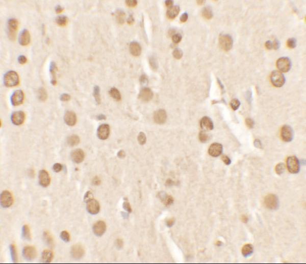
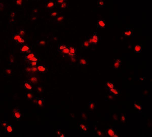
A02400 has rat-brain IHC and IF images (A02400 image captions). Human, Mouse, and Rat reactivity is listed (A02400 catalog reactivity).

Real IHC data Immunohistochemistry of Nucleobindin-2 in rat brain tissue with Nucleobindin-2 antibody at 2.5 μg/ml.
Anti-Nucleobindin-2 NUCB2 Antibody
Cat # A02400

A02400 has a rat-brain IHC image at 2.5 μg/ml (A02400 IHC image caption). A02400 also has a rat-brain IF image at 20 μg/ml; Human, Mouse, and Rat reactivity is listed (A02400 IF image caption; catalog reactivity).

Which to pick: Choose A02400 for paraffin-section tissue IHC: IHC-P is listed, and its own IHC image shows rat brain at 2.5 μg/ml; the fixative is unreported (A02400 applications; A02400 IHC image caption). For IF, A02400 has a rat-brain image at 20 μg/ml; ICC validation is unreported (A02400 IF image caption; A02400 applications). For cross-species work, A02400 lists Human, Mouse, and Rat reactivity, while the pictured IHC and IF data are from rat brain; its host is Rabbit and clonality is unreported (A02400 catalog reactivity/host/clone; A02400 image captions).

Each figure is that product's own IHC / IF validation image from its datasheet.

References

  1. UniProt Consortium. UniProt entry P80303 (NUCB2_HUMAN, Nucleobindin-2).
  2. Human Protein Atlas. NUCB2 tissue IHC expression (reliability: Enhanced).
  3. Human Protein Atlas. NUCB2 subcellular location (ICC-IF): Localized to the Golgi apparatus..
  4. Human Protein Atlas. NUCB2 antibody validation summary (3 antibodies).
  5. Prognostication of prostate cancer based on NUCB2 protein assessment: NUCB2 in prostate cancer. Journal of experimental & clinical cancer research : CR 2013 — PMC3852884.
  6. Nesfatin-1/Nucleobindin-2 Is a Potent Prognostic Marker and Enhances Cell Proliferation, Migration, and Invasion in Bladder Cancer. Disease markers 2018 — PMC6169241.
  7. High expression of NUCB2 promotes papillary thyroid cancer cells proliferation and invasion. OncoTargets and therapy 2019 — PMC6388962.
  8. High NUCB2 expression level represents an independent negative prognostic factor in Chinese cohorts of non-metastatic clear cell renal cell carcinoma patients. Oncotarget 2017 — PMC5471050.
  9. PubMed PMID:7811391 — UniProt-cited evidence.
  10. PubMed PMID:9639681 — UniProt-cited evidence.
  11. PubMed PMID:12087473 — UniProt-cited evidence.