OASL / 2'-5'-oligoadenylate synthase-like protein · IHC design guide

Design Immunohistochemistry for OASL

Plan chromogenic OASL IHC-P with catalog antibody A04123-1 at 1:50–1:100 (datasheet: IHC-P). Interpret cytoplasmic and nuclear staining in the context of the reported low agreement with RNA expression (HPA tissue IHC).

Evidence assembled Oct 2026 · For research use; verify linked source records and product datasheet before use
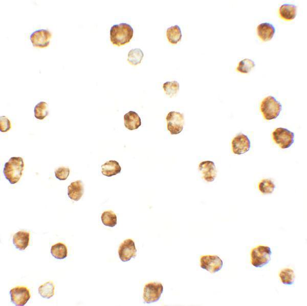
Immunohistochemistry protocol sheet for OASL (IHC for OASL): expected localisation Cytoplasm and nucleus in a few tissues (HPA tissue IHC), antibody A04123-1, validated IHC image, and IHC protocol steps
Printable OASL IHC protocol sheet — expected localisation Cytoplasm and nucleus in a few tissues (HPA tissue IHC), antibody A04123-1, controls and protocol steps. Open the full OASL IHC guide →

OASL Immunohistochemistry Experimental Design Guide

Expected localisation, validated protocols, controls and antibodies — the at-a-glance facts below, then the full design guide.

Must know before staining
Expected localisation Cytoplasm and nucleus in a few tissues (HPA tissue IHC)
Staining pattern Cytoplasmic and nuclear staining in a few tissues (HPA tissue IHC)
Antigen retrieval Tris-EDTA pH 9.0 HIER, 95–98 °C, 20 min (rule: nuclear antigen)
Positive control ⓘ Bone marrow+4 more · see all
Negative control ⓘ Adipose tissue+4 more · see all
Important caveats
Reasons your staining may differ from the expected pattern.
Fixation The selected-SKU tissue-IHC image shows formalin-fixed paraffin sections (selected-SKU IHC image A04123-1); Keep fixation conditions consistent across sections (standard IHC practice; not target-specific)
Caveat Staining shows low agreement with RNA expression (HPA tissue IHC)
Regulation No induction condition documented (UniProt record)
Isoform / epitope 3 isoforms; C-terminal epitope coverage is unknown (UniProt; datasheet: C-term)
Section 1

Recommended OASL IHC & IF Protocols

The catalog antibody protocol is accompanied by published OASL IHC methods for colorectal tumors and bladder tissue (PMC12639863; PMC9510761).

Recommended immunohistochemistry (IHC-P) protocol parameters
SampleFFPE human cancer tissue (datasheet A04123-1)
FixationImage formalin-fixed; duration unreported (datasheet A04123-1); verify before use.
Sectioning4–5 µm sections on charged slides (standard)
DeparaffinisationXylene, graded ethanol series to water (standard)
Antigen retrievalHeat-induced epitope retrieval in Tris-EDTA buffer, pH 9.0, 20 min at 95–98 °C (standard rule: nuclear antigen)
Peroxidase block3% H2O2, 10 min, room temperature (standard)
Blocking10% normal serum of the secondary host, 30 min, room temperature (standard)
Primary antibodyRabbit anti-OASL, 1:50-1:100 (datasheet A04123-1)
Primary incubationOvernight at 4 °C (standard)
DetectionHRP-polymer secondary, DAB chromogen 5–10 min (standard)
CounterstainHematoxylin, blue, dehydrate and mount (standard)
Expected resultOASL-positive staining in hematopoietic cells of bone marrow (HPA tissue IHC: Medium). HPA tissue profile: Cytoplasmic and nuclear expression in a few tissues. No signal in the no-primary control.
💡Decision noteStart with Tris-EDTA pH 9.0 retrieval at 95–98 °C for 20 min (page retrieval rule; UniProt Q15646: nucleus and nucleolus).
Section 2

What Is the Expected OASL Staining Pattern?

OASL can appear in cytoplasm and nuclei, including nucleoli (UniProt Q15646); HPA reports cytoplasmic and nuclear staining in a few tissues (HPA tissue IHC). In paraffin sections, assess the named positive cell populations rather than the whole tissue (HPA tissue IHC). OASL has no transmembrane segment (UniProt Q15646 topology). HPA rates its tissue staining Approved but reports low consistency with RNA expression (HPA tissue IHC).

What am I looking at on my slide?
Distinct cytoplasmic and/or nuclear staining in bone marrow hematopoietic cells; nearby cells may differ in intensity.This fits the reported compartment pattern and a medium staining call for hematopoietic cells (HPA tissue IHC). Score the named cells individually. The Approved rating supports use as a reference, while low consistency with RNA expression limits how strongly staining alone confirms OASL identity (HPA tissue IHC).
A sharp cell-surface rim is the dominant pattern, with little intracellular staining.A predominantly membrane-restricted result conflicts with the reported cytoplasmic and nuclear pattern (HPA tissue IHC) and the absence of a transmembrane segment (UniProt Q15646 topology). Check morphology and detection controls before interpreting the rim as OASL; localization alone cannot identify its cause.
Strong color appears in adipocytes or adrenal glandular cells, while the intended positive cells are unstained.Those cell populations were reported as not detected (HPA tissue IHC). Possible explanations include cross-reactivity or endogenous chromogenic activity; neither is established by the image alone. Compare an appropriate positive section, a no-primary control, and the distribution within each tissue as general IHC checks.
Weak, uniform color covers cells, stroma, and empty spaces without a clear intracellular pattern.This is difficult to score as specific OASL because HPA describes cytoplasmic and nuclear expression in a few tissues (HPA tissue IHC). As a general IHC check, inspect the no-primary control and review blocking, washing, and chromogen development; the supplied sources do not assign an OASL-specific cause.
No staining is visible in bone marrow hematopoietic cells despite intact tissue morphology.HPA reports medium staining in these cells, so an entirely blank section warrants a technical check (HPA tissue IHC). Confirm the correct cell population is present, then review the antibody, retrieval, detection, and counterstain records as general IHC practice. A negative slide alone cannot establish absent OASL expression.
💡Expected OASL appearanceA plausible positive is discernible cytoplasmic and/or nuclear color in named HPA-positive cells, such as medium staining in bone marrow hematopoietic cells (HPA tissue IHC); a dominant sharp membrane rim or uniform color across cell-free areas should prompt a control review (UniProt Q15646 topology; general IHC practice).
How each factor affects the staining
Compartment referenceUniProt lists nucleus, nucleolus, and cytoplasm (UniProt Q15646); HPA tissue IHC reports nuclear and cytoplasmic staining (HPA tissue IHC). Score compartment and cell identity together.
Tissue and cell selectionHPA reports medium staining in colon endothelial cells and testis elongated or late spermatids, but no detection in listed adipocytes (HPA tissue IHC). Select controls by cell population.
Antibody evidenceThe listed HPA001474 antibody has Approved IHC validation (HPA antibodies). HPA also notes low agreement between antibody staining and RNA expression, which limits a single-stain conclusion (HPA tissue IHC).
Isoforms and epitopeUniProt lists three OASL isoforms, p56, p30, and 3 (UniProt Q15646). Epitope coverage is not supplied; do not infer which isoform a stain detects or attribute a staining difference to isoforms.
Why is my staining missing, weak or wrong?
SituationLikely causeNext action
Expected positive cells stain weakly or remain blank.A failed staining run is possible; HPA reports medium staining in bone marrow hematopoietic cells (HPA tissue IHC).As general IHC practice, verify the positive control, antibody preparation, retrieval and detection settings, and reagent expiry; interpret a persistent negative result only after controls work.
Color appears throughout the section, including spaces without cells.Nonspecific background or excess chromogen development is possible (general IHC practice).Check a no-primary control and inspect washing, blocking, detection, and development time (general IHC practice). Score only color tied to identifiable cells and compartments.
A membrane rim dominates the stain.That distribution disagrees with HPA's intracellular pattern (HPA tissue IHC) and UniProt's lack of a transmembrane segment (UniProt Q15646 topology).Inspect section morphology and controls, then reassess the antibody signal before calling cells positive; the sources do not identify the rim's mechanism.
A supposedly negative tissue shows isolated positive cells.A tissue-level label may hide a different cell population; HPA reports staining by named cell type (HPA tissue IHC).Identify the stained cells before comparing them with HPA. Use a no-primary control to assess detection background as general IHC practice.
RNA data and IHC staining disagree.HPA explicitly reports low consistency between antibody staining and RNA expression (HPA tissue IHC).Report the disagreement with cell identity and compartment. Treat the Approved IHC rating as qualified evidence, not proof that every stained cell expresses OASL (HPA tissue IHC).
IF/ICC Q: What localization should I expect?HPA places OASL in nucleoplasm and cytosol in ICC-IF, with nucleoplasm approved and cytosol supported (HPA ICC-IF).A: Assess those intracellular compartments in the separate IF/ICC guide; use its controls to evaluate fluorescence background (HPA ICC-IF; general IF practice).

Sample controls for OASL IHC & IF

🧪Run bone marrow first and look for staining in hematopoietic cells (HPA: Medium). Use adipose tissue adipocytes as the negative tissue (HPA: Not detected); cells without specific staining on the positive slide should show only counterstain or background, but HPA does not identify a named internal negative cell type.
Positive control tissue: Bone marrow (Hematopoietic cells, HPA Medium)
Negative control tissue: Adipose tissue (HPA Not detected)
ICC-IF cell lines (HPA subcellular resource): HPA ICC-IF images show OASL in A-431, RT-4, U2OS, with annotated localisation: Nucleoplasm (approved), Cytosol (supported) (HPA subcellular).
Technical controls: Include a no-primary, secondary-only control, an isotype control matched to the primary antibody’s host species and immunoglobulin class as appropriate for its clonality, and OASL-knockout material as a biological negative (standard IHC practice). Block endogenous peroxidase and assess residual background in bone marrow before interpreting chromogenic staining (standard IHC practice).
⚠️Feasibility: The selected A04123-1 caption explicitly reports formalin-fixed, paraffin-embedded tissue with peroxidase and AEC detection; a target-specific fixation window or fixation effect is unreported (selected-SKU tissue-IHC caption). OASL’s antigen-retrieval dependency is unreported, so optimize retrieval using the positive and negative controls (supplied evidence; standard IHC practice). The supplied evidence does not establish whether frozen sections or IF are easier; marrow endogenous peroxidase can complicate chromogenic interpretation (standard IHC practice).

HPA tissue IHC evidence for OASL

Comprehensive Human Protein Atlas IHC scoring per tissue (reliability: Approved — Low consistency between antibody staining and RNA expression data.). Rows are taken directly from the HPA tissue chart — click any row's HPA link to view the source.

Positive expression · recommended positive controls

TissueCell typeLevelEvidenceSource
Bone marrow Hematopoietic cells Medium Protein (IHC) HPA →
Colon Endothelial cells Medium Protein (IHC) HPA →
Oral mucosa Squamous epithelial cells Medium Protein (IHC) HPA →
Placenta Trophoblastic cells Medium Protein (IHC) HPA →
Testis Elongated or late spermatids Medium Protein (IHC) HPA →

Undetected expression · recommended negative controls

TissueCell typeLevelEvidenceSource
Adipose tissue Adipocytes Not detected Protein (IHC) HPA →
Adrenal gland Glandular cells Not detected Protein (IHC) HPA →
Appendix Glandular cells Not detected Protein (IHC) HPA →
Breast Adipocytes Not detected Protein (IHC) HPA →
Caudate Glial cells Not detected Protein (IHC) HPA →
Section 3

Advanced OASL IHC Tips

Troubleshoot OASL staining in paraffin sections by checking retrieval, compartment, cell identity and controls before comparing signal across specimens.

How should I retrieve OASL in paraffin sections when nuclear staining is weak?
Start with Tris-EDTA at pH 9.0 for heat-induced retrieval at 95–98 °C for 20 min (page retrieval setting). If nuclear signal remains weak, compare adjacent sections with a shorter or longer heating time while keeping the buffer and detection conditions constant; inspect tissue preservation alongside staining (standard IHC practice). OASL can occupy the nucleus, nucleolus and cytoplasm, so assess both compartments rather than treating cytoplasmic staining alone as retrieval success (UniProt Q15646 subcellular location). Include a documented positive control section in each run, since retrieval changes can also increase nonspecific background (standard IHC practice).
Can formalin fixation explain inconsistent OASL staining between paraffin blocks?
The selected antibody has a formalin-fixed, paraffin-embedded cancer tissue IHC example, but that image does not establish OASL sensitivity to fixation duration or processing conditions (A04123-1 tissue-IHC caption). Target-specific fixation sensitivity is unknown from the supplied evidence (evidence scope). Record fixation and processing history for each block, then compare adjacent sections using identical retrieval, primary incubation and chromogen development (standard IHC practice). If signal differs, inspect morphology and a control stain before attributing the difference to OASL abundance; overfixation, uneven processing and section damage are general sources of IHC variation (standard IHC practice).
Should OASL appear in nuclei, cytoplasm or both on chromogenic sections?
Score nuclear and cytoplasmic staining separately: OASL is reported in the nucleus, nucleolus and cytoplasm (UniProt Q15646 subcellular location), while HPA reports cytoplasmic and nuclear tissue staining in a few tissues (HPA tissue IHC). HPA cell imaging places it in the nucleoplasm and cytosol, providing another compartment check rather than a required pattern for every tissue section (HPA subcellular). A diffuse nuclear or cytoplasmic deposit needs comparison with cell morphology, a no-primary control and the expected positive section (standard IHC practice). Document the fraction of cells showing each pattern before combining compartments into a single score (standard IHC practice).
Could an OASL isoform or hidden epitope explain patchy IHC staining?
OASL has 3 listed isoforms, including p56, p30 and isoform 3, so an antibody’s epitope determines which forms it can detect (UniProt Q15646 isoforms; standard antibody practice). The record identifies ubiquitin-like domains at residues 354–433 and 434–509, but it does not identify this antibody’s epitope (UniProt Q15646 domains; supplied antibody evidence). Check epitope documentation before assigning a negative cell to absent OASL, and compare matched sections after the page’s pH 9.0 retrieval (page retrieval setting; standard IHC practice). Preserve nuclear and cytoplasmic scores separately, because staining differences alone cannot establish which isoform is present (UniProt Q15646 subcellular location; standard interpretation).
How can IF help check cell identity and compartment when OASL IHC is ambiguous?
On an IF/ICC guide page, pair OASL with a validated marker for the expected cell population, such as a hematopoietic cell marker when investigating bone marrow, where HPA reports medium staining in hematopoietic cells (HPA tissue IHC; standard IF practice). Choose spectrally separated fluorophores and consider a far-red channel if the specimen has strong short-wavelength autofluorescence (standard IF practice). Because OASL is reported in the nucleus and cytoplasm and has no transmembrane segment, choose permeabilisation that permits antibody access to intracellular epitopes and verify the antibody’s epitope location when known (UniProt Q15646 subcellular location and topology; standard IF practice). Include single-stain and no-primary controls to assess bleed-through and background (standard IF practice).
What should I check when OASL chromogenic staining is widespread or granular?
First compare a no-primary section and the positive control, then inspect whether the deposit follows cell outlines or clusters at tissue folds and damaged edges (standard IHC practice). Block endogenous peroxidase when using peroxidase detection and review blocking, washes, antibody concentration and chromogen development time if diffuse colour persists (standard IHC practice). The selected IHC image used a peroxidase-conjugated secondary and AEC staining, which documents that detection combination but does not establish a universal optimal dilution (A04123-1 tissue-IHC caption). OASL has no transmembrane segment, so an exclusively crisp membrane rim warrants scrutiny for background or an unrelated antigen (UniProt Q15646 topology; standard interpretation).
How should I score heterogeneous OASL staining across tissue compartments? ⚠ ANSWER MARKED FOR VERIFICATION
Define the cell population and compartment before scoring, then report nuclear and cytoplasmic positivity separately because both locations are supported for OASL (UniProt Q15646 subcellular location; HPA tissue IHC). Record the percentage of positive cells and staining intensity; an H-score can combine intensity grades 0–3 with their respective percentages for a 0–300 scale (standard IHC practice). For spatial comparisons, report positive-cell density per mm² of viable, analysable tissue or normalise positive cells to all scored cells of the same type (standard IHC practice). Apply one threshold across matched sections and exclude folds, necrosis and edge artefacts before comparing groups (standard IHC practice).
How can I distinguish a credible OASL-positive cell from a staining artefact?
A credible signal should occupy plausible nuclear or cytoplasmic compartments, match intact cell morphology and exceed the no-primary control (UniProt Q15646 subcellular location; standard IHC practice). Give cell identity equal weight: HPA reports medium staining in bone marrow hematopoietic cells, among other listed populations, but describes low consistency between antibody staining and RNA expression (HPA tissue IHC). Treat staining restricted to cut edges, necrotic areas or sites of endogenous enzyme activity as suspect until controls resolve it (standard IHC practice). The selected cancer tissue image demonstrates IHC use; its caption explicitly says clinical relevance has not been evaluated (A04123-1 tissue-IHC caption).
Boster reagents

Best OASL / 2'-5'-oligoadenylate synthase-like protein IHC Antibodies

The catalog includes an IHC-P antibody with a human tissue image and an IF/ICC antibody with human HepG2 cell images; the latter also lists mouse and rat reactivity (catalog applications, reactivity, and image captions).

Real IHC data Formalin-fixed and paraffin-embedded human cancer tissue reacted with the primary antibody, which was peroxidase-conjugated to the secondary antibody, followed by AEC staining. This data demonstrates the use of this antibody for immunohistochemistry; clinical relevance has not been evaluated. BC = breast carcinoma; HC = hepatocarcinoma.
Anti-OASL Antibody (C-term)
Cat # A04123-1
Real IF data Immunocytochemistry of OASL1 in HepG2 cells with OASL1 antibody at 2.5 μg/mL.
Anti-OASL Antibody
Cat # A04123

A04123-1 is listed for IHC-P in human samples, with an AEC-stained image of formalin-fixed, paraffin-embedded breast carcinoma and hepatocarcinoma tissue (A04123-1 catalog and image caption). A04123 is listed for IF/ICC, with HepG2 cell images at 20 μg/mL for IF and 2.5 μg/mL for ICC (A04123 catalog and image captions).

Which to pick: Choose A04123-1 for human tissue IHC-P: it is polyclonal, has a listed dilution of 1:50–1:100, and its image documents formalin-fixed, paraffin-embedded tissue with AEC detection (A04123-1 catalog and image caption). Choose A04123 for IF/ICC in human cells because its images show those applications in HepG2 cells; it lists human, mouse, and rat reactivity, although the supplied images show human cells only (A04123 catalog and image captions).

Each figure is that product's own IHC / IF validation image from its datasheet.

References

  1. UniProt Consortium. UniProt entry Q15646 (OASL_HUMAN, 2'-5'-oligoadenylate synthase-like protein).
  2. Human Protein Atlas. OASL tissue IHC expression (reliability: Approved).
  3. Human Protein Atlas. OASL subcellular location (ICC-IF): Localized to the nucleoplasm and cytosol..
  4. Human Protein Atlas. OASL antibody validation summary (1 antibodies).
  5. OASL promotes immune evasion in pancreatic ductal adenocarcinoma by enhancing autolysosome-mediated degradation of MHC-I. Theranostics 2025 — PMC11840728.
  6. Sodium butyrate enhances sorafenib-induced ferroptosis and immunogenic cell death by modulating IRF2-Oasl2-cGAS pathway in colorectal cancer. Materials today. Bio 2025 — PMC12639863.
  7. Three Immune-Related Prognostic mRNAs as Therapeutic Targets for Pancreatic Cancer. Frontiers in medicine 2021 — PMC8047149.
  8. Values of OAS gene family in the expression signature, immune cell infiltration and prognosis of human bladder cancer. BMC cancer 2022 — PMC9510761.
  9. PubMed PMID:9722630 — UniProt-cited evidence.
  10. PubMed PMID:9826176 — UniProt-cited evidence.
  11. PubMed PMID:22531715 — UniProt-cited evidence.