This website uses cookies to ensure you get the best experience on our website.


- Table of Contents
Plan chromogenic OTUD5 IHC-P using 2.5 μg/mL as a starting antibody concentration (datasheet: 2.5 μg/mL). Compare cytoplasmic tissue staining (HPA tissue IHC) with the annotated nuclear location (UniProt), and use controls to assess specificity.

Expected localisation, validated protocols, controls and antibodies — the at-a-glance facts below, then the full design guide.
| Expected localisation | Cytoplasmic tissue staining (HPA tissue IHC); nuclear location annotated (UniProt) | |
| Staining pattern | Cytoplasmic staining in glandular cells and colon endothelium (HPA tissue IHC) | |
| Antigen retrieval | Tris-EDTA pH 9.0 HIER, 95–98 °C, 20 min (rule: nuclear antigen) | |
| Positive control | Appendix+4 more · see all | |
| Negative control | Epididymis+4 more · see all |
| Fixation | Matched tissue-IHC evidence does not establish the fixation claim. Validate the specimen-specific method before use. (selected-SKU IHC image A09744) | |
| Caveat | Observed cytoplasmic staining differs from the nuclear annotation (HPA tissue IHC; UniProt) | |
| Regulation | Expression regulation is not established (UniProt) | |
| Isoform / epitope | Five isoforms; epitope coverage is unresolved (UniProt) |
The catalog antibody’s IHC-P protocol is accompanied by published OTUD5 IHC methods from bladder cancer, liver, and tissue-section studies (PMC9463452; PMC10613150; PMC13559827).
| Sample | Tissue sections; selected-image fixative not specified (standard IHC workflow) |
| Fixation | Image fixative and duration unreported (datasheet A09744); verify before use. |
| Sectioning | 4–5 µm sections on charged slides (standard) |
| Deparaffinisation | Xylene, graded ethanol series to water (standard) |
| Antigen retrieval | Heat-induced epitope retrieval in Tris-EDTA buffer, pH 9.0, 20 min at 95–98 °C (standard rule: nuclear antigen) |
| Peroxidase block | 3% H2O2, 10 min, room temperature (standard) |
| Blocking | 10% normal serum of the secondary host, 30 min, room temperature (standard) |
| Primary antibody | Rabbit anti-OTUD5, 2.5 μg/mL (datasheet A09744) |
| Primary incubation | Overnight at 4 °C (standard) |
| Detection | HRP-polymer secondary, DAB chromogen 5–10 min (standard) |
| Counterstain | Hematoxylin, blue, dehydrate and mount (standard) |
| Expected result | OTUD5-positive staining in glandular cells of appendix (HPA tissue IHC: Medium). HPA tissue profile: General cytoplasmic expression. No signal in the no-primary control. |
For paraffin-section IHC, expect mainly cytoplasmic OTUD5 staining, including medium staining in glandular cells of appendix, breast, cervix, duodenum, endometrium, and gallbladder (HPA tissue IHC). OTUD5 has no transmembrane segment (UniProt Q96G74 topology). UniProt lists the nucleus, while HPA reports general cytoplasmic IHC expression; HPA rates its tissue IHC evidence Approved, with medium staining–RNA consistency and external verification pending (UniProt Q96G74; HPA tissue IHC).
| Cytoplasmic chromogen in glandular cells of appendix or duodenum, with discernible cell boundaries and limited background (HPA tissue IHC). | This fits the reported compartment and a medium-staining cell population (HPA tissue IHC). Compare cells within the same section before scoring: scattered pale deposits outside cell boundaries should not be counted as OTUD5-positive cells (general IHC practice). |
| A crisp membrane rim is the dominant signal, with little cytoplasmic staining. | A membrane-only pattern conflicts with general cytoplasmic tissue staining and the absence of a transmembrane segment (HPA tissue IHC; UniProt Q96G74 topology). Treat it as a possible artefact and review antibody concentration, detection controls, and tissue morphology before interpreting it as OTUD5 (general IHC practice). |
| Strong staining appears in cardiomyocytes or smooth muscle cells, which HPA lists as not detected (HPA tissue IHC). | An unexpected cell-type pattern raises concern for cross-reactivity or endogenous detection activity; it does not by itself identify the cause (general IHC practice). Compare a known-positive cell population and a primary-antibody omission control under the same detection conditions (HPA tissue IHC; general IHC practice). |
| Brown precipitate covers stroma, empty spaces, and cell surfaces without a clear cellular pattern. | Diffuse deposition is background rather than a scoreable cytoplasmic pattern (general IHC practice; HPA tissue IHC). Check the primary-antibody omission control, washing, blocking, and chromogen development; assign a cell-level score only where staining can be localized (general IHC practice). |
| Glandular cells in appendix or duodenum show no signal, despite medium staining reported there (HPA tissue IHC). | First check whether another expected-positive section stained in the same run (HPA tissue IHC; general IHC practice). Review retrieval conditions, antibody dilution, detection reagents, and tissue preservation as general workflow variables; this record does not establish OTUD5-specific fixation sensitivity (general IHC practice). |
| Compartment evidence | HPA describes general cytoplasmic tissue IHC staining, whereas UniProt lists nuclear localization (HPA tissue IHC; UniProt Q96G74). Record nuclear signal separately instead of assuming that either source proves every nuclear or cytoplasmic deposit specific (general IHC practice). |
| Tissue and cell selection | Appendix and duodenal glandular cells are reported at medium intensity; cardiomyocytes and smooth muscle cells are listed as not detected (HPA tissue IHC). These provide practical comparison populations, although a not-detected HPA entry is not proof that every specimen will be blank (HPA tissue IHC; general IHC practice). |
| Strength of IHC evidence | The HPA tissue profile is Approved, but staining and RNA have medium consistency and external verification is pending (HPA tissue IHC). The listed HPA017375 antibody is IHC Approved, not IHC Enhanced (HPA antibodies). Interpret an unexpected pattern with controls rather than treating approval as independent confirmation in every tissue (general IHC practice). |
| IF/ICC Q: Where should OTUD5 appear? | A: HPA reports mainly cytosolic staining, with additional nucleoplasm and nucleoli fibrillar-center localization in ICC-IF; its main and additional locations are Approved (HPA subcellular ICC-IF). This describes IF/ICC images and does not replace the cytoplasmic tissue-IHC expectation (HPA tissue IHC). |
| Isoform coverage | UniProt lists five OTUD5 isoforms and an OTU domain at residues 213–341 (UniProt Q96G74). The supplied evidence gives no antibody epitope or isoform-recognition map, so a staining difference cannot be assigned to a particular isoform from this record (UniProt Q96G74; HPA antibodies). |
| Situation | Likely cause | Next action |
|---|---|---|
| Expected-positive glandular cells remain blank (HPA tissue IHC). | A failed IHC run or weak detection is possible; the supplied sources do not identify an OTUD5-specific retrieval requirement (general IHC practice). | Check a same-run positive section, reagent integrity, antibody dilution, and retrieval conditions using the applicable antibody instructions; document any change before comparing intensity (general IHC practice). |
| Every compartment has faint, even chromogen. | Nonspecific primary or detection background may obscure the reported cytoplasmic pattern (HPA tissue IHC; general IHC practice). | Compare primary-antibody omission and detection controls; review blocking, washes, and development time, then score only localized cellular staining (general IHC practice). |
| Nuclear staining dominates while cytoplasm is blank. | UniProt lists nuclear localization, and ICC-IF includes nuclear sites, but HPA tissue IHC describes general cytoplasmic expression (UniProt Q96G74; HPA subcellular ICC-IF; HPA tissue IHC). | Record the compartment and assess a positive tissue and detection controls; do not score nuclear-only IHC as the expected cytoplasmic pattern without further validation (HPA tissue IHC; general IHC practice). |
| Unexpected strong staining appears in an HPA not-detected cell population (HPA tissue IHC). | Cross-reactivity or endogenous detection activity is possible, particularly when the signal appears independently of the primary antibody (general IHC practice). | Run a primary-antibody omission control and, for peroxidase detection, check endogenous peroxidase blocking; compare morphology and the expected-positive cells (general IHC practice; HPA tissue IHC). |
| Signal forms a sharp membrane outline. | The pattern conflicts with HPA's general cytoplasmic IHC profile and UniProt's lack of a transmembrane segment (HPA tissue IHC; UniProt Q96G74 topology). | Inspect the omission control, tissue edges, chromogen development, and primary-antibody dilution before assigning OTUD5 positivity (general IHC practice). |
| Adjacent sections give different apparent intensities. | Cell composition and staining-run variation can change the comparison; HPA lists medium staining in selected cell populations rather than a uniform tissue-wide signal (HPA tissue IHC; general IHC practice). | Compare the same cell type and compartment across sections, retain a same-run control, and report intensity separately from the fraction of positive cells (HPA tissue IHC; general IHC practice). |
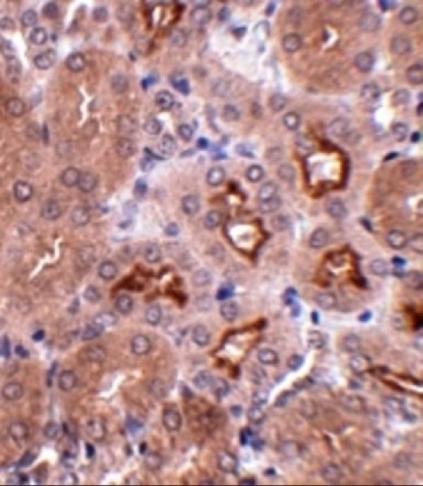
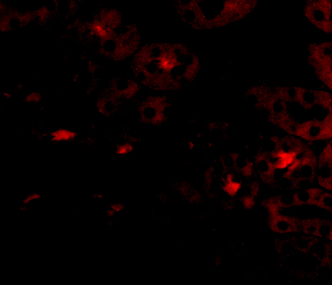
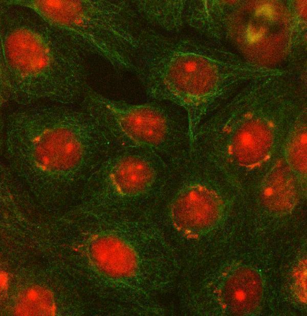


Comprehensive Human Protein Atlas IHC scoring per tissue (reliability: Approved — Medium consistency between antibody staining and RNA expression data. Pending external verification.). Rows are taken directly from the HPA tissue chart — click any row's HPA link to view the source.
| Tissue | Cell type | Level | Evidence | Source |
|---|---|---|---|---|
| Appendix | Glandular cells | Medium | Protein (IHC) | HPA → |
| Breast | Glandular cells | Medium | Protein (IHC) | HPA → |
| Cervix | Glandular cells | Medium | Protein (IHC) | HPA → |
| Colon | Endothelial cells | Medium | Protein (IHC) | HPA → |
| Duodenum | Glandular cells | Medium | Protein (IHC) | HPA → |
| Tissue | Cell type | Level | Evidence | Source |
|---|---|---|---|---|
| Epididymis | Glandular cells | Not detected | Protein (IHC) | HPA → |
| Fallopian tube | Ciliated cells (cell body) | Not detected | Protein (IHC) | HPA → |
| Heart muscle | Cardiomyocytes | Not detected | Protein (IHC) | HPA → |
| Parathyroid gland | Glandular cells | Not detected | Protein (IHC) | HPA → |
| Seminal vesicle | Glandular cells | Not detected | Protein (IHC) | HPA → |
Troubleshoot OTUD5 staining in paraffin sections by checking retrieval, cellular distribution, controls and scoring before interpreting chromogenic signal.
Validated anti-OTUD5 antibodies have IHC data from mouse kidney tissue and IF data from human kidney cells and A431 cells (catalog image captions).
A09744 supports IHC-P and IF, with an IHC image from mouse kidney tissue and an IF image from human kidney cells (catalog applications and image captions). A09744-1 supports ICC/IF, with an IF image from A431 cells (catalog applications and IF image caption).
Which to pick: Choose A09744 for paraffin-section tissue IHC: its application list includes IHC-P, and its own IHC image shows mouse kidney tissue at 2.5 μg/mL; the fixative is unreported (catalog applications; A09744 IHC image caption). Choose A09744-1 for ICC/IF of human cells: its application list includes ICC/IF, and its own image shows A431 cells at 5 μg/mL (catalog applications; A09744-1 IF image caption). For broader listed species reactivity, choose A09744, which lists human, mouse and rat; A09744-1 lists human and rat (catalog reactivity).