This website uses cookies to ensure you get the best experience on our website.


- Table of Contents
Plan PARK7 staining in paraffin sections using cytoplasmic and nuclear tissue staining as the expected pattern (HPA tissue IHC). Compare cases with consistent fixation and include a positive tissue control, such as pancreas with high exocrine glandular cell staining (HPA tissue IHC).

Expected localisation, validated protocols, controls and antibodies — the at-a-glance facts below, then the full design guide.
| Expected localisation | Cytoplasmic and nuclear staining in most tissues (HPA tissue IHC) | |
| Staining pattern | Most tissues show cytoplasmic and nuclear cell staining (HPA tissue IHC) | |
| Antigen retrieval | EDTA pH 8.0 HIER, heat-mediated (datasheet M00757-4) | |
| Positive control | Adrenal gland+4 more · see all | |
| Negative control | None in HPA (detected in all 44 tissues); use no-primary + isotype controls |
| Fixation | Matched tissue-IHC evidence does not establish the fixation claim. Validate the specimen-specific method before use. (selected-SKU IHC image M00757-4) | |
| Caveat | Oxidative stress can shift PARK7 toward mitochondria and nucleus (UniProt) | |
| Regulation | No expression induction is specified (UniProt) | |
| Isoform / epitope | No isoforms or signal peptide are annotated; epitope is unspecified (UniProt) |
The catalog antibody’s IHC-P protocol (datasheet M00757-4) is accompanied by three published PARK7 IHC protocols (PMC6377712; PMC3260266; PMC11538469).
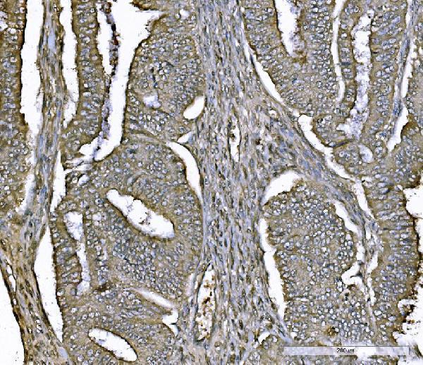
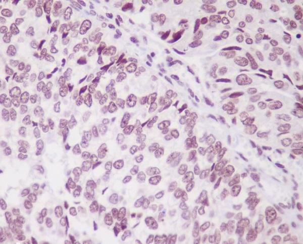
| Sample | Paraffin-embedded human endometrial carcinoma tissue; fixative not specified (datasheet M00757-4) |
| Fixation | Image fixative and duration unreported (datasheet M00757-4); verify before use. |
| Sectioning | 4–5 µm sections on charged slides (standard) |
| Deparaffinisation | Xylene, graded ethanol series to water (standard) |
| Antigen retrieval | Heat retrieval: EDTA pH 8.0 (datasheet M00757-4); 20 min, 95–100 °C (standard) |
| Peroxidase block | 3% H2O2, 10 min, room temperature (standard) |
| Blocking | 10% goat serum (datasheet M00757-4) |
| Primary antibody | Mouse monoclonal (clone 4B10) anti-PARK7, 2-5 µg/ml (datasheet M00757-4) |
| Primary incubation | Overnight at 4 °C (datasheet M00757-4) |
| Detection | HRP-conjugated secondary, DAB chromogen (datasheet M00757-4) |
| Counterstain | Hematoxylin, blue, dehydrate and mount (standard) |
| Expected result | PARK7-positive staining in glandular cells of adrenal gland (HPA tissue IHC: High). HPA tissue profile: Cytoplasmic and nuclear expression in most tissues. No signal in the no-primary control. |
PARK7 staining is expected mainly in the cytoplasm, with nuclear staining in many tissues (HPA: tissue IHC profile; UniProt Q99497: predominantly cytoplasmic under normal conditions). Assess the named cell population within each tissue: HPA reports High staining in several glandular and other cell types, and Low staining in others (HPA: tissue IHC). The tissue IHC profile has Supported reliability, with medium consistency between staining and RNA data (HPA: tissue IHC reliability). PARK7 has no transmembrane segment (UniProt Q99497: topology).
| Cytoplasmic staining, with some nuclear staining, appears in glandular cells or other documented positive cells. | This fits the broad tissue pattern (HPA: cytoplasmic and nuclear expression in most tissues). For a stronger reference, examine an HPA High population such as pancreatic exocrine glandular cells or hippocampal glial cells (HPA: tissue IHC). Compare intensity within the identified cell population, because HPA assigns levels to cells within tissues. |
| Staining appears exclusively at cell borders or in an unexpected isolated compartment, without the expected cytoplasmic signal. | Treat this as a localization discrepancy to investigate, rather than a PARK7 positive result: the routine pattern is mainly cytoplasmic with some nuclear staining (UniProt Q99497: subcellular location; HPA: tissue IHC profile). UniProt lists additional locations, including mitochondria and membrane rafts, but those annotations alone do not validate a dominant cell-border pattern in a particular paraffin section (UniProt Q99497: subcellular location). |
| Strong staining appears chiefly in a cell population reported as Low, while documented High cells show little signal. | Check cell identification and assay specificity. Examples of Low populations include cardiomyocytes and vaginal squamous epithelial cells (HPA: tissue IHC). Unexpected chromogen can also reflect cross-reactivity or endogenous detection activity (general IHC practice). Low is a relative HPA category, not a requirement for complete absence of stain (HPA: tissue IHC levels). |
| Pale, widespread color obscures cell boundaries or appears over tissue and empty slide areas. | Interpret this as background until the staining run is checked (general IHC practice). Compare the negative reagent control, blocking, washes and chromogen development (general IHC practice). A true PARK7 pattern should remain interpretable by cell and compartment: cytoplasmic and nuclear expression is reported across most tissues (HPA: tissue IHC profile). |
| A documented High cell population is unstained. | Consider an unsuccessful staining run or antibody conditions before concluding biological absence (general IHC practice). Repeat alongside a documented positive population, such as breast glandular cells or lung alveolar type I cells (HPA: High tissue IHC). HPA reports no negative reference population in this payload; an unstained section therefore cannot by itself establish a valid PARK7 negative pattern (HPA: tissue IHC list). |
| Tissue and cell selection | Choose a named High population for a positive reference, such as duodenal glands of Brunner or pancreatic exocrine glandular cells (HPA: tissue IHC). Do not treat an entire organ as uniformly high: HPA levels refer to specified cells, and UniProt also reports higher expression in pancreatic islets than surrounding exocrine tissue (UniProt Q99497: tissue specificity). |
| Antibody validation | HPA lists Supported IHC staining for HPA004190 and CAB005870 (HPA: antibody validation). This supports use of their observed tissue patterns as context, while the tissue profile itself has medium consistency with RNA data (HPA: tissue IHC reliability). Validation of those antibodies does not establish the specificity of a different catalog antibody. |
| Localization and biological state | Under normal conditions PARK7 is predominantly cytoplasmic, with lesser nuclear and mitochondrial localization (UniProt Q99497: subcellular location). Oxidative stress can drive mitochondrial and subsequent nuclear translocation (UniProt Q99497: subcellular location, PubMed:18711745); compartment shifts therefore need sample context and cannot be inferred from chromogen intensity alone. |
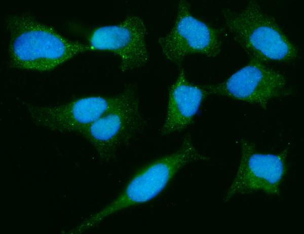
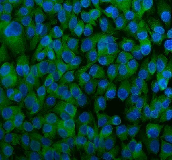
| IF/ICC Q: What pattern should an IF image show? | A: HPA reports nucleoplasm and cytosol as Supported main locations; flagellar centriole and mid piece are additional Approved locations (HPA: subcellular ICC-IF). HPA004190 has Supported ICC validation (HPA: antibodies). These IF observations guide localization comparison; they are not an IHC-P protocol or a guarantee of identical appearance in paraffin sections. |
| Situation | Likely cause | Next action |
|---|---|---|
| No signal in a documented High reference population. | The staining run, antibody dilution or retrieval conditions may need checking (general IHC practice); target-specific fixation sensitivity is unreported in the supplied sources. | Run a known High cell population alongside the sample and review the catalog antibody's IHC-P instructions, including its stated retrieval and dilution, before changing conditions (HPA: tissue IHC levels; general IHC practice). |
| The section has broad haze or deposit that masks compartments. | Excess background may come from blocking, washing or chromogen development conditions (general IHC practice). | Review the negative reagent control; optimize blocking and washes, and shorten development if the control also colors (general IHC practice). Score PARK7 only where cell boundaries and localization remain clear (HPA: tissue IHC profile). |
| Color persists in the negative reagent control. | Endogenous enzyme activity or nonspecific detection reagents can produce chromogen independently of the primary antibody (general IHC practice). | Check the detection chemistry and apply the corresponding endogenous activity control or quench, then repeat the negative reagent control (general IHC practice). |
| Signal is predominantly membranous, with little cytoplasmic staining. | The pattern conflicts with the usual predominant cytoplasmic location (UniProt Q99497: subcellular location); a border artifact or nonspecific stain is possible (general IHC practice). | Compare adjacent cells and the negative reagent control, then verify the pattern with an independently validated IHC antibody if available (general IHC practice; HPA: HPA004190 and CAB005870 IHC Supported). |
| A low-staining cell type looks brighter than the chosen High reference. | Cell identification, cross-reactivity or endogenous detection activity may explain the mismatch (general IHC practice); HPA categories are cell specific (HPA: tissue IHC). | Recheck tissue morphology and controls, then compare like cell populations rather than whole sections (general IHC practice; HPA: tissue IHC levels). |
| Nuclear staining is stronger than expected. | Nuclear PARK7 is documented, and oxidative stress can alter localization (HPA: tissue IHC profile; UniProt Q99497: subcellular location, PubMed:18711745). The image alone does not establish the cause. | Record cytoplasmic and nuclear staining separately, confirm the cell identity and assay controls, and interpret any proposed stress association against independent sample information (HPA: tissue IHC profile; general IHC practice). |


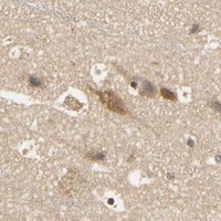


Comprehensive Human Protein Atlas IHC scoring per tissue (reliability: Supported — Medium consistency between antibody staining and RNA expression data.). Rows are taken directly from the HPA tissue chart — click any row's HPA link to view the source.
| Tissue | Cell type | Level | Evidence | Source |
|---|---|---|---|---|
| Adrenal gland | Glandular cells | High | Protein (IHC) | HPA → |
| Breast | Glandular cells | High | Protein (IHC) | HPA → |
| Duodenum | Glands of Brunner | High | Protein (IHC) | HPA → |
| Epididymis | Glandular cells | High | Protein (IHC) | HPA → |
| Fallopian tube | Glandular cells | High | Protein (IHC) | HPA → |
| Tissue | Cell type | Level | Evidence | Source |
|---|---|---|---|---|
| None in HPA: PARK7 is detected in all 44 scored tissues. Use a no-primary (secondary-only) and an isotype control instead. | ||||
Troubleshoot PARK7 staining in paraffin sections by checking retrieval, tissue handling, compartment patterns, controls and scoring before interpreting biological differences.
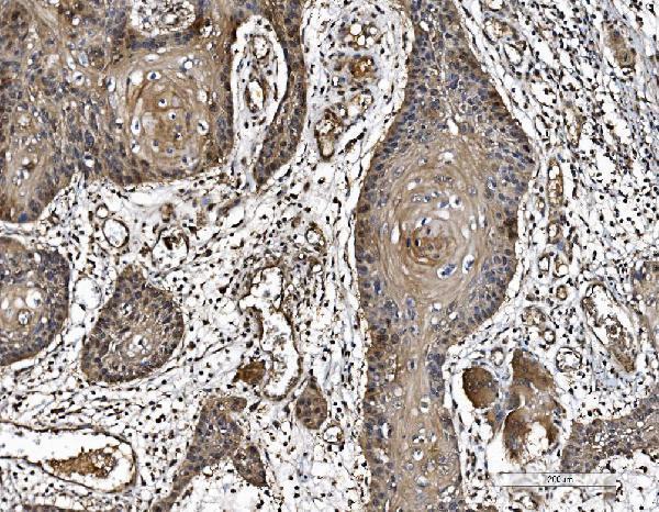
Validated anti-PARK7 antibodies have paraffin-section IHC images from human carcinomas and mouse and rat pancreas, plus IF/ICC images from HeLa and SK-OV-3 cells (catalog IHC/IF captions).
M00757-4 shows paraffin-section IHC in human endometrial carcinoma and IF/ICC in HeLa cells; M00757 shows paraffin-section IHC in human bladder cancer (respective image captions). PB9308 shows IHC in mouse and rat pancreas and human pancreatic cancer, plus IF/ICC in SK-OV-3 cells (PB9308 image captions).
Which to pick: For human tissue IHC, choose mouse monoclonal M00757-4 when its documented paraffin-section workflow is useful: its caption reports EDTA pH 8 retrieval and 2 μg/ml primary antibody (catalog host/clone; M00757-4 IHC caption). For IF/ICC, M00757-4 and rabbit PB9308 each have a cell image and list 5 μg/ml; M00757 lists IF/ICC but has no IF image in the payload (catalog applications, IF dilutions and image captions). For cross-species tissue IHC, PB9308 lists human, mouse and rat reactivity and has an IHC image for each; the fixative is unreported in all three SKUs’ IHC captions (catalog reactivity; respective IHC captions).