PAX6 / Paired box protein Pax-6 · IHC design guide

Design Immunohistochemistry for PAX6

Plan PAX6 IHC in paraffin sections around nuclear staining in retinal ganglion cells and the cerebellar granular layer (HPA tissue IHC). This guide covers fixation, antigen retrieval, antibody dilution, chromogenic detection and cell-level scoring.

Evidence assembled Oct 2026 · For research use; verify linked source records and product datasheet before use
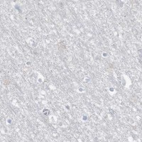
Immunohistochemistry protocol sheet for PAX6 (IHC for PAX6): expected localisation Nuclear in retina and cerebellar granular layer (HPA tissue IHC), antibody PB9768, validated IHC image, and IHC protocol steps
Printable PAX6 IHC protocol sheet — expected localisation Nuclear in retina and cerebellar granular layer (HPA tissue IHC), antibody PB9768, controls and protocol steps. Open the full PAX6 IHC guide →

PAX6 Immunohistochemistry Experimental Design Guide

Expected localisation, validated protocols, controls and antibodies — the at-a-glance facts below, then the full design guide.

Must know before staining
Expected localisation Nuclear in retina and cerebellar granular layer (HPA tissue IHC)
Staining pattern Retinal ganglion and cerebellar granular cell nuclei (HPA tissue IHC)
Antigen retrieval EDTA pH 8.0 HIER, heat-mediated (datasheet PB9768)
Positive control ⓘ Appendix+4 more · see all
Negative control ⓘ Adipose tissue+4 more · see all
Important caveats
Reasons your staining may differ from the expected pattern.
Fixation Keep fixation consistent across paraffin sections. (standard IHC practice; not target-specific)
Caveat Pancreatic endocrine cells show low staining (HPA tissue IHC)
Regulation TRIM11-linked turnover (by similarity) (UniProt)
Isoform / epitope Three isoforms (1, 5a, 3); epitope coverage is unknown (UniProt)
Section 1

Recommended PAX6 IHC & IF Protocols

Compare the catalog antibody’s EDTA pH 8.0 IHC-P protocol (datasheet PB9768) with four published PAX6 IHC protocols (PMC9606524; PMC7695321; PMC2699344; PMC5023747).

Recommended immunohistochemistry (IHC-P) protocol parameters
SampleParaffin-embedded mouse pancreas tissue; fixative not specified (datasheet PB9768)
FixationImage fixative and duration unreported (datasheet PB9768); verify before use.
Sectioning4–5 µm sections on charged slides (standard)
DeparaffinisationXylene, graded ethanol series to water (standard)
Antigen retrievalHeat retrieval: EDTA pH 8.0 (datasheet PB9768); 20 min, 95–100 °C (standard)
Peroxidase block3% H2O2, 10 min, room temperature (standard)
Blocking10% goat serum (datasheet PB9768)
Primary antibodyRabbit anti-PAX6, 0.5-1μg/ml (datasheet PB9768)
Primary incubationOvernight at 4 °C (datasheet PB9768)
DetectionStreptavidin-biotin complex (SABC), DAB chromogen (datasheet PB9768)
CounterstainHematoxylin, blue, dehydrate and mount (standard)
Expected resultPAX6-positive staining in endocrine cells of appendix (HPA tissue IHC: High). HPA tissue profile: Distinct nuclear expression mainly in retina and cerebellar granular layer. No signal in the no-primary control.
💡Decision noteStart with heat-mediated EDTA pH 8.0 for the catalog antibody (datasheet PB9768). Use a paper’s retrieval conditions when reproducing its protocol (PMC9606524; PMC7695321; PMC2699344; PMC5023747).
Section 2

What Is the Expected PAX6 Staining Pattern?

PAX6 is a nuclear transcription factor with no transmembrane segment (UniProt P26367). In paraffin-section IHC, expect nuclear staining chiefly in retinal ganglion cells and cerebellar granular cells, with staining also reported in intestinal endocrine cells (HPA tissue IHC). HPA rates the tissue pattern Enhanced, while reporting medium consistency between antibody staining and RNA expression; interpret individual sections in that context (HPA tissue IHC).

What am I looking at on my slide?
Nuclear chromogen in retinal ganglion cells or the cerebellar granular layer, with limited staining in surrounding cells.This matches the reported PAX6 compartment and prominent tissue pattern (UniProt P26367; HPA tissue IHC). Judge the named cell population and its nuclei; a positive section does not imply that every cell should stain (HPA tissue IHC).
Predominantly cytoplasmic or membrane-like staining, with little nuclear signal.That distribution conflicts with the nuclear localization reported by UniProt and the nucleoplasmic localization seen by HPA ICC-IF (UniProt P26367; HPA subcellular). Treat it as suspect staining and review morphology and detection controls before scoring it as PAX6.
Strong staining in cells listed as undetected, such as adipocytes or bronchial respiratory epithelial cells.HPA reports no detection in those specified cells (HPA tissue IHC). Cross-reactivity or endogenous chromogenic activity is a possible assay explanation, not an established cause; confirm the cell identity and examine a detection-only control (standard IHC practice).
Diffuse chromogen across tissue, including spaces between cells, without a discernible nuclear pattern.A diffuse field cannot support a PAX6-positive call based on its expected nuclear localization (UniProt P26367). General IHC causes include excess background from detection reagents or insufficient blocking or washing; assess those with appropriate controls (standard IHC practice).
No nuclear staining in a section containing identifiable retinal ganglion cells or cerebellar granular cells.Those are reported high-staining populations, so an entirely blank result warrants an assay check (HPA tissue IHC). Verify that the expected cells are present and assess section handling, retrieval and detection controls before interpreting absence as biology (standard IHC practice).
💡Expected PAX6 appearanceCall a result consistent with PAX6 when distinct nuclear staining appears in the expected retinal ganglion or cerebellar granular cells at HPA-reported high levels; diffuse nonnuclear chromogen is suspect (HPA tissue IHC; UniProt P26367).
How each factor affects the staining
Compartment: nucleus without a transmembrane segment (UniProt P26367).Score staining in nuclei. Membrane-like staining lacks support from the supplied localization and topology; nuclear counterstaining helps distinguish chromogen in a nucleus from signal in adjacent cytoplasm (UniProt P26367; standard IHC practice).
Cell distribution varies within and between tissues (HPA tissue IHC).HPA reports high levels in retinal ganglion cells, cerebellar granular cells and several intestinal endocrine populations, but low levels in pancreatic endocrine cells (HPA tissue IHC). Select and read controls by cell population, rather than treating a whole tissue as uniformly positive.
Tissue evidence is Enhanced, with medium antibody-to-RNA consistency (HPA tissue IHC).HPA lists HPA030775 and CAB034143 as Enhanced for IHC; this supports the reported pattern but does not validate every unexpected stained cell (HPA antibodies; HPA tissue IHC). Use localization, morphology and assay controls when interpreting a discordant field.
Three isoforms are listed: 1, 5a and 3 (UniProt P26367).The supplied record gives no antibody epitope or isoform coverage. An IHC result therefore cannot establish which isoform contributed to the stain, and a negative result cannot establish that every isoform is absent (UniProt P26367; supplied antibody evidence).
No signal peptide, propeptide or annotated cleavage is listed (UniProt P26367).The supplied processing record offers no basis to expect a shed or secreted staining pattern (UniProt P26367). It also gives no target-specific fixation or retrieval sensitivity; choose and evaluate those conditions as general IHC workflow variables (standard IHC practice).
Why is my staining missing, weak or wrong?
SituationLikely causeNext action
Expected positive nuclei are blank in retina or cerebellum.The expected cell population may be absent from the section, or the IHC workflow may have failed; HPA reports high staining in the named populations (HPA tissue IHC).Confirm the cells on the counterstained slide, then check a working positive control and the retrieval and detection steps (standard IHC practice). Avoid calling the tissue negative from an unverified blank field.
Staining is mainly cytoplasmic or outlines cell borders.That compartment differs from the reported nuclear and nucleoplasmic locations (UniProt P26367; HPA subcellular). Background or nonspecific antibody binding remains possible (standard IHC practice).Compare chromogen with nuclear counterstain and a detection-only control. Score PAX6 only where cell identity and nuclear localization are clear (UniProt P26367; standard IHC practice).
Many HPA-listed negative cells stain strongly.The result conflicts with the specified negative cell populations; cross-reactivity or endogenous detection activity are possible explanations (HPA tissue IHC; standard IHC practice).Verify morphology and run appropriate detection-only and endogenous-activity controls for the chromogenic system (standard IHC practice). Reassess any residual, cell-specific nuclear staining against HPA's cell-level pattern.
The section has broad, hazy chromogenic background.Background can obscure the nuclear pattern expected for PAX6 (UniProt P26367). In general IHC, blocking, washing or detection-reagent conditions can contribute (standard IHC practice).Inspect control sections and optimize those general workflow conditions; read only discrete nuclear signal after background is controlled (standard IHC practice; UniProt P26367).
Pancreatic endocrine cells look weaker than retinal ganglion cells.HPA lists pancreatic endocrine cells as Low and retinal ganglion cells as High; equal staining is not the expected comparison (HPA tissue IHC).Interpret each identified cell population against its own reported level, with an appropriate positive section in the same run (HPA tissue IHC; standard IHC practice).
Does an IF/ICC image need to match this paraffin-section IHC result?HPA ICC-IF reports mainly nucleoplasmic PAX6, but it is a different application from tissue IHC (HPA subcellular; HPA tissue IHC).Use the IF/ICC guide for that assay. Here, compare only the supported localization expectation; do not infer an IF/ICC protocol or tissue staining level from its images (HPA subcellular).

Sample controls for PAX6 IHC & IF

🧪Run cerebellum first: granular cell nuclei should stain strongly (HPA: High in cerebellar granular cell nuclei; UniProt P26367: nucleus). Use adipose tissue as the negative, focusing on adipocytes (HPA: Not detected in adipocytes); on the cerebellum slide, assess neighboring cells without granular cell morphology as candidate internal negatives rather than assuming every other cell type is negative (HPA: High in granular cells).
Positive control tissue: Appendix (Endocrine cells, HPA High)
Negative control tissue: Adipose tissue (HPA Not detected)
ICC-IF cell lines (HPA subcellular resource): HPA ICC-IF images show PAX6 in HEK293, U-251MG, U2OS, HeLa BAC 5495, HeLa , with annotated localisation: Nucleoplasm (enhanced) (HPA subcellular).
Technical controls: Include a no-primary, secondary-only control and a concentration-matched rabbit IgG isotype control matched to the primary antibody’s clonality (caption: rabbit anti-PAX6); use a validated PAX6 knockout specimen or an immunizing-peptide block as a biological specificity control. For chromogenic detection, quench endogenous peroxidase and check endogenous biotin background because the caption uses a biotinylated secondary and streptavidin–biotin detection (caption: SABC with DAB).
⚠️Feasibility: A target-specific fixation window or fixation effect is unreported, and the selected PB9768 caption does not state a fixative (caption: fixative not stated). The paraffin-section example used heat-mediated EDTA retrieval at pH 8.0, but retrieval dependency is unreported (caption: mouse pancreas IHC). The supplied evidence does not establish whether frozen sections or IF are easier; for cerebellar IHC, assess background from the biotin-based detection system alongside the nuclear signal (caption: SABC with DAB; HPA: cerebellar granular cell nuclei High).

HPA tissue IHC evidence for PAX6

Comprehensive Human Protein Atlas IHC scoring per tissue (reliability: Enhanced — Medium consistency between antibody staining and RNA expression data.). Rows are taken directly from the HPA tissue chart — click any row's HPA link to view the source.

Positive expression · recommended positive controls

TissueCell typeLevelEvidenceSource
Appendix Endocrine cells High Protein (IHC) HPA →
Cerebellum Granular cells - nucleus High Protein (IHC) HPA →
Colon Endocrine cells High Protein (IHC) HPA →
Duodenum Endocrine cells High Protein (IHC) HPA →
Rectum Endocrine cells High Protein (IHC) HPA →

Undetected expression · recommended negative controls

TissueCell typeLevelEvidenceSource
Adipose tissue Adipocytes Not detected Protein (IHC) HPA →
Adrenal gland Glandular cells Not detected Protein (IHC) HPA →
Bone marrow Hematopoietic cells Not detected Protein (IHC) HPA →
Breast Adipocytes Not detected Protein (IHC) HPA →
Bronchus Respiratory epithelial cells Not detected Protein (IHC) HPA →
Section 3

Advanced PAX6 IHC Tips

Troubleshoot PAX6 staining by checking retrieval, nuclear localisation, cell identity and assay controls before comparing signal across sections (UniProt P26367; HPA tissue IHC).

What retrieval should I use when PAX6 nuclear staining is weak?
Start with heat-mediated retrieval in EDTA at pH 8.0 for paraffin sections (datasheet PB9768). The catalog antibody produced a mouse-pancreas IHC image after this retrieval, followed by 1 μg/ml primary antibody overnight at 4°C (datasheet PB9768). If staining remains weak, compare a longer heat exposure or citrate at pH 6.0 on adjacent sections as a troubleshooting trial, while holding detection constant (standard IHC practice). Score nuclear signal in a known positive cell population and monitor tissue damage, because stronger chromogen alone does not establish better PAX6 detection (UniProt P26367 subcellular; standard IHC practice).
How should I troubleshoot PAX6 staining across differently fixed specimens?
Target-specific sensitivity to fixation is unknown from the supplied evidence (datasheet PB9768: fixative not stated). The selected image documents a paraffin-embedded mouse-pancreas section, but its caption does not identify the fixative or fixation duration (datasheet PB9768). Record fixative, time before fixation, fixation duration and section age, then compare adjacent sections with identical EDTA pH 8.0 retrieval and detection conditions (datasheet PB9768; standard IHC practice). If signal varies, use a concurrently processed positive control to separate batch effects from specimen effects, and avoid treating an HPA tissue pattern as evidence for a preferred fixative (HPA tissue IHC; standard IHC practice).
Where should convincing PAX6 staining appear in a tissue section?
Prioritise nuclear chromogen in morphologically intact cells: PAX6 is nuclear, and the HPA subcellular profile places it mainly in the nucleoplasm (UniProt P26367 subcellular; HPA subcellular). The HPA tissue profile describes distinct nuclear expression in retina and the cerebellar granular layer (HPA tissue IHC). Use the cell annotation as well as compartment: HPA reports high staining in cerebellar granular-cell nuclei and retinal ganglion cells, while pancreatic endocrine cells are listed as low (HPA tissue IHC). Diffuse cytoplasmic or extracellular colour should prompt checks of counterstain, chromogen deposition and section quality before it is assigned to PAX6 (UniProt P26367 subcellular; standard IHC practice).
Could PAX6 isoforms explain inconsistent nuclear staining?
PAX6 has 3 listed isoforms, designated 1, 5a and 3 (UniProt P26367 isoforms). The supplied catalog caption does not map the antibody epitope or establish which isoforms it detects, so isoform coverage cannot be inferred from a positive IHC image (datasheet PB9768). PAX6 has no annotated transmembrane segment, signal peptide or glycosylation sites in this record; those annotations do not identify the antibody-binding sequence (UniProt P26367 topology, processing and glycosylation). For discordant specimens, obtain epitope mapping or isoform validation for the antibody and compare nuclear staining under identical retrieval and detection conditions (standard IHC practice).
How can I examine PAX6 by multiplex immunofluorescence?
Treat IF as a separate assay: the supplied PB9768 image documents chromogenic IHC on a paraffin section, without an IF validation result (datasheet PB9768). Pair nuclear PAX6 with a validated marker for the expected cell population, such as retinal ganglion cells or cerebellar granular cells, and include a nuclear counterstain for segmentation (HPA tissue IHC; standard IF practice). Select fluorophores after checking tissue autofluorescence and use single-stain controls to measure channel bleed-through (standard IF practice). Because the expected epitope is on a nuclear protein rather than across a membrane, test a mild permeabilisation condition after fixation and verify retained nuclear morphology (UniProt P26367 subcellular and topology; standard IF practice).
How do I distinguish background from weak PAX6 nuclear signal?
Begin with a no-primary control and inspect whether colour follows tissue edges, damaged regions or widespread cytoplasm rather than nuclei (UniProt P26367 subcellular; standard IHC practice). The PB9768 image used 10% goat-serum blocking, a biotinylated secondary antibody, streptavidin-biotin detection and DAB, so secondary binding and endogenous biotin deserve attention in that workflow (datasheet PB9768; standard IHC practice). Include an endogenous-peroxidase block for DAB detection and compare the no-primary section before increasing primary-antibody concentration (standard IHC practice). Use the reported low staining in pancreatic endocrine cells and the HPA nuclear pattern as context, without requiring every pancreatic cell to be positive (HPA tissue IHC).
What should I score when PAX6 staining varies among sections? ⚠ ANSWER MARKED FOR VERIFICATION
Define the eligible cell population and score nuclei, because the expected PAX6 compartment is nuclear (UniProt P26367 subcellular; HPA subcellular). Report the percentage of positive nuclei or an H-score from 0–300, using fixed intensity thresholds across the comparison (standard IHC scoring practice). For spatial comparisons, positive nuclei per mm² can supplement those measures when the analysed tissue area is explicitly recorded (standard IHC scoring practice). Normalise positive counts to all evaluable nuclei of the same cell type or to its sampled area, and exclude folds, edges and necrotic regions by a prespecified rule (standard IHC scoring practice).
Which staining patterns support a true PAX6-positive result?
A credible result places chromogen in intact nuclei within an expected cell population and exceeds the matched no-primary control (UniProt P26367 subcellular; HPA tissue IHC; standard IHC practice). HPA reports high staining in cerebellar granular-cell nuclei and retinal ganglion cells, but low staining in pancreatic endocrine cells; interpret intensity in its tissue context (HPA tissue IHC). Cytoplasmic colour, edge-restricted staining, necrotic deposits or signal persisting without primary antibody warrants investigation of artefact or endogenous enzyme activity (UniProt P26367 subcellular; standard IHC practice). The mouse-pancreas PB9768 image supports that antibody's stated retrieval and detection workflow, while its unstated fixative limits conclusions about fixation effects (datasheet PB9768).
Boster reagents

Best PAX6 / Paired box protein Pax-6 IHC Antibodies

PAX6 antibodies have IHC images from human brain, human pancreatic cancer, mouse brain and pancreas, and rat brain and pancreas (catalog image captions); IF is illustrated in HeLa cells (A00273-1 image caption).

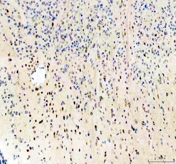
Real IHC data IHC analysis of PAX6 using anti-PAX6 antibody (PB9768). PAX6 was detected in a paraffin-embedded section of mouse pancreas tissue. Heat mediated antigen retrieval was performed in EDTA buffer (pH 8.0, epitope retrieval solution). The tissue section was blocked with 10% goat serum. The tissue section was then incubated with 1 μg/ml rabbit anti-PAX6 Antibody (PB9768) overnight at 4°C. Biotinylated goat anti-rabbit IgG was used as secondary antibody and incubated for 30 minutes at 37°C. The tissue section was developed using Strepavidin-Biotin-Complex (SABC) (Catalog # SA1022) with DAB as the chromogen.
Anti-PAX6 Antibody ®
Cat # PB9768
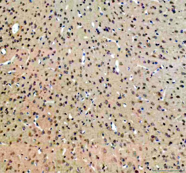
Real IHC data IHC analysis of PAX6 using anti-PAX6 antibody (M00273-3). PAX6 was detected in a paraffin-embedded section of mouse brain tissue. Heat mediated antigen retrieval was performed in EDTA buffer (pH 8.0, epitope retrieval solution). The tissue section was blocked with 10% goat serum. The tissue section was then incubated with 1:50 rabbit anti-PAX6 Antibody (M00273-3) overnight at 4°C. Peroxidase Conjugated Goat Anti-rabbit IgG was used as secondary antibody and incubated for 30 minutes at 37°C. The tissue section was developed using HRP Conjugated Rabbit IgG Super Vision Assay Kit (Catalog # SV0002) with DAB as the chromogen.
Anti-PAX6 Rabbit Monoclonal Antibody
Cat # M00273-3
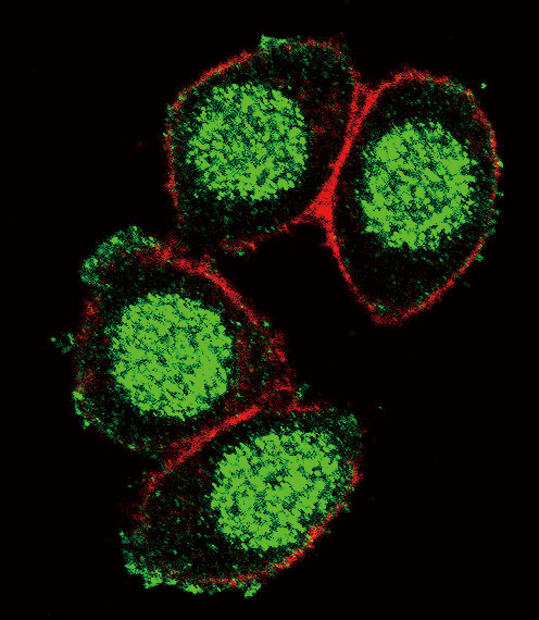
Real IHC data Formalin-fixed and paraffin-embedded human brain tissue with PAX6 Antibody (Center), which was peroxidase-conjugated to the secondary antibody, followed by DAB staining. This data demonstrates the use of this antibody for immunohistochemistry; clinical relevance has not been evaluated.
Anti-PAX6 Antibody (Center)
Cat # A00273-1
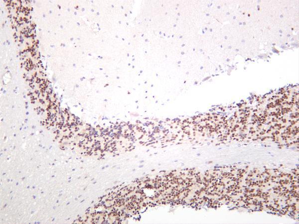
Real IHC data Mouse brain was stained with anti-PAX6 rabbit antibody
Anti-PAX6 Rabbit Monoclonal Antibody
Cat # M00273-5

PB9768 has paraffin-section IHC images from mouse and rat pancreas and human pancreatic cancer (PB9768 image captions); M00273-3 has paraffin-section IHC images from mouse and rat brain (M00273-3 image captions). A00273-1 has an IHC image from formalin-fixed, paraffin-embedded human brain and an IF image from HeLa cells (A00273-1 image captions); M00273-5 has a mouse-brain staining image, with processing unreported (M00273-5 image caption).

Which to pick: For tissue IHC, choose PB9768 for pancreas at 0.5–1 μg/ml (PB9768 datasheet; PB9768 image captions) or M00273-3 for brain at 1:50 (M00273-3 catalog dilution; M00273-3 image captions); both captions show paraffin sections but do not report the fixative (PB9768 and M00273-3 image captions). For IF/ICC, choose M00273-3 for its listed ICC/IF application across human, mouse and rat (M00273-3 catalog), or A00273-1 for its illustrated human-cell IF application (A00273-1 catalog and IF image caption). For cross-species tissue IHC, PB9768 and M00273-3 list human, mouse and rat reactivity (PB9768 and M00273-3 catalogs); M00273-3 is monoclonal (M00273-3 catalog), while PB9768 has no clone specified (PB9768 catalog).

Each figure is that product's own IHC / IF validation image from its datasheet.

References

  1. UniProt Consortium. UniProt entry P26367 (PAX6_HUMAN, Paired box protein Pax-6).
  2. Human Protein Atlas. PAX6 tissue IHC expression (reliability: Enhanced).
  3. Human Protein Atlas. PAX6 subcellular location (ICC-IF): Mainly localized to the nucleoplasm..
  4. Human Protein Atlas. PAX6 antibody validation summary (2 antibodies).
  5. PAX6 is frequently expressed in ependymal tumours and associated with prognostic relevant subgroups. Journal of clinical pathology 2022 — PMC9606524.
  6. Dynamic Enhancement Pattern on CT for Predicting Pancreatic Neuroendocrine Neoplasms with Low PAX6 Expression: A Retrospective Observational Study. Diagnostics (Basel, Switzerland) 2020 — PMC7695321.
  7. Normal ventral telencephalic expression of Pax6 is required for normal development of thalamocortical axons in embryonic mice. Neural development 2009 — PMC2699344.
  8. Analysis of compound heterozygotes reveals that the mouse floxed Pax6 (tm1Ued) allele produces abnormal eye phenotypes. Transgenic research 2016 — PMC5023747.
  9. PubMed PMID:1684738 — UniProt-cited evidence.
  10. PubMed PMID:1345175 — UniProt-cited evidence.
  11. PubMed PMID:17974005 — UniProt-cited evidence.