Click image to see more details
Product Info Summary
| SKU: | A00273-1 |
|---|---|
| Size: | 80 µl |
| Reactive Species: | Human |
| Host: | Rabbit |
| Application: | Flow Cytometry, IF, IHC-P, WB |
Customers Who Bought This Also Bought

Product info
Product Name
Anti-PAX6 Antibody (Center)
SKU/Catalog Number
A00273-1
Size
80 µl
Form
Liquid
Description
Boster Bio Anti-PAX6 Antibody (Center) (Catalog # A00273-1). Tested in WB, Flow Cytometry, IF, IHC-P application(s). This antibody reacts with Human.
Storage & Handling
Maintain refrigerated at 2-8°C for up to 2 weeks. For long-term storage, store at -20°C in small aliquots to prevent freeze-thaw cycles.
Cite This Product
Anti-PAX6 Antibody (Center) (Boster Biological Technology, Pleasanton CA, USA, Catalog # A00273-1)
Host
Rabbit
Contents
Purified polyclonal antibody supplied in PBS with 0.09% (W/V) sodium azide.
Clonality
Polyclonal
Isotype
Rabbit IgG
Immunogen
This PAX6 antibody is generated from rabbits immunized with a KLH conjugated synthetic peptide between 183-210 amino acids from the Central region of human PAX6.
Cross-reactivity
No cross reactivity with other proteins.
Reactive Species
A00273-1 is reactive to PAX6 in Human
Calculated molecular weight
46.7 kDa
Background of PAX6
PAX6 is paired box gene 6, one of many human homologs of the Drosophila melanogaster gene prd. In addition to the hallmark feature of this gene family, a conserved paired box domain, the encoded protein also contains a homeo box domain. Both domains are known to bind DNA, and function as regulators of gene transcription.
Antibody Validation
Boster validates all antibodies on WB, IHC, ICC, Immunofluorescence, and ELISA with known positive control and negative samples to ensure specificity and high affinity, including thorough antibody incubations.
Application & Images
Applications
A00273-1 is guaranteed for Flow Cytometry, IF, IHC-P, WB Boster Guarantee
Recommend Dilution
IF: 1:10-1:50
WB: 1:1000
IHC-P: 1:10-1:50
FC: 1:10-1:50
Validation Images & Assay Conditions
Click image to see more details
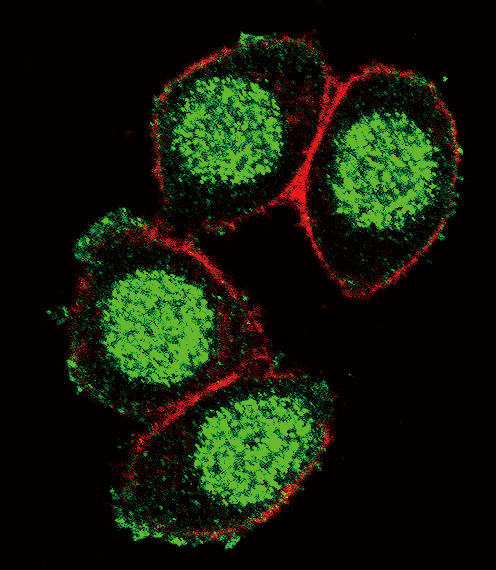
Confocal immunofluorescent analysis of PAX6 Antibody (Center) with Hela cell followed by Alexa Fluor 488-conjugated goat anti-rabbit lgG (green). Actin filaments have been labeled with Alexa Fluor 555 phalloidin (red).

Click image to see more details
PAX6 Antibody (Center) western blot analysis in U251 cell line lysates (35ug/lane).This demonstrates the PAX6 antibody detected the PAX6 protein (arrow).

Click image to see more details
Formalin-fixed and paraffin-embedded human brain tissue with PAX6 Antibody (Center), which was peroxidase-conjugated to the secondary antibody, followed by DAB staining. This data demonstrates the use of this antibody for immunohistochemistry; clinical relevance has not been evaluated.

Click image to see more details
Flow cytometric analysis of hela cells using PAX6 Antibody (Center)(bottom histogram) compared to a negative control cell (top histogram). FITC-conjugated goat-anti-rabbit secondary antibodies were used for the analysis.
Specific Publications For Anti-PAX6 Antibody (Center) (A00273-1)
Loading publications
Recommended Resources
Here are featured tools and databases that you might find useful.
- Boster's Pathways Library
- Protein Databases
- Bioscience Research Protocol Resources
- Data Processing & Analysis Software
- Photo Editing Software
- Scientific Literature Resources
- Research Paper Management Tools
- Molecular Biology Software
- Primer Design Tools
- Bioinformatics Tools
- Phylogenetic Tree Analysis
Customer Reviews
Have you used Anti-PAX6 Antibody (Center)?
Share your experimental results or join a short interview to earn up to $1,000 in product credits or other rewards.
0 Reviews For Anti-PAX6 Antibody (Center)
Customer Q&As
Have a question?
Find answers in Q&As, reviews.
Can't find your answer?
Submit your question
